Brief Report
226 Ann Dermatol
Received January 19, 2018, Revised March 7, 2018, Accepted for publication March 14, 2018
Corresponding author: Hyang-Suk You, Department of Dermatology, Pusan National University Hospital, 179 Gudeok-ro, Seo-gu, Busan 49241, Korea. Tel:
82-51-240-7338, Fax: 82-51-245-9467, E-mail: [email protected] ORCID: https://orcid.org/0000-0002-1697-397X
This is an Open Access article distributed under the terms of the Creative Commons Attribution Non-Commercial License (http://creativecommons.org/
licenses/by-nc/4.0) which permits unrestricted non-commercial use, distribution, and reproduction in any medium, provided the original work is properly cited.
Copyright © The Korean Dermatological Association and The Korean Society for Investigative Dermatology
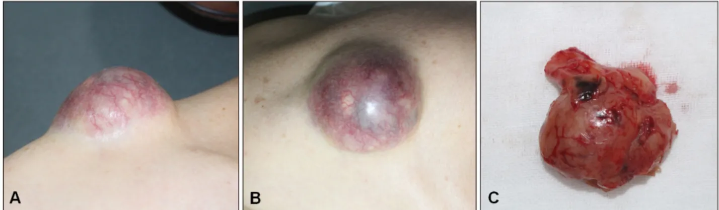
Fig. 1. (A, B) A 5.0×4.3 cm-sized protruding mass on the right shoulder and (C) gross features of the excised tumor.
https://doi.org/10.5021/ad.2019.31.2.226
A Case of Malignant Solitary Fibrous Tumor of the Skin
Kihyuk Shin
1, Tae-Wook Kim
1, Hyun-Joo Lee
1,2, Sung-Min Park
1, Hyunju Jin
1, Woo-Haing Shim
1, Gun-Wook Kim
1,2, Hoon-Soo Kim
1,2, Hyun-Chang Ko
1, Byung-Soo Kim
1,2, Moon-Bum Kim
1,2, Hyang-Suk You
1,21Department of Dermatology, Pusan National University School of Medicine, 2Biomedical Research Institute, Pusan National University Hospital, Busan, Korea
Dear Editor:
Solitary fibrous tumor (SFT) is uncommon fibroblastic mes- enchymal tumor that commonly affects pleura and peri- toneum1. Despite multiple reports of SFT in various or- gans, cutaneous SFT is rare and few case reports have been issued. Furthermore, malignant cutaneous SFT is ex- ceptional2. To our knowledge, this is only the second re- port of malignant SFT of the skin. We received the pa- tient’s consent form about publishing all photographic materials.
A 59-year-old man presented with a protruding mass on his right shoulder, which had grown slowly over 10 years.
The 5.0×4.3 cm-sized tumor was relatively hard and te- langiectasia was observed on its surface (Fig. 1A, B). The
patient did not complain of lesion-associated pain or itch- ing. Other than the skin lesion, a physical examination re- vealed no other abnormality. On ultrasonography, a heter- ogeneous echogenic mass with internal vascularity was observed. An excisional biopsy was performed, and dur- ing excision no connection was observed between the tu- mor and other organs.
Grossly the tumor was well-circumscribed (Fig. 1C), and histopathologic examination revealed an unencapsulated hypercellular tumor composed of a haphazard pattern of proliferating spindle cells embedded in fibrotic stroma (Fig. 2A, B). Cells were characterized by pleomorphism and a mitotic rate of 34 mitotic figures per 10 high-power fields (HPFs) (Fig. 2C). On immunohistochemical exami-
Brief Report
Vol. 31, No. 2, 2019 227 Fig. 2. Histopathologic findings of the tumor mass. (A) The tumor was well circumscribed and unencap- sulated (H&E, ×40). (B) Image show- ing proliferation of spindle cells in a haphazard manner (H&E, ×100).
(C) Photomicrograph showing mi- totic activity and cellular atypia (H&E, ×400). The tumor cells were strongly positive for (D) CD34 (×200) and (E) vimentin (×200), and negative for (F) S100 (×200).
nation, tumor cells were strongly positive for CD34 and vimentin, and negative for cytokeratin, desmin, S100, and smooth muscle actin (Fig. 2D∼F). These clinico-histopath- ologic findings were consistent with malignant SFT of the skin and the patient was transferred to our oncology de- partment for chemotherapy.
Although SFT is a spindle cell tumor which were initially described as pleural tumors, and most involve visceral pleura, though extrapleural involvement has been well de- scribed3. SFT is a type of spindle cell tumor, and cuta- neous SFT is extremely rare. Cutaneous SFT has been de- scribed as a superficial, painless mass often misdiagnosed as a lipoma or epidermal cyst clinically. Histologically, SFT is classified as storiform, herring-bone, hemangioper- icytic, neural-type palisading, or diffuse sclerosing, and the spindle cells of SFTs exhibited “patternless” prolifera- tion with thick collagen bundles arranged in the stroma4. The varied histologic features of SFT can generate a broad histologic differential diagnoses, which include dermatofi- broma, dermatofibrosarcoma protuberans, smooth muscle tumors, spindle cell lipoma, hemangiopericytoma, benign peripheral nerve sheath tumors, melanoma, and cutane- ous myofibroma. Immunohistochemical studies are help- ful because SFTs are typically reactive for CD34 and vi- mentin, but not for cytokeratins, smooth muscle actin, CD31, S100, and CD685.
Most SFTs are clinically benign, but approximately 5%∼10%
show local recurrence and/or metastasis2. Although the histologic criteria for malignant SFTs are controversial, tu- mor size >5 cm, dense cellularity, infiltrative growth, ple- omorphism, mitotic indices >4 per 10 HPFs, and necrosis
are generally considered worrisome. In the current case, the tumor showed all of these features. Histopathologi- cally malignant SFT is extremely rare, and to date, only one report on histopathologically malignant SFT of the skin has been issued2. Prognosis and treatment options for cutaneous malignant SFTs remain uncertain, therefore, fur- ther clinicopathologic studies are required on more cases with longer clinical follow ups.
CONFLICTS OF INTEREST
The authors have nothing to disclose.
ORCID
Kihyuk Shin, https://orcid.org/0000-0001-8955-9828 Tae-Wook Kim, https://orcid.org/0000-0002-8138-7993 Hyun-Joo Lee, https://orcid.org/0000-0002-1696-0976 Sung-Min Park, https://orcid.org/0000-0002-4915-8111 Hyunju Jin, https://orcid.org/0000-0002-0343-1629 Woo-Haing Shim, https://orcid.org/0000-0002-5182-5294 Gun-Wook Kim, https://orcid.org/0000-0003-1599-7045 Hoon-Soo Kim, https://orcid.org/0000-0002-7649-1446 Hyun-Chang Ko, https://orcid.org/0000-0002-3459-5474 Byung-Soo Kim, https://orcid.org/0000-0003-0054-8570 Moon-Bum Kim, https://orcid.org/0000-0003-4837-0214 Hyang-Suk You, https://orcid.org/0000-0002-1697-397X
REFERENCES
1. Mena H, Ribas JL, Pezeshkpour GH, Cowan DN, Parisi JE.
Brief Report
228 Ann Dermatol
Received December 19, 2017, Revised February 13, 2018, Accepted for publication March 21, 2018
Corresponding author: Young Kwan Sung, Department of Immunology, School of Medicine, Kyungpook National University, 680 Gukchaebosang-ro, Jung-gu, Daegu 41944, Korea. Tel: 82-53-420-4874, Fax: 82-53-661-3133, E-mail: [email protected]
ORCID: https://orcid.org/0000-0002-5122-4616
This is an Open Access article distributed under the terms of the Creative Commons Attribution Non-Commercial License (http://creativecommons.org/
licenses/by-nc/4.0) which permits unrestricted non-commercial use, distribution, and reproduction in any medium, provided the original work is properly cited.
Copyright © The Korean Dermatological Association and The Korean Society for Investigative Dermatology Hemangiopericytoma of the central nervous system: a review
of 94 cases. Hum Pathol 1991;22:84-91.
2. Creytens D, Ferdinande L, Van Dorpe J. Histopathologically malignant solitary fibrous tumor of the skin: a report of an unusual case. J Cutan Pathol 2016;43:629-631.
3. Chan JK. Solitary fibrous tumour--everywhere, and a diag- nosis in vogue. Histopathology 1997;31:568-576.
4. Hardisson D, Cuevas-Santos J, Contreras F. Solitary fibrous tumor of the skin. J Am Acad Dermatol 2002;46(2 Suppl Case Reports):S37-S40.
5. Soldano AC, Meehan SA. Cutaneous solitary fibrous tumor:
a report of 2 cases and review of the literature. Am J Der- matopathol 2008;30:54-58.
https://doi.org/10.5021/ad.2019.31.2.228
Impairment of Hair-Inducing Capacity of
Three-Dimensionally Cultured Human Dermal Papilla Cells by the Ablation of STAT5
Chang Hoon Seo, Mi Hee Kwack
1, Moon Kyu Kim
1, Jung Chul Kim
1, Young Kwan Sung
1New Drug Development Center, Daegu-Gyeongbuk Medical Innovation Foundation, 1Department of Immunology, School of Medicine, Kyungpook National University, Daegu, Korea
Dear Editor:
The dermal papilla (DP) of hair follicle is essential for hair morphogenesis, growth, and regeneration. Therefore, DP cells are considered to be an optimal cell source for gen- esis of new hair follicles1,2. However, there remains an ex- perimental challenge resulting from DP cells gradually los- ing their hair-inductive capacity when cultured two-di- mensionally (2D)3. Three-dimensional (3D) spheroid cul- turing was successfully employed to overcome the loss of hair inductivity of 2D-cultured human DP cells4-6.
STAT5 is a signal transducer and activator of transcription (STAT). Recently, Legrand et al.7 reported that the actived form of STAT5 (phospho-STAT5; P-STAT5) is restricted to the DP cells and activation of STAT5 in the DP plays an important role in hair growth phase induction. They show- ed that hair-inductive capacity of mouse DP-derived multi-
potent stem cells, skin-derived precursors, is significantly enhanced by adenoviral overexpression of STAT5A or STAT5B7. In line with this, STAT5 deletion impaired for- mation of de novo hair follicles in skin-derived precur- sors7. In contrast, Harel et al.8 reported that the hair induc- tivity of human DP spheres is enhanced by the treatment with tofacitinib, a STAT signaling inhibitor. These con- troversial reports prompted us to investigate role of STAT5 in the hair inductivity of human DP cells. We performed STAT5 knock-down in human DP spheres and the spheres were implanted into the back of the nude mice together with mouse epidermal cells.
The Medical Ethical Committee of the Kyungpook National University Hospital (Daegu, Korea) approved all of the de- scribed studies (IRB no. KNUH 2013-02-001-007). Student’s t-test was used to analyze differences between groups us-