INTRODUCTION
Solitary fibrous tumor (SFT) is a rare collagen-rich, spin- dle-cell, mesenchymal-origin neoplasm. Klempere and Rabin first reported pleural SFT in 1931. Several cases have been re- ported at pleural and extrapleural sites, including the pericar- dium, peritoneum, lung, liver, upper respiratory tract, medi- astinum, thyroid gland, parotid glands, and central nervous system (CNS). Extrapleural SFT, especially CNS SFT, is very rare. CNS SFT has been reported at the cerebellopontine an- gle, spinal dura, parasagittal region, meninges, and the intra- ventricular region [1-3]. SFT is thought to arise from the fi- broblast and needs to be differentiated from some tumors, like fibrous meningioma and hemangiopericytoma, and from myxoid variants like myxochordoid meningioma and myxoid peripheral nerve sheath tumor [3-6]. Reported herein is a case of CNS SFT in a 63-year-old female patient.
Solitary Fibrous Tumor of Central Nervous System:
A Case Report
Jang Hoon Kim1, Kook Hee Yang1, Pyeong Ho Yoon2, Jeong Hae Kie3
Departments of 1Neurosurgery, 2Radiology, 3Pathology, National Health Insurance Service Ilsan Hospital, Goyang, Korea
Received May 26, 2015 Revised July 28, 2015 Accepted August 11, 2015 Correspondence Kook Hee Yang
Department of Neurosurgery, National Health Insurance Service Ilsan Hospital, 100 Ilsan-ro, Ilsandong-gu, Goyang 10444, Korea
Tel: +82-31-900-0114 Fax: +82-31-900-0049 E-mail: [email protected]
Solitary fibrous tumor (SFT) is a rare neoplasm of mesenchymal origin, especially in the central nervous system (CNS). Reported herein is a case of SFT of CNS in a 63-year-old female patient who had con- fused mentality, without other neurological deficit. The brain MRI showed an ovoid mass in the right frontal lobe. The tumor was surgically removed grossly and totally, and the pathologic diagnosis was SFT. At 55 months after the surgery, the tumor recurred at the primary site and at an adjacent area. A second operation was thus done, and the tumor was again surgically removed grossly and totally. The pathologic diagnosis was the same as the previous, but the Ki-67 index was elevated. Ten months later, two small recurring tumors in the right frontal skull base were found in the follow-up MRI. It was decid- ed that radiation therapy be done, and MRI was done again 3 months later. In the follow-up MRI, the size of the recurring mass was found to have decreased, and the patient did not manifest any significant symptom. Follow-up will again be done 18 months after the second surgery.
Key Words Solitary fibrous tumors; Central nervous system.
CASE REPORT
A 63-year-old female patient visited hospital with headache and intermittent confused mentality for 1 month. She had a history of coronary artery occlusive disease. In the neurologic examination, the patient was found to have had disorienta- tion to time, without other neurological abnormalities. The brain magnetic resonance imaging (MRI) showed an 8.0×
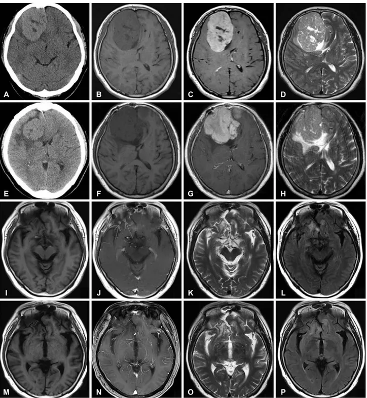
5.2×6.4 cm ovoid mass in the right frontal convexity, with peritumoral edema. The tumor showed intermediate-low sig- nal intensity in the T1-weighted image (T1WI), and slightly increased signal intensity in T2-weighted image (T2WI). The upper and medial portions of the mass showed heterogenous and relatively low signal intensity in T2WI, and suggested a fibrotic mass. The mass showed strong enhancement in the gadolinium-enhanced T1 image. The initial impression was meningioma (Fig. 1A-D).
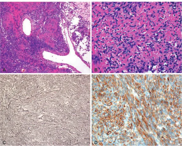
Gross total resection was done. The tumor was a well-en- capsulated and whitish solid, round, and firm mass. In the his- tologic examination, the tumor showed mixed hypercellular and hypocellular areas, with multifocal intervening collagen laid down, and scattered vessels. The tumor cells were short and spindle-shaped and showed diffuse, strong immunoreac-
This is an Open Access article distributed under the terms of the Creative Commons Attribution Non-Commercial License (http://creativecommons.org/licenses/by-nc/3.0) which permits unrestricted non-commercial use, distribution, and reproduction in any medium, provided the original work is properly cited.
Copyright © 2015 The Korean Brain Tumor Society, The Korean Society for Neuro- Oncology, and The Korean Society for Pediatric Neuro-Oncology
tivity for CD34. The mitosis was less than 1/10 high power field (HPF), with an about 1.60% Ki-67 labelling index, and there was no evidence of necrosis. Sparse reticulin fibers were
observed among the tumor cells on special stain. With these results, hemangiopericytoma was ruled out, and a diagnosis of SFT was made (Fig. 2A-D).
A
E
I
M
B
F
J
N
C
G
K
O
D
H
L
P
Fig. 1. Preoperative image shows about 8.0×5.2×6.4 cm3 sized ovoid mass in the right frontal convexity. A: CT shows iso to high density with parenchyma. B: T1WI shows intermediate-low signal intensity. C: T2WI shows intermediate-low signal intensity and in upper and medial por- tion of this mass, there are lesions with relatively low signal intensity. D: Gd-enhanced T1WI shows relatively strong enhancement. Postop- erative 55 months MRI shows recurrence of tumor. E: CT shows isodensity mass with around low density with parenchyma. F: T1WI shows intermediate-low signal intensity. G: T2WI shows intermediate-high signal intensity. H: Gd-enhanced T1WI shows relatively strong enhance- ment. After 10 months second surgery, MRI shows a few small recurred nodule in right frontal base. I: T1WI. J: Gd-enhanced T1WI shows small nodule (arrows) in right frontal base. K: T2WI. L: FLAIR. After 3 months IMRT, MRI shows decreased size of nodule. M: T1WI. N: Gd- enhanced T1WI. O: T2WI. P: FLAIR. T1WI, T1-weighted image; T2WI, T2-weighted image; FLAIR, fluid attenuated inversion recovery; IMRT, intensity-modulated radiation therapy.
After the surgery, the patient was symptom-free, and the MRI showed no recurrence at 6 months, 1 year, and 36 months after the surgery. During the follow-up visit, at 55 months af- ter the surgery, the patient revisited the hospital due to con- fusion for a few weeks. Brain MRI was again conducted, and the tumor was shown to have recurred at the primary site and at an adjacent area (Fig. 1E-H). Reoperation was thus done, and the tumor was grossly and totally removed. The patholog- ic findings were relatively the same as the previous ones, but the tumor was more cellular, with increased mitosis (about 2/10 HPF) and Ki-67 labelling index (12.85%). There was no evidence of necrosis. Still, the tumor cells showed diffuse strong immunoreactivity for CD34, and sparse reticulin fiber (Fig. 2E-H). After the operation, the patient recovered and be- came symptom-free again. Brain MRI was again conducted 10 months later; three small (5×5×5 mm) recurring nodules were found in the right frontal base, and the patient did not show any symptom (Fig. 1I-L). As the affected area was too small, it was difficult to perform surgery again therein; as such, radiation therapy was planned. The radiation dose totaled 5,940 cGy and was fractionated 33 times. Three months after
the radiation therapy, follow-up brain MRI was done, and it was found that the nodules in the right frontal base had be- come smaller (Fig. 1M-P). Outpatient follow-up brain MRI will again be conducted.
DISCUSSION
SFT is an uncommon tumor that mostly occurs in the vis- ceral pleura. SFT has been reported to occur outside the tho- racic cavity, including the pericardium, peritoneum, lung, liv- er, upper respiratory tract, mediastinum, thyroid glands, and parotid glands. It has also been reported to occur in the CNS, including the cerebellopontine angle, spinal dura, parasagit- tal region, meninges, and the intraventricular region. It is th- ought that SFT is a mesenchymal-origin tumor and should thus be differentiated from fibrous meningioma, fibrosarcoma, meningeal sarcoma, meningeal myofibroblastoma, schwanno- ma, neurofibroma, and gastrointestinal stromal tumor [1-4].
Patients may present several non-specific symptoms related with increased intracranial pressure or with the location of the tumor, such as headache, dizziness, gait disturbance, hemipa-
Fig. 2. Immunohistochemical features of first operation. A: On histologic examination of lower power view, mixed hypercellular and hypocellu- lar area are seen with multifocally laid down collagen and occasional staghorn-like vessels (H&E, ×100). B: On high power field, relatively homogenous short spindle tumor cells in intervening collagens are seen (H&E, ×400). C: Sparse reticulin fibers are observed among tumor cells contrary to hemangiopericytoma (reticulin stain, ×200). D: The tumor cells show diffuse strong reactivity for CD34 on immunohistochem- ical stain (CD34, ×400). H&E, hematoxylin and eosin.
A
C
B
D
resis or hemiplegia, hearing loss, and mental change [7].
Immunohistochemically, although it is still being debated on, SFT has fibroblastic or myofibroblastic differentiation, which means that it has a mesenchymal fibroblast origin rath- er than a meningothelial origin. In an immunochemical study, CD34 was found to be strongly expressed in most SFT cases;
Bcl-2 in 80–100% of SFT cases; and CD99 in 75–100% of SFT cases. Generally, its diagnosis is confirmed through positive staining for CD34 and negative staining for S-100 [2,3,8,9], as distinguished from that of meningioma (usually negative for CD34 and S-100) and neurinoma (usually negative for CD34 and positive for S-100), and it should have a differential diag- nosis with hemangiopericytoma. Most hemangiopericytoma cases, in contrast to SFT cases, are hypercellular and are oval to round rather than spindle-shaped, and immunohistochemi- cally, they are reactive with CD34 and Bcl-2 only in a few cases [4-6].
Radiologically, it is difficult to differentially diagnose SFT through image study. SFT has variable signal intensities in MRI. T1WI and T2WI show hypointensity and/or hyperinten- sity with enhancement diffusely or heterogeneously [1,7,8,10].
The authors’ first impression was meningioma as the mass was seen as a dural-based enhanced mass with heterogenous and relatively low signal intensity in T2WI, and a fibrotic mass was suggested, but the final diagnosis was changed to SFT. As such, there is a need to consider a differential-diagnosis fibrotic mass, especially meningioma, hemanigompericytoma, and SFT.
The treatment of choice is complete resection. Even though SFT is considered benign, it should be followed up regularly due to a possible recurrence [4,9,11]. Due to the rarity of the tumor, its adjuvant therapy and prognosis have not been well studied, and the indications of adjuvant therapy are uncer- tain [2,12]. Adjuvant radiotherapy was not done in the case re- ported herein, but several reports recommend it [13]. As such, close follow-up without radiation therapy was decided. As the tumor recurred at the primary site, however, radiation thera- py was eventually done.
In conclusion, SFT is a rare mesenchymal-origin neoplasm especially occurring in the CNS. Image study and immuno- histochemistry are crucial for its correct diagnosis. Surgery is the treatment of choice, and some reports recommend adju- vant radiotherapy. SFT is generally considered benign, but re- Fig. 2. Immunohistochemical features of second operation. E: On histologic examination of recurred mass, the tumor is also composed of short spindle cells but it shows more homogenous and cellular features compared to original tumor mass (H&E, ×200). F: The recurred tu- mor shows increased Ki-67 labelling index compared to original tumor (not shown) (Ki-67, ×200). G: Still sparse reticulin fibers are observed among tumor cells contrary to hemangiopericytoma (reticulin stain, ×200). H: The tumor cells also show diffuse strong reactivity for CD34 on immunohistochemical stain (CD34, ×400). H&E, hematoxylin and eosin.
E
G
F
H
currences have been reported. Thus, long-term follow-up is necessary.
Conflicts of Interest
The authors have no financial conflicts of interest.
REFERENCES
1. Musyoki FN, Nahal A, Powell TI. Solitary fibrous tumor: an update on the spectrum of extrapleural manifestations. Skeletal Radiol 2012;41:5-13.
2. Hu SW, Tsai KB, Yang SF, Lee KS, Chai CY. Unusual solitary fibrous tumors in the central nervous system: a report of two cases. Kaohsiung J Med Sci 2005;21:179-84.
3. Brunori A, Cerasoli S, Donati R, Giangaspero F, Chiappetta F. Solitary fibrous tumor of the meninges: two new cases and review of the litera- ture. Surg Neurol 1999;51:636-40.
4. Bisceglia M, Dimitri L, Giannatempo G, et al. Solitary fibrous tumor of the central nervous system: report of an additional 5 cases with com- prehensive literature review. Int J Surg Pathol 2011;19:476-86.
5. Ambrosini-Spaltro A, Eusebi V. Meningeal hemangiopericytomas and hemangiopericytoma/solitary fibrous tumors of extracranial soft tissues:
a comparison. Virchows Arch 2010;456:343-54.
6. Park MS, Araujo DM. New insights into the hemangiopericytoma/soli- tary fibrous tumor spectrum of tumors. Curr Opin Oncol 2009;21:327-31.
7. Wang XQ, Zhou Q, Li ST, Liao CL, Zhang H, Zhang BY. Solitary fibrous tumors of the central nervous system: clinical features and imaging findings in 22 patients. J Comput Assist Tomogr 2013;37:658-65.
8. Weon YC, Kim EY, Kim HJ, Byun HS, Park K, Kim JH. Intracranial solitary fibrous tumors: imaging findings in 6 consecutive patients.
AJNR Am J Neuroradiol 2007;28:1466-9.
9. Caroli E, Salvati M, Orlando ER, Lenzi J, Santoro A, Giangaspero F. Soli- tary fibrous tumors of the meninges: report of four cases and literature review. Neurosurg Rev 2004;27:246-51.
10. Ginat DT, Bokhari A, Bhatt S, Dogra V. Imaging features of solitary fi- brous tumors. AJR Am J Roentgenol 2011;196:487-95.
11. Fargen KM, Opalach KJ, Wakefield D, Jacob RP, Yachnis AT, Lister JR.
The central nervous system solitary fibrous tumor: a review of clinical, imaging and pathologic findings among all reported cases from 1996 to 2010. Clin Neurol Neurosurg 2011;113:703-10.
12. Münks S. [Solitary fibrous tumor (SFT) of the nasal mucosa]. Laryn- gorhinootologie 2003;82:655-8.
13. Macfarlane RG, Galloway M, Plowman PN, Thomas DG. A highly vas- cular intracranial solitary fibrous tumor treated with radiotherapy and toremifene: case report. Neurosurgery 2005;56:E1378.