57
◇ 증 례 ◇
접수:2009년 2월 3일, 승인:2009년 2월 27일 책임저자:이경훈, 705-718, 대구시 남구 대명4동 3056-6
대구가톨릭대학교 의과대학 소아청소년과학교실 Tel: 053-650-4246, Fax: 053-622-4240
E-mail: [email protected]
위 내 이물로 오인한 석회화 섬유 종양 1예
대구가톨릭대학교 의과대학 소아과학교실, *병리과학교실 정지은ㆍ이경훈ㆍ성현정*ㆍ조창호*
Calcifying Fibrous Tumor Mimicking a Foreign Body of the Stomach: A Case Report
Ji-Eun Jeong, M.D., Kyung-Hun Lee, M.D., Hyun Jung Sung, M.D.* and Chang-Ho Cho, M.D.*
Departments of Pediatrics and *Pathology, School of Medicine, Catholic University of Daegu, Daegu, Korea Calcifying fibrous tumors (CFTs) are unusual benign tumors of childhood, located primarily in soft tissues, pleura, and peritoneum. The cause and pathogenesis are unclear. We report a rare case of a CFT in a 2-year-old boy who presented with vomiting and abdominal distension. An abdominal X-ray showed an elliptical, calcific shadow in the LUQ area mimicking a foreign body. An internally protruding mass along the lesser curvature of the gastric body was an incidental finding during upper endoscopy, biopsies of which were negative. Abdominal CT showed a 4.5×3.2 cm soft tissue mass of the gastric wall with calcifications. A diagnosis of gastric submucosal mass was suspected and a wedge resection of the stomach was performed. On microscopic examination, the tumor was composed of whorls of dense hyalinized collagen bundles with a few fibroblasts. There were also amorphous dystrophic calcifications and nodular aggregates of mononuclear inflammatory cells. Immunohistochemically, spindle cells did not stain for anaplastic lymphoma kinase-1 (ALK-1), CK, smooth muscle actin (SMA), or desmin. Taken together, the mass was compatible with a CFT of the gastric wall. This is the first reported case of CFT in a Korean child. (Korean J Pediatr Gastroenterol Nutr 2009; 12: 57∼63)
Key Words: Calcifying fibrous tumor, Gastric wall, Calcifications, Child
서 론
석회화 섬유 종양(calcifying fibrous tumor, CFT)은 주
로 소아와 젊은 성인에서 발생하고 아직까지 발생 기전 이 밝혀지지 않은 대단히 드문 양성 질환이다1). 주로 피하와 심부 연 조직 또는 흉막과 복막과 같은 장막하 조직에 국한된 결절 형태를 보인다2,3).
1988년에 소아에서 각각 허벅지와 전박에 모래종체 (psammoma body)를 가진 섬유 종양 2예가 처음 기술되 었고3), 이후 Fetsch 등1)에 의하여 명확하게 밝혀지게 되 었다. 현재까지 국내에서 위벽 내에서의 석회화 섬유 종양에 대한 보고는 없었다.
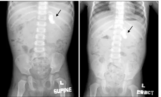
Fig. 1. A plain abdominal X-ray reveals an elliptical cal- cifications (arrow) in the LUQ area.
증 례
환 아: 임○○, 2세, 남아
주 소: 내원 3일 전부터 시작한 구토와 복부 팽만 현병력: 최근 환아의 복부가 불러 보이던 중 내원 3일 전부터 간헐적인 구토가 있어 외래를 방문하였다.
과거력: 38주 4일, 2.83 kg으로 건강하게 출생하였으 며 생후 12일경 신생아 황달로 입원하여 광선 치료를 받았으며, 내원 2개월 전 마이코플라즈마 폐렴으로 입 원하여 치료받았다.
가족력: 특이 사항은 없었다.
진찰 소견: 내원 당시 체중은 13.5 kg (50∼75 백분위 수), 키는 90 cm (75∼90 백분위수)였다. 활력 징후는 혈압 94/55 mmHg, 맥박수 120회/분, 호흡수 26회/분, 체 온 36.5oC였고, 의식은 명료하였다. 환아는 전반적으로 양호해 보였으며 두경부 및 흉부 진찰에서 특이 소견은 보이지 않았다. 복부는 부드러웠고 약간 팽만된 소견을 보였으며 장음은 정상 소견이었다. 간과 비장은 만져지
mEq/L, Cl 101 mEq/L, 생화학 검사에서 BUN 8.0 mg/dL, creatinine 0.6 mg/dL, AST 27 IU/L, ALT 33 IU/L, 총 단백 7.3 g/dL, 알부민 4.3 g/dL였다. 적혈구 침 강 속도는 7 mm/hr, C-반응 단백 2.4 mg/L였다.
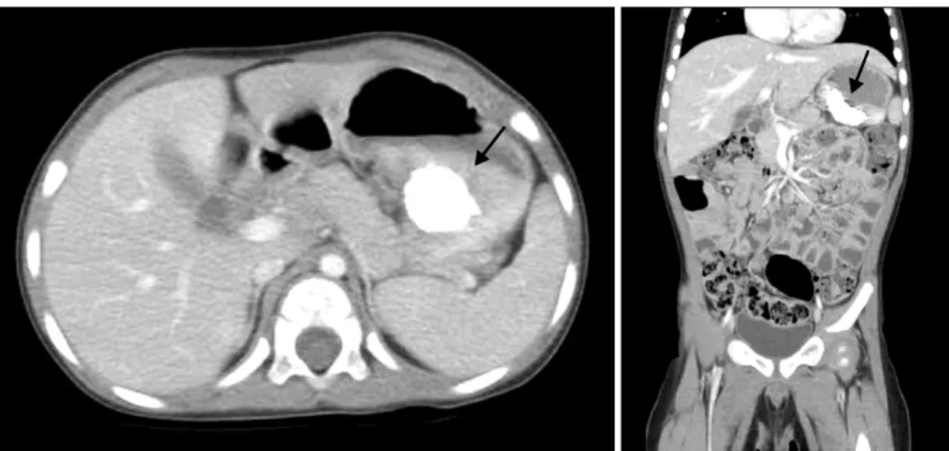
방사선 소견: 복부 방사선 사진에서 좌상부에 단일 타원형의 석회화가 관찰되었다(Fig. 1). 복부 전산화 단 층 촬영에서 위 체부에 4.5×3.2 cm 크기의 내부 석회화 를 보이는 연 조직 종괴 관찰되고, 다수의 장간막 림프 절 들이 보였다(Fig. 2).
내시경 소견: 식도에서는 특이 소견 관찰되지 않았 으며, 위 내부에 소량의 커피 양상의 액체가 관찰되었 다. 위 체부의 소만부에서 표면에 혈행성이 발달한 4 cm 가량의 단일 종괴가 관찰되었다(Fig. 3). 점막의 궤 양 소견은 보이지 않았다.
수술 소견: 상복부 가로 절개를 5 cm 시행 후, 위 체 부 후벽 부위에서 5×4 cm 크기의 단단한 종괴를 쐐기 절제술을 시행하였다.
병리조직학 소견: 육안 검사에서 덩어리는 3.5×2×2 cm 크기로 잘 경계가 구분 되었지만 피막화되어 있지
Fig. 2. Abdominal computed tomography (CT) shows a 4.5×3.2 cm sized soft tissue mass (arrow) of the gastric wall with calcifications.
Fig. 3. Upper endoscopy shows a protruding mass covered by mucosa with well developed vasculature along the lesser curvature of the gastric body.
Fig. 4. The surgical specimen reveals a 3.5×2×2 cm well demarcated but unencapsulated white firm gritty tumor.
는 않았다. 잘랐을 때 표면은 전체적으로는 회색빛을 띠는 흰색이었고 군데군데 모래와 같은 양상을 보였으 며 불규칙적인 석회화가 관찰되었다(Fig. 4). 현미경적
으로 수많은 형성 장애 석회화를 가진 전반적이고 광범 위한 석회화가 있었다(Fig. 5). 림프구와 형질 세포로 이루어진 다양한 염증세포의 침윤과 저세포성, 유리질 화, 섬유 경화를 보였다(Fig. 6, 7). Streptavidin-biotiny- lated peroxydase 방법을 이용하여 조직 표본에 면역 조 직학적 검사를 시행했다. CD34, 데스민, 평활근 액틴, anaplastic lymphoma kinase (ALK-1)에 대한 항체를 이 용한 면역조직학적 염색에 양성 반응을 보이지 않았다 (Fig. 8). 최종적으로 위 석회화 섬유 종양으로 진단하 였다.
치료 및 경과: 수술 후 음식물 경구 섭취에 별다른 이상 없고 체중 증가가 잘 이루어지고 있다.
고 찰
석회화 섬유 종양은 이전에는 가짜종양(pseudotumor)
Fig. 7. Scattered throughout the tumors were singly and aggregated lymphocytes in the germinal center (A) (H&E, ×200). Diffuse but sparse plasma cells infiltrates (B) (H&E, ×400).
Fig. 6. Histologically, it shows proliferating spindle shaped fibroblasts in a densely hyalinized collagenous matrix (H&E,
×100).
Fig. 5. Histologically, it shows scattered amorphous dystro- phic calcifications of variable size and shape with dense collageneous stroma after the tissue was decalcified (H&E,
×40).
mesothelium) 세포로부터의 기원이 제시되고 있다 . 현재까지 세계적으로 보고된 70여 개의 증례 중에 단 지 12개가 위장관에 발생한 경우인데, 이 경우 주로 장 간막, 그물막, 위장관의 장막하에 발생한다. 환아의 경
동, 후복막강, 부신, 정삭에 발생한 4예의 보고가 있다 . 발생하는 위치로는 흉부, 복강, 사지, 골반을 포함한 다양한 해부학적인 위치에서 발생할 수 있다. 발생 형 태로 주로 피하와 심부 연 조직 또는 흉막과 복막과 같
Fig. 8. Immunohistochemically, CFT spindle cells did not stain for ALK, CK, SMA and Desmin (×200, ×400).
은 장막하 조직에 국한된 결절을 보인다. 종격동, 흉막, 장간막에 다발성 병변 보고가 있지만 발생하는 위치에 따른 차이는 없다11).
소화기 증상으로 내시경을 시행하거나 다른 목적으 로 시행한 수술 중 우연히 발견되는 경우가 많으나2,5) 드물게 복막염, 장중첩증으로 인한 급성 복통으로 인하 여 발견되는 경우도 있다8,12). 증상의 유무는 종괴의 위 치와 크기에 따라 달라질 수 있다5). 위 벽에 발생된 성 인 증례의 경우 위식도 역류로 인한 장기간의 심와부 통증을 호소한 경우와13), 체중 감소 및 심와부 통증을 호소한 경우가 있었다6). 위 내 종괴의 크기가 클수록 증상의 발현이 높아, 본 증례의 경우는 종괴의 크기로 인하여 복부 팽만과 구토가 발생되었다.
임상적으로 특별한 진단 방법이 있지는 않으나, 일반 방사선 사진과 전산화 단층 사진에서 띠 혹은 구두점
모양의 석회화를 보인다14). 위 벽에 위치한 경우 소화 기 증상을 호소하는 경우가 많아 환아처럼 내시경시 종 괴가 발견되는 경우도 있다13). 초음파 내시경 시행시 중심부에 뚜렷한 저음영을 보이는 점막하 병변을 보인 다13). 환아의 경우 초음파 내시경을 하기에는 나이가 어려 내시경 직경에 따른 부담으로 시행을 하지 못하였 다. 자기 공명 영상장치에서 병변은 T1에는 동신호 (isosignal), T2에는 저신호(hyposignal)로 나타난다13). 따라서 이 질환은 병리학적으로 진단하는 것이 가장 중요하다. 육안적으로 잘 국한된 0.6∼25 cm 크기의 형 성 장애 또는 모래종체 석회화를 가진 비피막화된 덩어 리를 보인다. 현미경적으로 림프구와 형질 세포로 이루 어진 다양한 염증세포의 침윤과 저세포성, 유리질화, 섬유 경화를 보인다. 환아의 경우도 현미경적으로 동일 한 양상을 보였다. 면역 조직학적으로 vimentin과 factor
다르다. 위장관에 발생한 경우 감별 할 것으로 인대 모 양 종양(desmoids tumor)과 위장관 간질 종양(gastro- intestinal stromal tumor, GIST)과 같은 다른 방추 세포 종양과 조직학적으로 잘 구분하여야 한다15). 인대 모양 종양은 모래종체 석회화가 보이지 않고, 면역조직학적 염색에서 전반적으로 베타 카테닌에 양성 반응을 보인 다. 또한 위장관 간질 종양은 일반적으로 석회화 섬유 종양에서 보이는 저세포성, 층판 콜라겐 다발, 만성 염 증, 모래종체 등이 보이지 않는다. 그리고 면역조직학 적 방법으로 CD117이나 혈소판 유래 성장 인자 수용체 를 이용하여 감별 할 수 있다2).
그 외 염증 근섬유모세포 종양, 단독 섬유 종양(soli- tary fibrous tumor), 반응 결절 섬유 가짜 종양(reactive nodular fibrous pseudotumor)도 감별 해야 한다. 석회화 섬유 종양과는 달리 염증 근섬유모세포 종양은 ALK-1, CD34와 액틴에 양성 반응을 보이고 석회화를 거의 포 함하지 않는다16). Van Dorpe 등17)은 석회화 섬유 종양 과 염증 근섬유모세포 종양(inflammatory myofibro- blastic tumor)은 별개의 질환이 아니고, 석회화 섬유 종 양의 조직 발생은 염증 근섬유모세포 종양의 경화성 말 기 단계를 나타난다고 가설하였지만, 두 질환 사이의 뚜렷한 차이로 지지를 얻지 못하였다2,16). 단독 섬유 종 양은 석회화 섬유 종양 보다 고세포 지역을 보이고, CD99와 bcl-2에 양성 반응을 보인다.
치료로 병변의 충분한 제거가 필요하다. 위에서 발생 한 경우 크기에 따라 환아의 경우처럼 쐐기 절제술을 시행하면 된다6). 문헌에 따르면 17∼30%의 국소 재발 보고도 있지만1,2,18,19) 일단 충분히 제거되면 예후는 좋 은 것으로 알려져 있다.
드물기는 하지만 위 중간엽 종괴의 감별 진단으로서 석회화 섬유 종양을 고려하여야 한다.
내시경과 복부 전산화 단층 사진을 시행하였다. 술 후 병리 검사에서 상당히 빈도가 드문 석회화 섬유 종양을 발견하였기에 보고하는 바이다.
참 고 문 헌
1) Fetsch JF, Montgomery EA, Meis JM. Calcifying fibrous pseudotumor. Am J Surg Pathol 1993;17:502-8.
2) Nascimento AF, Ruiz R, Hornick JL, Fletcher CD.
Calcifying fibrous ‘pseudotumor’: clinicopathologic study of 15 cases and analysis of its relationship to inflam- matory myofibroblastic tumor. Int J Surg Pathol 2002;
10:189-96.
3) Kocova L, Michal M, Sulc M, Zamecnik M. Calcifying fibrous pseudotumour of visceral peritoneum. Histo- pathology 1997;31:182-4.
4) Rosenthal NS, Abdul-Karim FW. Childhood fibrous tumor with psammoma bodies. Clinicopathologic features in two cases. Arch Pathol Lab Med 1988;112:798-800.
5) Elpek GO, Kupesiz GY, Ogus M. Incidental calcifying fibrous tumor of the stomach presenting as a polyp.
Pathol Int 2006;56:227-31.
6) Puccio F, Solazzo M, Marciano P, Benzi F. Laparoscopic resection of calcifying fibrous pseudotumor of the gastric wall. A unique case report. Surg Endosc 2001;15:1227.
7) Chatelain D, Lauzanne P, Yzet T, Guernou M, Del- censerie R, Regimbeau JM, et al. Gastric calcifying fibrous pseudotumor, a rare mesenchymal tumor of the stomach. Gastroenterol Clin Biol 2008;32:441-4.
8) Liang HH, Chai CY, Lin YH, Lee CH, Wu CH, Chang CC. Jejunal and multiple mesenteric calcifying fibrous pseudotumor induced jejunojejunal intussusception. J Formos Med Assoc 2007;106:485-9.
9) Chen KT. Familial peritoneal multifocal calcifying fibrous tumor. Am J Clin Pathol 2003;119:811-5.
10) Jeong HS, Lee GK, Sung R, Ahn JH, Song HG.
Calcifying fibrous pseudotumor of mediastinum-a case report. J Korean Med Sci 1997;12:58-62.
11) Dumont P, de Muret A, Skrobala D, Robin P, Toumieux B. Calcifying fibrous pseudotumor of the mediastinum.
Ann Thorac Surg 1997;63:543-4.
12) Ben-Izhak O, Itin L, Feuchtwanger Z, Lifschitz-Mercer B, Czernobilsky B. Calcifying fibrous pseudotumor of mesentery presenting with acute peritonitis: case report with immunohistochemical study and review of literature.
Int J Surg Pathol 2001;9:249-53.
13) Delbecque K, Legrand M, Boniver J, Lauwers GY, de Leval L. Calcifying fibrous tumour of the gastric wall.
Histopathology 2004;44:399-400.
14) Erasmus JJ, McAdams HP, Patz EF Jr, Murray JG, Pinkard NB. Calcifying fibrous pseudotumor of pleura:
radiologic features in three cases. J Comput Assist Tomogr 1996;20:763-5.
15) Lee YS, Sen BK. Dystrophic and psammomatous calcifications in a desmoid tumor. A light microscopic
and ultrastructural study. Cancer 1985;55:84-90.
16) Hill KA, Gonzalez-Crussi F, Chou PM. Calcifying fibrous pseudotumor versus inflammatory myofibroblastic tumor:
a histological and immunohistochemical comparison. Mod Pathol 2001;14:784-90.
17) Van Dorpe J, Ectors N, Geboes K, D’Hoore A, Sciot R.
Is calcifying fibrous pseudotumor a late sclerosing stage of inflammatory myofibroblastic tumor. Am J Surg Pathol 1999;23:329-35.
18) Mito K, Kashima K, Daa T, Kondoh Y, Miura T, Kawahara K, et al. Multiple calcifying fibrous tumors of the pleura. Virchows Arch 2005;446:78-81.
19) Maeda A, Kawabata K, Kusuzaki K. Rapid recurrence of calcifying fibrous pseudotumor (a case report). Anticancer Res 2002;22:1795-7.