Copyright © 2012, the Korean Surgical Society J Korean Surg Soc 2012;83:56-59
http://dx.doi.org/10.4174/jkss.2012.83.1.56
CASE REPORT
Journal of the Korean Surgical Society
JKSS
pISSN 2233-7903ㆍeISSN 2093-0488
Received January 9, 2012, Reviewed January 19, 2012, Accepted February 2, 2012 Correspondence to: Ho Sung Park
Department of Pathology, Chonbuk National University Medical School, 567 Baekje-daero, Deokjin-gu, Jeonju 561-756, Korea Tel: +82-63-270-3073, Fax: +82-63-270-3135, E-mail: [email protected]
cc Journal of the Korean Surgical Society is an Open Access Journal. All articles are distributed under the terms of the Creative Commons Attribution Non-Commercial License (http://creativecommons.org/licenses/by-nc/3.0/) which permits unrestricted non-commercial use, distribution, and reproduction in any medium, provided the original work is properly cited.
Calcifying fibrous tumor of the stomach: a case report
Kyu Yun Jang
1,2,3, Ho Sung Park
1,2,3, Woo Sung Moon
1,2,3, Ho Lee
2,4, Chan Young Kim
2,51Department of Pathology, Chonbuk National University Medical School, Jeonju, 2Research Institute of Clinical Medicine, 3Research Institute for Endocrine Sciences, Chonbuk National University Medical School, Jeonju, Departments of 4Forensic Medicine and
5Surgery, Chonbuk National University Medical School, Jeonju, Korea
Calcifying fibrous tumor (CFT) is a rare, benign mesenchymal tumor usually affecting children and young adults, and it shows a predilection for the soft tissue and the abdominal cavity. Intrinsic visceral CFT is extremely rare and we present here- in the case of a 59-year-old man with an asymptomatic gastric lesion, incidentally detected 1 month before this presentation.
Thus, gastric endoscopy revealed a polypoid submucosal mass in the fundus, covered by an erythematous mucosa. The pol- ypoid mass was a 3.9 × 2.7 cm-sized well-defined tumor located in the proper muscle, with extension to the subserosa. The tu- mor showed characteristic hypocellular sclerosis with coarse collagen, mononuclear inflammatory infiltrates, sparse fibro- blastic spindle cells and occasional, psammomatous or dystrophic calcifications. Immunohistochemically, the spindle cells were negative for CD117, CD34, platelet-derived growth factor receptor-alpha, S100, smooth muscle actin, desmin and ana- plastic lymphoma kinase.
Key Words: Stomach, Gastrointestinal stromal tumors, Calcifying fibrous tumor
INTRODUCTION
Calcifying fibrous tumor (CFT) is a rare, benign fibrous lesion, usually affecting children and young adults, and originally described as childhood fibrous tumor with psammoma bodies [1]. CFT is composed of hyalinized fi- brous tissue with interspersed bland fibroblastic spindled cells, scattered psammomatous, and/or dystrophic calcifi- cations, and variably prominent mononuclear inflamma- tory infiltrate. CFTs show a predilection for the soft tissue and the abdominal cavity. However, CFT of the stomach is very rare [2-5]. Herein, we present a rare case of gastric CFT, with a review of the literature and differential
diagnosis.
CASE REPORT
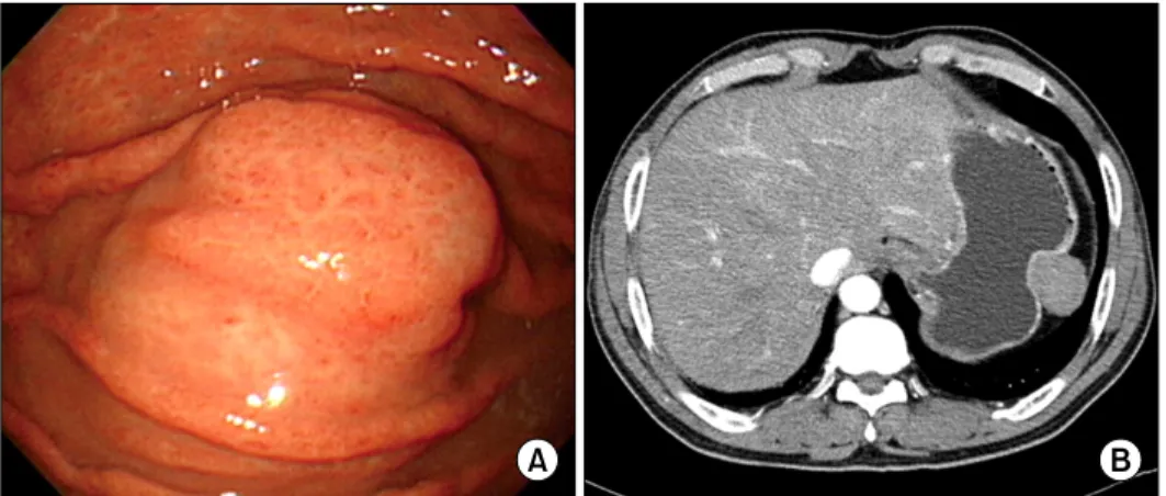
A 59-year-old man was admitted to Chonbuk National University Hospital for the evaluation of an asymptomatic gastric lesion that had been detected incidentally 1 month earlier. Physical examination of the abdomen was un- remarkable and laboratory findings, including tumor markers, were found within normal limit. The patient un- derwent an endoscopic evaluation, which revealed a poly- poid submucosal mass, with erythematous mucosa, at the
Gastric calcifying fibrous tumor
thesurgery.or.kr 57
Fig. 1. (A) Endoscopic evaluation reveals the polypoid submucosal mass in the greater curvature of the gastric fundus with mild erythema- tous mucosa. (B) Contrast-enhanced computed tomography scan reveals the well-circumscribed, non-enhan- ced, homogeneous, round mass at the great curvature of the gastric fundus.
greater curvature of the gastric fundus (Fig. 1A).
Contrast-enhanced computed tomography scan revealed a well-circumscribed, non-enhanced, and homogeneous round mass, at the great curvature of the gastric fundus (Fig. 1B). Surgery was determined as the best treatment option, and the patient underwent laparoscopic wedge re- section of the gastric fundus. Macroscopically, the mass was covered by intact mucosa. Cut-sections revealed that it was a relatively well-defined tumor, measuring 3.9 × 2.7 cm, and located in the proper muscle, with extension to the subserosa. Sections showed a homogeneous yellow to white, firm surface (Fig. 2A). Microscopically, the tumor was well-circumscribed, but not encapsulated, and was surrounded by a peripheral cuff of lymphoid aggregates (Fig. 2B). The tumor showed characteristic hypocellular sclerosis, with wavy, vaguely storiform coarse collagen, and either scattered or patchy mononuclear inflammatory infiltrates, composed of plasma cells, lymphocytes and mast cells. Small nodular lymphoid aggregates were com- mon, and usually located at the periphery of the tumor.
Sparse spindle cells were dispersed among thick collagen bundles. These cells had ovoid, vesicular nuclei with fine chromatin, and did not show any cellular atypia or mitotic activity (Fig. 2C). Occasional psammomatous and dystro- phic calcifications were found (Fig. 2D). Some of the calci- fications revealed minute lumina, suggesting calcified vascular channels. Immunohistochemically, the spindle cells were negative for CD117 (Fig. 2E), CD34, plate- let-derived growth factor receptor-alpha, S100, smooth muscle actin (SMA) (Fig. 2F), desmin and anaplastic lym- phoma kinase (ALK). However, there were scattered
CD117-positive mast cells and immunoglobulin G4-posi- tive plasma cells. Two months after the operation, the pa- tient remained in good condition, with no signs or symp- toms of tumor recurrence.
DISCUSSION
CFTs are rare, benign lesions consisting of well circum- scribed, un-encapsulated, paucicellular, hyalinized fibro- sclerotic tissue, with a variable inflammatory infiltrate, consisting of lymphocytes and plasma cells. Lymphoid ag- gregates may be present. Calcifications, both psammoma- tous and dystrophic, are scattered throughout the tumor.
CFTs show a predilection for the soft tissue and the ab- dominal cavity, especially subserosal location involving either the pleura or the visceral peritoneum [1]. However, intra-abdominal CFT occurring as an intrinsic, visceral le- sion is rare. In addition, it has been shown that gastric CFTs are different from their soft tissue counterpart in their smaller tumor size, higher mean age at presentation and no tendency for local recurrence. These findings sug- gest different pathogenic pathways between gastric CFTs and their soft tissue counterpart, regardless of morpho- logic similarity.
CFT is thought to represent a true neoplasm, with a ten- dency for nondestructive local recurrence, rather than a re- active process resulting from abnormal tissue healing.
In the literature, gastric CFTs have been documented no predominance of both sex in occurrence, with a mean age of 52.5 years [2-5]. Mean age was slightly higher for wom-
Kyu Yun Jang, et al.
58 thesurgery.or.kr
Fig. 2. (A) Cut-section reveals the well-defined tumor measuring 3.9 × 2.7 cm covered by intact mucosa. The tumor is located in the proper muscle with extension to the subserosa and shows a homogeneous yellow to white, firm surface. (B) The tumor is well-circumscribed, but not encapsulated, and located in the proper muscle (H&E, ×20). (C) The tumor shows characteristic hypocellular sclerosis, with wavy collagen and scattered mononuclear inflammatory infiltrates (H&E, ×100). Note the sparse spindle cells dispersed among thick collagen bundles and some plasma cells (inset, H&E, ×400). (D) Occasional psammomatous and dystrophic calcifications are found (H&E, ×100). (E) Spindle cells show no immunoreactivity for CD117 (CD117, ×100). Note the immunoreactive mast cells, as the internal positive control (inset, CD117, ×400).
(F) Spindle cells show no immunoreactivity for smooth muscle actin (SMA, ×200). Note the immunoreactive vascular walls, as the internal positive control.
en than for men (i.e., 55.6 years vs. 49.5 years). Tumor mean size was 1.9 cm. Eight tumors were originated in the body of the stomach, 2 in the lesser curvature, 1 in the antrum, 1
in the antrum junction, and 2 in unspecified gastric sites.
None of the 9 patients with mean follow-up of 51 months developed recurrence. The clinical features of the present
Gastric calcifying fibrous tumor
thesurgery.or.kr 59
case are somewhat different from the previous cases, in that the age of the patient was older (59 years) than the re- ported mean age of male patients. In addition, the tumor size was larger (3.9 cm) than the reported mean size and lo- cated in the greater curvature of the fundus.
The differential diagnosis of gastric CFT includes gas- trointestinal stromal tumor (GIST), inflammatory fibroid polyp (IFP), schwannoma, sclerosing leiomyoma, in- flammatory myofibroblastic tumor (IMT) and plexiform fibromyxoma. Gastric GISTs are frequently hyalinized, with dystrophic calcifications in approximately 50% of cases [6]. However, psammomatous calcifications and lymphoplasmacytic infiltrates are not features of regress- ing GIST [6]. In addition, in sclerosing GIST, immunor- eactivity for CD117 and CD34 is helpful in differentiation.
Unlike gastric CFTs, gastric IFPs are located mostly in the antrum, are submucosally-based lesions with eosinophil- dominant inflammatory cells, higher cellularity and on- ion-skin pattern of the stromal cell arrangement [7].
Gastric schwannoma may reveal the residual schwanno- ma tissue and a strong immunoreactivity for S100 [8].
Sclerosing leiomyoma may also reveal residual smooth muscle tumor cells, which are immunoreactive for both SMA and desmin. IMTs reveal infiltrative growth of either fibroblasts or myofibroblasts, which are immunoreactive for both SMA and ALK [9]. Plexiform fibromyxomas are uniformly larger (>5 cm) tumors of the antrum, with prominent capillary network, fibromyxoid stroma, high cellularity and a common expression of SMA [10].
In general, gastric CFTs are under-recognized because they are unfamiliar with the surgeons and pathologists and because their small size, bland looking histology and benign course attract less attention from the clinicians.
Awareness of the characteristic morphology of gastric CFTs will help to distinguish them from other gastric scle- rosing stromal tumors.
CONFLICTS OF INTEREST
No potential conflict of interest relevant to this article was reported.
REFERENCES
1. Rosenthal NS, Abdul-Karim FW. Childhood fibrous tumor with psammoma bodies: clinicopathologic features in two cases. Arch Pathol Lab Med 1988;112:798-800.
2. Puccio F, Solazzo M, Marciano P, Benzi F. Laparoscopic re- section of calcifying fibrous pseudotumor of the gastric wall: a unique case report. Surg Endosc 2001;15:1227.
3. Elpek GO, Kupesiz GY, Ogus M. Incidental calcifying fi- brous tumor of the stomach presenting as a polyp. Pathol Int 2006;56:227-31.
4. Attila T, Chen D, Gardiner GW, Ptak TW, Marcon NE.
Gastric calcifying fibrous tumor. Can J Gastroenterol 2006;20:487-9.
5. Agaimy A, Bihl MP, Tornillo L, Wunsch PH, Hartmann A, Michal M. Calcifying fibrous tumor of the stomach: clin- icopathologic and molecular study of seven cases with lit- erature review and reappraisal of histogenesis. Am J Surg Pathol 2010;34:271-8.
6. Agaimy A, Wunsch PH, Hofstaedter F, Blaszyk H, Rummele P, Gaumann A, et al. Minute gastric sclerosing stromal tumors (GIST tumorlets) are common in adults and frequently show c-KIT mutations. Am J Surg Pathol 2007;31:113-20.
7. Kolodziejczyk P, Yao T, Tsuneyoshi M. Inflammatory fib- roid polyp of the stomach: a special reference to an im- munohistochemical profile of 42 cases. Am J Surg Pathol 1993;17:1159-68.
8. Daimaru Y, Kido H, Hashimoto H, Enjoji M. Benign schwannoma of the gastrointestinal tract: a clinicopatho- logic and immunohistochemical study. Hum Pathol 1988;
19:257-64.
9. Shi H, Wei L, Sun L, Guo A. Primary gastric inflammatory myofibroblastic tumor: a clinicopathologic and immuno- histochemical study of 5 cases. Pathol Res Pract 2010;
206:287-91.
10. Miettinen M, Makhlouf HR, Sobin LH, Lasota J. Plexiform fibromyxoma: a distinctive benign gastric antral neoplasm not to be confused with a myxoid GIST. Am J Surg Pathol 2009;33:1624-32.