Mitochondrial Dysfunction and Cancer
Yu-Seon Han, Myeong-Eun Jegal and Yung-Jin Kim*
Department of Molecular Biology, College of Natural Science, Pusan National University, Busan 46241, Korea Received July 30, 2019 /Revised September 3, 2019 /Accepted September 11, 2019
The mitochondria is the major cellular organelle of energy metabolism for the supply of cellular en- ergy; it also plays an important role in controlling calcium regulation, reactive oxygen species (ROS) production, and apoptosis. Mitochondrial dysfunction causes various diseases, such as neurodegenerative diseases, Lou Gehrig’s disease, cardiovascular disease, mental disorders, diabetes, and cancer. Most of the diseases are age-related diseases. In this review, we focus on the roles of mitochondrial dysfunc- tion in cancer. Mitochondrial dysfunction induces carcinogenesis and is found in many cancers. The factors that cause mitochondrial dysfunction differ depending on the types of carcinoma, and those factors could cause cancer malignancy, such as resistance to therapy and metastasis. Mitochondrial dysfunction is caused by a lack of mitochondria, an inability to provide key substances, or a dysfunc- tion in the ATP synthesis machinery. The main factor associated with cancer malignancy is mtDNA depletion. Mitochondrial dysfunction would leads to malignancy through changes in molecular activ- ity or expression, but it is not known in detail which changes lead to cancer malignancy. In order to explore the relationship between mitochondrial dysfunction and cancer malignancy in detail, mi- tochondria dysfunctional cell lines are constructed using chemical methods such as EtBr treatment or gene editing methods, including shRNA and CRISPR/Cas9. Those mitochondria dysfunctional cell lines are used in the study of various diseases caused by mitochondrial dysfunction, including cancer.
Key words : Cancer, metastasis, mitochondria, mitochondrial dysfunction, therapy resistance
*Corresponding author
*Tel : +82-50-510-2176, Fax : +82-513-9258
*E-mail : [email protected]
This is an Open-Access article distributed under the terms of the Creative Commons Attribution Non-Commercial License (http://creativecommons.org/licenses/by-nc/3.0) which permits unrestricted non-commercial use, distribution, and reproduction in any medium, provided the original work is properly cited.
Journal of Life Science 2019 Vol. 29. No. 9. 1034~1046 DOI : https://doi.org/10.5352/JLS.2019.29.9.1034
서 론
초반 미토콘드리아 연구는 ATP 생성과 고분자 생성을 위한 대사 물질 생성 기전과같은 대사과정에 초점을 두었지만[121], 이후 생물 대사부터 신경, 면역을 넘어 암과 같은 질병에서도 미토콘드리아가 관여하는 것으로 밝혀졌다. 미토콘드리아는 ATP 생산을 넘어 세포의 운명과 기능을 조절할 수 있다는 것을 보여주었다[26, 113, 122, 127, 135]. 예를 들어, ATP 생산 에 관여하는 cytochrome c가 세포 생존이 아닌 죽음에 관여하 고[76], 미토콘드리아로부터 발생하는 활성 산소가 세포 죽음 이나 손상에 영향을 줄 뿐만 아니라 hypoxia-inducible factor (HIF)의 표적 유전자인 erythropoietin (EPO)의 발현을 야기하 여 골수에서 적혈구 생성을 촉진한다[14]. 최근, 미토콘드리아 에서 발생하는 활성 산소가 생리적 조건에서 발생할 수 있으 며, 세포 증식과 분화, 대사 적응, 면역 기능과 같은 다양한 과정에 관여한다고 알려졌다[23, 27, 71, 98]. 더 나아가, 미토콘 드리아는 세포 기능을 조절하기 위해 대사 물질, mtDNA와
미토콘드리아 유래 소포체의 분비와 같이 세포와 소통할 수 있는 다양한 방법을 가지고 있다[6, 13]. 이렇게 분비되는 물질 들이 염색질과 DNA 변형을 조절하여 유전자 발현을 변화 시 킨다[10]. 따라서, 미토콘드리아는 신호 전달에도 중요하게 작 용하며, 다양한 질병과도 연관성을 보이고 있어 많은 연구가 진행 중에 있다. 본 총설에선 여러 연구를 통해 밝혀진 미토콘 드리아와 암 간의 상관 관계에 대해 정리하고자 한다.
미토콘드리아의 기능
미토콘드리아는 과거 α-proteobacterium의 유입으로 인해 발 생한 소기관으로 사료된다[24]. 이러한 유입은 첫 진핵 세포의 진화로, 유산소 호흡이 가능해질 뿐만 아니라 다세포 생물로 의 진화에 중요한 역할을 하였다[116]. 이렇게 발생한 미토콘 드리아는 이중막 구조를 가지며[33] 다른 세포 소기관과는 달 리 자체 DNA를 가지고 있다[33]. 미토콘드리아는 2-10개의 mtDNA 복사본을 가진다[33]. 인간 mtDNA는 16.6 kb의 이중 가닥으로 원형 구조를 가지며, 13개의 호흡 효소 복합체와 미 토콘드리아 단백질 합성에 필요한 22개의 tRNA, 2개의 rRNA 를 암호화하고 있다[1, 129]. mtDNA는 핵 DNA와는 달리 히스톤과 결합하고 있지 않아, 유리기(free radical)에 의한 손 상을 받을 수 있으나, 자체적 수선 기전을 통한 수선이 가능하 다[22, 87].
미토콘드리아는 생물 에너지 대사와 세포 항상성에 관여하 - Review -
며, 전자 전달과 산화적 인산화를 통한 ATP 생산, TCA 회로 (tricarboxylic acid cycle)에 의한 대사 산물의 산화와 베타 산 화에 의한 지방산의 이화 작용, 활성 산소 생산, 세포 사멸의 개시와 실행과 같은 역할을 한다[34, 131].
에너지 대사
세포는 에너지의 90% 이상을 미토콘드리아에서 산화적 인 산화를 통해 생산한다[12]. 산화적 인산화 과정은 TCA 회로와 전자 전달계 두가지 대사 과정을 이용한다. TCA 회로는 탄수 화물과 지방을 일부 ATP로 전환시키지만, 보다 중요한 것은 이후 전자 전달 과정에서 사용될 NADH와 FADH와 같은 보 효소(coenzyme)를 만들어내는 일이다. NADH와 FADH는 산 소로 전자를 전달시켜, 산소의 전자와 양성자(수소)를 기질에 서 내외막 안쪽 공간으로 이동시킨다. 이렇게 생성된 전기 화 학 기울기에 의해 ATP 합성 효소가 ATP를 합성한다[8, 9].
세포 에너지 요구량은 각각의 세포에 존재하는 미토콘드리 아 수와 연관되어 있다. 하나의 체세포는 200-2,000개의 미토 콘드리아를 가지지만[9, 37], 인간 생식 세포는 정자의 경우 16개만을 가지고 난자는 100,000개 이상 가진다[125]. 골격근, 심근, 간, 뇌 등 대사가 활발히 일어나는 세포에서 가장 많은 수의 미토콘드리아가 관찰되며, 인간 세포 중 성숙한 적혈구 에서만 미토콘드리아가 존재하지 않는다[18].
칼슘 조절
미토콘드리아는 칼슘 조절에도 중요한 역할을 한다. 세포는 일반적으로 역수송체(antiporter)나 펌프를 이용하여 칼슘 이 온을 조절하나, 세포내 소기관도 선택적 펌프나 교환체 (exchanger) 등을 통해 세포와 소기관 내의 칼슘 이온 농도를 조절할 수 있다. 미토콘드리아는 에너지 의존적 방법과 칼슘 이온 방출이 가능한 역수송체를 통해 칼슘 이온을 조절하며, 미토콘드리아 주요 기능은 기질 내 칼슘 이온 농도에 매우 많은 영향을 받는 것으로 알려져 있다. 또한, 세포내 칼슘 이온 이 미토콘드리아 내막 외부 표면에 위치한 대사 관련 미토콘 드리아 효소 활성에 영향을 줄 수 있다고 알려져 있다[19].
활성 산소 생성
활성 산소는 독성을 띄는 대사 부산물로서 인간 질병의 유 발 요인으로 여겨진다. 세포내의 활성 산소는 미토콘드리아 산화 대사 중 원치 않게 발생하는 부산물로 여겨지며, 미토콘 드리아는 최소 10군데에서 활성 산소 발생이 가능하다. 미토 콘드리아 활성 산소가 면역 반응과 자식 작용(autophagy)을 조절하는 신호 전달에 관여한다는 증거가 발견되고 있으며, 대사 중추로서 세포의 대사 상태에 영향을 미칠 수도 있다.
미토콘드리아의 활성 산소 생산은 세포 에너지 상태, 대사 산 물 농도, 다른 상위 신호 발생을 통합시키고, 세포 스트레스, 줄기 세포 집단(population), 세포 생존, 암으로의 변이에 주요
한 영향을 준다[23, 43, 117].
세포 사멸
세포 사멸의 많은 주요 현상들이 미토콘드리아에서 발생하 기 때문에 미토콘드리아는 세포 사멸 활성에 중요한 역할을 한다. 주요 현상들로는 caspase 활성 인자 방출, 전자 전달계의 변화, 미토콘드리아 막 전위 감소, 세포의 산화-환원 변화, pro-/antiapoptotic Bax, Bak, Bcl-2, Bcl-xL과 같은 세포 사멸 관련 단백질들의 조절 등이 있다. 이러한 현상들을 기반으로 세포를 분화 시키고, 사멸한 세포를 효율적으로 처리하는 식 균 작용을 일으킨다. 특히, 세포 사멸은 질병을 억제하는 역할 이 있어 중요하다. 예를 들어 암의 발생부터 암의 진행 과정을 제어할 수 있다[39, 77, 132].
미토콘드리아 기능 이상의 발생 원인과 관련 질환
미토콘드리아는 세포 에너지 생산 외에도 다양한 기능에 관여하는 핵심 소기관으로서, 미토콘드리아 기능 이상은 다양 한 질병의 원인이 된다. 특히 노화 관련 질병에 많은 영향을 미치는데, 미토콘드리아 기능 이상으로 인한 ATP 합성 감소 는 노화에서 나타나는 특징으로 특히 만성 질환에서 자주 나 타난다. 미토콘드리아 기능 이상은 미토콘드리아 수 부족, 주 요 물질 제공 불능, 혹은 전자 전달과 ATP 합성 기전의 이상으 로부터 발생한다[94]. 이는 주로 미토콘드리아에서 부산물로 생성되는 활성 산소가 축적되어 mtDNA를 파괴시켜 나타나 는 것으로 알려져 있다[29, 134].
대부분의 활성 산소는 복합체 I과 III(complex I, III)에서 생성되는데, 이는 전자 전달계에서 NADH와 FADH에 의해 전자가 방출되기 때문이다[45]. 미토콘드리아는 ATP 생산에 세포가 사용하는 산소의 85%를 소모하며, 산화적 인산화로 사용한 산소의 0.4-4.0%가 과산화물(superoxide; O2-) 라디칼 (radical)이 된다[106]. 이렇게 생성된 과산화물 라디칼은 해독 효소(detoxification enzyme)인 MnSOD (manganese super- oxide dismutase), Cu/Zn SOD (copper/zinc superoxide dis- mutase)에 의해 H2O2로 바뀌고[130] 이후, GPX (glutathione peroxidase), PRX III (peroxidredoxin III)에 의해 H2O로 전환 된다[40]. 이러한 효소들이 과산화물 라디칼와 같은 활성 산소 를 H2O로 빠르게 전환시키지 못하면 미토콘드리아에 산화적 손상이 발생되고 축적된다[108]. 미토콘드리아 내에는 지질, 단백질, 산화적 인산화 효소, mtDNA와 같은 유리기에 취약한 물질들이 존재한다[119]. 미토콘드리아 단백질의 직접적인 손 상은 기질 혹은 보효소와의 친화도를 감소시키고, 미토콘드리 아 단백질의 기능을 감소시킨다[75]. 미토콘드리아 손상은 피 드포워드(feed-forward) 과정과 같아, 미토콘드리아 단백질 기 능 감소로 인한 미토콘드리아 손상은 미토콘드리아 수선을 위 한 필요 에너지 증가를 야기하여 추가적인 손상을 유발한다[3].
Fig. 1. Mitochondrial DNA [123].
Fig. 2. mtDNA mutations in cancer [78].
미토콘드리아 질병은 Luft와 그 연구진들에 의해 1962년에 처음 언급되었다. 갑상선 기능이 정상인 한 35세 여성에서 근 질환, 과도한 땀, 열 과민증, 다뇨증을 동반한 다갈증, 약 2배 정도(180%)의 기초 대사량 증가 등이 나타났고[79], 이 환자는 산화적 인산화 분리를 겪고 있음이 밝혀졌다. 산화적 인산화 분리는 ATP의 생성 없이 열 발생을 유도하여 앞서 언급한 증상들의 원인이 되는 미토콘드리아 기능 장애이다[9].
고혈당은 활성 산소 생산을 유도하여 미토콘드리아 기능 이상 질환의 원인이 된다. 당뇨가 있을 시 혈관 세포 미토콘드 리아의 과산화물 생산으로 심혈관 질병 같은 당뇨 합병증을 일으킨다[28, 40]. 또한 내피 세포의 과산화물 생산은 아테롬성 동맥 경화증, 고혈압, 심부전, 노화, 패혈증, 허혈 재관류 손상, 과콜레스테롤혈의 원인이 된다[70].
그 외 미토콘드리아 손상으로 인한 미토콘드리아 기능 이상
질환으로는 신경퇴행질환(알츠하이머병, 파킨슨병, 헌팅턴병, 루게릭병, 프리드라이히 운동실조증)[55, 103, 118], 심혈관계 질환(아테롬성 동맥경화증, 심장과 혈관 상태)[72, 126], 당뇨 병, 신진 대사 장애[80, 93] , 자가 면역 질환(다발성 경화증, 전신 홍반성 루프스, 제 1형 당뇨병)[30, 81, 84], 신경행동 질환 과 정신 의학 질병(자폐 범주성 장애, 조현병, 조울증, 감정장 애)[61, 96, 100], 위장 장애[17], 만성 피로 증후군[89], 근골격 질환(섬유근육통, 골격근 비대/위축)[21, 102], 암[114], 만성 감염[2] 등이 있다.
미토콘드리아 기능 이상과 암의 상관관계
지난 수 십 년간의 연구를 통해, 조절되지 않는 세포 에너지 론은 최근 암 특징들 중 하나로 추가되었다[44]. 정상 세포와 달리 암 세포에서 산소가 존재함에도 해당 과정의 증가와 미 토콘드리아 호흡의 감소가 나타나는 현상을 Otto Warburg가 가장 먼저 제안하여, 이러한 현상을 Warburg 효과라고 부른 다[128].
세포내 핵심 소기관으로서 미토콘드리아의 정상적인 기능 을 유지하기 위하여 온전한 mtDNA는 아주 중요하다. mtDNA 돌연변이의 축적이나 mtDNA 복사본 수의 감소는 에너지 생 산과 세포 생존에 영향을 미치고, 활성 산소 생성을 증가 시키 게 되는데 이러한 과정들은 암과 관계된다[63, 128].
암에서 가장 일반적인 mtDNA 변화는 점 돌연변이(point mutation)와 복사본 수의 변화이다[63, 66]. 20종 암의 66%에 서 최소 한 군데 이상의 점 돌연변이가 나타난다. 대부분의 돌연변이는 mtDNA의 D-loop 부위에서 가장 많이 발생하며, 그 다음으로는 단백질 암호화 부위에서 많이 발생한다. 낮은 확률로 rRNA와 tRNA에서도 발견된다[64].
대부분의 mtDNA 점 돌연변이는 미토콘드리아 기능 이상
Table 1. mtDNA mutation region in cancer [78]
Cancer types Mutation region
Parathyroid gland tumor Head and neck cancer Thyroid tumor
ND1,ND2, ND3, ND4, ND4L, ND5, ND6, Cyt b, COⅠ,COⅡ, COⅢ, ATPase6, ATPase8, D-loop, tRNA, 12S rRNA, 16S rRNA
Colorectal cancer ND1, ND3, ND4, ND4L, ND5, ND6, Cyt b, COⅠ,COⅡ, COⅢ, ATPase6, ATPase8, D-loop, tRNA, 12S rRNA, 16S rRNA
Lung cancer ND1,ND2, ND3, ND4, ND4L, ND5, ND6, Cyt b, COⅠ,COⅡ, COⅢ, ATPase6, D-loop, tRNA, 16S rRNA
Gastrointestinal tract tumor ND1,ND2, ND3, ND4, ND4L, ND5, ND6, Cyt b, COⅠ,COⅡ, COⅢ, ATPase8, tRNA, 12S rRNA, 16S rRNA
Breast cancer ND1,ND2, ND4, ND5, ND6, Cyt b, COⅠ,COⅡ, COⅢ, ATPase6, ATPase8, D-loop Ovarian cancer ND1, ND4, ND4L, ND6, Cyt b, COⅠ,COⅡ, COⅢ, ATPase8, D-loop, tRNA, 12S rRNA, 16S
rRNA
Prostate cancer ND1,ND2, ND4, ND4L, ND5, ND6, Cyt b, COⅠ, ATPase8, D-loop, tRNA, 12S rRNA, 16S rRNA
Skin cnacer ND1,ND2, ND4L, ND5, Cyt b, COⅠ,COⅡ, COⅢ, ATPase6, D-loop, tRNA, 16S rRNA Leukemia ND1, ND4, ND5, ND6, Cyt b, COⅡ, ATPase6, D-loop, tRNA, 12S rRNA, 16S rRNA Renal cancer ND1,ND2, ND3, ND4, ND5, ND6, D-loop, tRNA, 12S rRNA, 16S rRNA
Brain tumor ND2, ND4, ND4L, Cyt b, COⅠ, COⅢ, D-loop, tRNA, 16S rRNA Cervical tumor ND1,ND2, ND3, ND4, ND4L, ND5, ND6, D-loop
Pancreatic cancer ND2, ND5, ND6, Cyt b, COⅠ, ATPase8, tRNA
Glioma ND2, ND5, ND6, Cyt b, COⅠ, D-loop
Bladder cancer ND3, ND4, ND5, Cyt b, D-loop, 16S rRNA
Oral cancer ND2, ND3, ND4, COⅢ, D-loop
Hepatocellular cancer COⅠ, ATPase8, D-loop, tRNA Nasopharyngeal carcinoma Cyt b, D-loop, tRNA, 12S rRNA Endometrial tumor D-loop, 12S rRNA, 16S rRNA
Renal oncocytoma ND1
Esophageal cancer ND5
Gastric tumor ATPase6
Stomach cancer Uterine carcinoma Astrocytoma Barrett's cancer Osteosarcoma Ewing's sarcoma Gallbladder carcinoma
D-loop
Splenic lymphoma tRNA
을 야기하며, 몇몇 점 돌연변이는 암 발생에 관여한다[50, 86, 99]. mtDNA 돌연변이가 많이 나타나는 암종으로는 유방암, 결장암, 자궁암, 위암, 간암, 췌장암, 전립선암, 폐암, 신장암, 갑상선암, 뇌종양 등이 있으며, 각 암 종에 따라 돌연변이가 발생하는 위치가 다르다(Fig. 1) [16].
암에서 mtDNA 복사본 수의 변화는 조직별로 다양한 형태 로 나타난다[66, 67] 간암, 위암, 유방암에서는 주로 mtDNA 복사본 수가 감소하고[65, 120, 137], 이러한 변화는 발암, 암 진행, 전이 등을 촉진시킨다[139]. 반대로, 두경부암, 식도암, 자궁암에서는 mtDNA 복사본 수가 증가되어 있으며, 조직 병
리학적 단계가 높을수록 복사본 수가 더 많았다[52, 59, 73, 74, 133]. 하지만, 암 발생 부위에 따른 mtDNA 복사본 수의 차이와 암 발생과의 상관 관계에 대해서는 아직 정확하게 알 려진 바가 없다.
Succinate dehydrogenase (SDH) [46, 60], fumarate hydra- tase (FH) [11, 62], isocitrate dyhydrogenase (IDH) [54, 111]와 같은 미토콘드리아 효소의 손상은 가족력이 있는 암과 산발적 형태의 암 발생 모두에 관여한다. SDH, FH, IDH와 같은 대사 관련 효소가 손상되면, 미토콘드리아에서 방출하는 succinate, fumarate, 2-hydroxyglutarate와 같은 대사물질들이 축적되는
A
B
C D
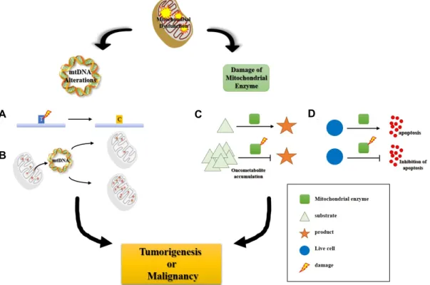
Fig. 3. Schematic representation of correlation between mitochondrial dysfunction and cancer. a) point mutation. b) alternation of mtDNA copy number. c) accumulation of oncometabolite due to damaged mitochondrial enzyme. d) inhibition of apoptosis due to damaged mitochondrial enzyme.
데, 이러한 대사물질들은 발암 활성에도 중요한 역할을 하기 에 oncometabolite라고도 불린다. 축적된 oncometabolite는 암 발생에 관여하는 유전자 발현에 후성적 변화를 야기한다 [95].
조절되지 않는 sirtuin 3 (Sirt 3) 역시 세포 대사 변화와 산화 스트레스를 통해 암을 촉진시킨다[5, 138]. Sirt 3는 미토콘드리 아 단백질 기능을 조절하며[42, 47, 101], 다양한 암에서 종양 억제 유전자로서 기능한다고 알려져 있다[32, 58]. Sirt3가 감소 하면 활성 산소가 증가하여 HIF-1과 같은 종양 유전자를 활성 화시키고, 세포 에너지 대사 과정을 Warburg 효과로 변화시 켜 발암을 촉진한다[32]. 이처럼, HIF-1, p53과 같은 종양 유전 자 혹은 종양 억제 유전자의 변화로 인한 미토콘드리아 호흡 과 세포 대사의 손상은 암 발생과 진행을 유도할 수 있다[57, 105].
미토콘드리아 기능 이상에 의한 암 악성화
앞에서 언급한 바와 같이, mtDNA 돌연변이나 미토콘드리 아 효소 결함에 의한 미토콘드리아 기능 이상은 세포 에너지 대사에 혼란을 일으키고, 미토콘드리아에서 나오는 활성 산 소, 칼슘 이온, 저분자 대사 산물의 변화는 암 발생을 촉진시키 는 결과를 야기한다[7].
암세포에서 mtDNA의 전구물질 고갈 또는 미토콘드리아
호흡 사슬 손상에 의한 미토콘드리아 기능 이상은 항암 내성 또는 침윤성 표현형과 같은 암 진행 또한 촉진시킨다[63, 66].
미토콘드리아 손상은 암 발생과 진행 과정에서 미토콘드리아 가 핵내 유전자 발현을 변화시키는 역행 신호 전달(retrograde signaling) 과정으로 세포내 신호 전달을 활성화시킨다. 뿐만 아니라, 미토콘드리아에 의해 조절되는 세포 내 활성 산소, 칼슘 이온 또는 종양 대사 산물의 양 변화 또한 미토콘드리아 역행 신호에 중요하다[41].
이처럼 미토콘드리아 손상에 의한 기능 이상은 항암제 내 성, 전이와 같은 암 악성화의 원인이 되며, 기능 이상을 야기한 원인 혹은 기능 이상으로 나타나는 변화에 따라 각기 다른 기전으로 암 악성화 표현형을 나타낸다.
항암제 내성
HeLa (cervix) 세포에 mtDNA 감소가 일어나면 미토콘드 리아 복합체 I의 손상이 발생하여 doxorubicin의 활성을 막아 항암제 내성이 나타난다. Doxorubicin은 복합체 I에 의해 qui- none에서 semiquimone으로 하나의 전자가 환원되어야 활성 을 가지며, 생성된 semiquinone은 활성 산소 생성을 증가시켜 세포를 죽인다. 미토콘드리아 기능 이상은 doxorubicin의 활 성화와 활성 산소 생성을 모두 방해함으로써 항암제 내성을 야기한다[110]. SK-Hep1 (liver) 세포에서 mtDNA 감소가 일 어나면, 미토콘드리아 기능 이상 세포주로 변화하는 과정에서
증가하는 활성 산소를 억제하기 위하여 MnSOD와 항산화 효 소 발현이 증가하게 된다. 이로 인해, 산화 스트레스나 항암제 에 대응할 수 있는 범위가 증가되어 항암제 내성이 나타난다 [97]. 또한, HCT-8 (colon), 143B (bone), SK-Hep1 세포에서 mtDNA 감소는 다약제 내성을 일으키는 유전자인 MDR1의 발현 증가를 통해 항암제 내성이 유도됨이 보고되어 있다[31, 36, 68]. Hydroxytamoxifen은 mtDNA 합성과 topoisomerase 를 억제하여 mtDNA를 감소 시켜 미토콘드리아 기능 이상 세포를 유도할 수 있다. 이렇게 유도된 미토콘드리아 기능 이 상 MCF-7 (breast) 세포는 hydroxytamoxifen과 ICI 182, 780에 내성을 보인다[90]. 이 외에도, HepG2 (liver) 세포와 NCI-H1299 (lung) 세포에서, chloramphenicol 처리로 미토콘드리아 번역 과정을 억제하여 미토콘드리아 스트레스를 유발하면 mito- mycin에 의한 세포 사멸이 p21 발현 증가를 통해 억제된다 [69]. 미토콘드리아 기능 이상 결장암 세포에서는 ATP syn- thase의 α subunit의 감소로 ATP synthase 활성이 낮아지면 5-fluorouracil에 저항성을 획득한다[107]. 미토콘드리아와 핵 사이의 커뮤니케이션도 중요하여, 둘 사이의 커뮤니케이션의 손상으로 인한 항암제 내성도 유발 될 수 있다. 예를 들어, 에너지 대사를 해당과정부터 미토콘드리아 호흡까지 이용하 는 간암 세포와 해당과정만을 사용하는 간암 세포를 비교해보 면 해당과정을 이용하는 간암 세포가 항암제에 더 내성을 가 지는 것을 확인할 수 있다. 아직 에너지 대사 변화와 간암 세포 의 항암제 내성과의 정확한 기전이 알려진 바는 없으나, 미토 콘드리아를 이용하지 않음으로 인해 발생하는 세포의 성질 변화가 이를 유도하는 것으로 보인다[48].
암세포 전이
A549 (lung) 세포에서는 일부 mtDNA 감소나 미토콘드리 아 호흡 사슬의 손상이 나타날 경우, 세포외기질(extracellular matrix: ECM) 리모델링 유전자의 전사 조절로 인해 침윤성 표현형이 나타난다[124]. LNCaP (prostate)와 MCF-7 세포에 서 mtDNA 감소가 나타나면, TGF-β와 Raf/MAPK 신호 기전 이 활성화되어 상피-간엽 전이(EMT)가 유도되어 상피-간엽 전이로 인한 침윤성 표현형이 나타난다[91]. 유방암 세포의 63%에서는 미토콘드리아 DNA 중합효소 γ(POLG) 돌연변이로 인한 mtDNA 감소가 나타난다. POLG 돌연변이는 핵산말단가 수분해효소(exonuclease), 연결부(linker), 중합효소(polymerase) 에 해당하는 세 도메인 모두에서 발생하며, mtDNA의 수와 미 토콘드리아 활성, 미토콘드리아 막 포텐셜을 낮추고 ROS 발 생 증가와 함께 침윤성을 증가 시킨다[109]. SC-M1 (stomach) 세포에서는 미토콘드리아 호흡 사슬의 손상으로 인한 활성 산소의 증가로 인해 integrin β5 발현 증가로 유도되어 세포 이동성이 증가한다[49]. 활성 산소뿐만 아니라 미토콘드리아 에 의해 조절되는 칼슘 이온에 의해서도 악성화 표현형이 나 타난다. HepG2 세포의 경우, 칼슘 이온 증가와 활성 산소 과생
성으로 amphiregulin(EGF member) 과발현과 활성화를 통해 암세포의 항암제 내성과 이동성이 증가된다[15]. 또한, EtBr 처리를 통해 mtDNA 감소를 유도하면, 세포내 칼슘 이온이 증가되고, 그로 인해 cathepsin L과 TGF-β 같은 침윤성 표지 자의 발현 뿐만 아니라 침윤성 표현형 또한 같이 나타남을 보였다[124].
이처럼 미토콘드리아 기능 이상은 종양 성장 또는 세포 사 멸 내성, 항암제 내성, 침윤성 표현형과 같은 암 악성화를 다양 한 기전을 통해 향상시킨다. 뿐만 아니라 미토콘드리아 변화 는 미토콘드리아에서 핵으로 새포내 신호 기전을 활성화 시킬 수 있고, 최종적으로 암 악성화가 되도록 핵 유전자 발현을 변화시킨다. 하지만 여전히 어떠한 mtDNA 돌연변이가 암 발 생과 아울러 암 악성화를 진행시키는 지에 대해 구체적으로 알려진 바가 없어 더 연구가 필요한 부분이다.
미토콘드리아 기능 이상 연구 방법
미토콘드리아 기능에 대한 연구 초기에 주로 EtBr(Ethi- dium bromide)을 이용하여 연구를 진행하였다[104, 112]. 질 병과 미토콘드리아 관계에 대한 연구를 위해서는 대부분 환자 조직 일부를 이용하여 연구하며, 암의 경우 채취한 조직을 이 용하여 mtDNA 양을 확인하거나, mtDNA 중 돌연변이가 일 어난 유전자를 확인하는 연구가 주를 이루었다[66, 82]. 하지 만, 환자의 조직을 이용하여 mtDNA에 의한 병의 발생이나 진행에 대한 영향을 연구하기에는 임상병리학적으로 제한적 인 부분들이 많다[85, 141]. 미토콘드리아 돌연변이와 관련이 있는 다른 질병의 경우, 환자로부터 직접적인 조직을 얻는 것 이 힘들기 때문에 연구 진행에 어려움이 있어왔다[85]. 이러한 문제점들을 보완하기 위해서 in vitro에서 EtBr을 처리하거나 gene editing을 이용하여 환자에게서 나타나는 현상을 재현할 수 있는 미토콘드리아 기능 이상 세포를 제작하여 연구를 진 행하였다[141].
EtBr을 이용한 mtDNA 감소
EtBr은 원형인 mtDNA 염기 사이에 들어가서 구조를 뒤틀 리게 하거나[142] linear DNA로 만들어[92] mtDNA 복제와 전사를 억제하는 기능을 한다. EtBr을 척추동물 세포에 짧은 기간 처리 시에는 mtDNA 양이 감소 되지만, 이 후 EtBr을 제거하면 mtDNA 양이 다시 회복된다. EtBr 처리 기간이 길수 록 mtDNA 복제는 현저히 감소되며, 이 후 EtBr을 제거하여도 mtDNA 양이 회복되지 않는 것이 확인 되었다[104, 112]. 특히, EtBr을 장시간 처리했을 시(35~52 days), mtDNA가 완전히 사라져 회복되지 않았으며, cytochrome c oxidase 활성 또한 감소한다[25].
이러한 연구들을 토대로 미토콘드리아 기능 이상 세포 제작 이 가능 해졌으며, 그에 관한 연구 또한 활발하게 진행되고
Table 2. Construction of mitochondria dysfunctioned cell line using EtBr
Cell type Treatment period EtBr concentration Reference
human line VA2-B 3 days 20 ng/ml [136]
Mouse and hamster cell 4 days 0.1~5 μg/ml [56]
Primary chicken Embryo Fibroblasts ~43 days 0.4 μg/ml [25]
A549,SPC-A1, H322 7 days 250 ng/ml [138]
Breast cancer cell 40 days 50 ng/ml [140]
Hep3B, HEK293, 143B Hep3B - 3 weeks (100 ng/ml)
HEK293 - 2 weeks (50 ng/ml) 50 or 100 ng/ml [83]
HeLa7 70 passage 10 ng/ml [88]
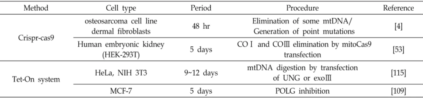
Table 3. Construction of mitochondria dysfunctioned cell line using gene editing
Method Cell type Period Procedure Reference
Crispr-cas9
osteosarcoma cell line
dermal fibroblasts 48 hr Elimination of some mtDNA/
Generation of point mutations [4]
Human embryonic kidney
(HEK-293T) 5 days COⅠ and COⅢ elimination by mitoCas9
transfection [53]
Tet-On system HeLa, NIH 3T3 9~12 days mtDNA digestion by transfection
of UNG or exoⅢ [115]
MCF-7 5 days POLG inhibition [109]
있다. 예를 들어, mtDNA 돌연변이에 의한 미토콘드리아 기능 이상과 관련 질환 발병 원인과의 연관성이나, mtDNA 감소 정도와 질환과의 상관 관계에 대한 연구가 진행되고 있다. 암 과 미토콘드리아 기능 이상의 연관성을 연구하는 방법으로는 기능 이상에 주요한 영향을 미칠 것으로 사료되는 일부 유전 자(COX2, ND1, ND2, D-loop)의 발현이 사라진 세포주를 이 용하거나, mtDNA 발현이 전부 사라진 세포주를 이용하는 방 법이 있다. 이러한 세포주는 암 발생과 암 악성화 연구에 이용 된다. 현재는 미토콘드리아 기능 이상 세포주를 이용하여, mtDNA 변화에 의한 핵 유전자 발현 조절과 같은 역행 신호 전달에 대한 연구도 많이 진행되고 있다.
유전자 수선(gene editing)을 이용한 mtDNA 감소 유전자 수선에 관한 연구 방법 진화에 따라, 미토콘드리아 기능 이상 세포주 제작에 기존에 주로 사용되던 EtBr 방법 외 에 새로운 방법이 제시되고 있다. siRNA, shRNA를 이용한 핵 DNA에 존재하는 미토콘드리아 단백질의 발현 억제를 통 해 그 기능을 연구하거나, mtDNA의 복제 및 수선에 관여하는 POLG를 표적으로 하는 siRNA를 통해 mtDNA를 감소시키는 방법으로 기능을 연구하기도 하였다. 또한, POLG 우성음성 (dominant negative) 돌연변이인 POLG D1135A를 세포주에 삽입하면, mtDNA 기능 이상 세포를 제작할 수 있다[20, 38, 51, 109]. 최근에는 CRISPR/Cas9이라는 새로운 실험 기법이 도입됨에 따라, 우성음성 POLG를 사용하지 않고 siRNA, shRNA 방법보다 더 효과적으로 POLG 유전자를 없애는 방법
을 활용할 수 있게 되었다[35]. 한편 mtDNA로부터 만들어지 는 단백질의 기능에 대한 연구는 돌연변이를 가진 세포를 이 용한 방법으로 제한적이었으나, CRISPR/Cas9 유전자 조작법 의 발달은 이 부분에도 상당한 진보를 가져오게 되었다. 미토콘 드리아로 이동하도록 조작된 CRISPR/Cas9을 이용하여 mtDNA 유전자(cox1, cox3)를 직접 제어하여, 특정 미토콘드리아 유전 자의 발현을 제어할 수 있는 방법이 최근에 보고되었다[53].
하지만, 유전자 수선 방법을 이용한 미토콘드리아 기능 이상 연구는 효율성 문제와 다수의 유전자를 동시에 수선하기에는 부족한 기술력 등의 문제점들로 여전히 더 많은 연구가 필요 한 실정이다.
결 론
미토콘드리아는 에너지 대사, 세포 사멸, 칼슘 이온 조절과 같은 여러 역할을 하는 세포 주요 소기관이다. 미토콘드리아 에 발생하는 기능 이상은 신경퇴행 질환, 심혈관계 질환, 대사 장애, 자가 면역 질환, 암과 같은 다양한 질병을 야기하며, 대 부분이 노화 관련 질병이다. 미토콘드리아 기능 이상은 대부 분 자체 수선 능력을 넘어선 손상 축적으로 발생하기에, 노화 될수록 많은 손상에 노출된다. 이들 질병 치료를 위한 많은 연구들이 진행 중에 있으며, 이를 토대로 본 논문에서는 미토 콘드리아 기능 이상과 암과의 상관 관계에 대해 고찰하였다.
많은 암 종에서 mtDNA 돌연변이와 미토콘드리아 손상에 의 한 에너지 대사에 변화가 관찰되며, 이러한 변화가 암 발생
과정과 악성화 현상을 촉진시킨다. 미토콘드리아 기능 이상에 의한 암 발생과 진행에 대한 연구를 위해, 주로 미토콘드리아 기능 이상 세포주를 제작하여 활용한다. 그 방법으로는 EtBr과 같은 화학 물질을 이용하거나 우성음성 POLG 돌연변이를 도 입하거나, gene editing을 이용하는 방법이 있다. 현재까지 많 은 연구들이 진행되어 왔으나, 미토콘드리아 기능 이상 세포 주 제작 부분과 기능 이상과 암 생성, 암 악성화간의 직접적이 고 명확한 기전에 대한 연구는 여전히 요구된다. 이러한 연구 들이 기반이 된다면 새로운 항암 치료법을 제시할 수 있을 것이다.
감사의 글
이 논문은 부산대학교 기본연구지원사업(2년)에 의하여 연 구되었음.
References
1. Anderson, S., Bankier, A. T., Barrell, B. G., de Bruijn, M.
H., Coulson, A. R., Drouin, J., Eperon, I. C., Nierlich, D.
P., Roe, B. A., Sanger, F., Schreier, P. H., Smith, A. J., Staden, R. and Young, I. G. 1981. Sequence and organization of the human mitochondrial genome. Nature 290, 457-465.
2. Ashida, H., Mimuro, H., Ogawa, M., Kobayashi, T., Sasaka- wa, C., Sanada, T. and Kim, M. 2011. Cell death and in- fection: A double-edged sword for host and pathogen survival. J. Cell. Biol. 195, 931-942.
3. Aw, T. Y. and Jones, D. P. 1989. Nutrient supply and mi- tochondrial function. Annu. Rev. Nutr. 9, 229-251.
4. Bacman, S. R., Williams, S. L., Pinto, M., Peralta, S. and Moraes, C. T. 2013. Specific elimination of mutant mitochon- drial genomes in patient-derived cells by mitoTALENs. Nat.
Med. 19, 1111-1113.
5. Bell, E. L., Emerling, B. M., Ricoult, S. J. and Guarente, L.
2011. SirT3 suppresses hypoxia inducible factor 1alpha and tumor growth by inhibiting mitochondrial ROS production.
Oncogene 30, 2986-2996.
6. Bohovych, I. and Khalimonchuk, O. 2016. Sending out an SOS: mitochondria as a signaling hub. Front. Cell. Dev. Biol.
4, 109.
7. Boland, M. L., Chourasia, A. H. and Macleod, K. F. 2013.
Mitochondrial dysfunction in cancer. Front. Oncol. 3, 1-28.
8. Bralley, J. and Lord, R. 2001. Laboratory Evaluations in Molecular Medicine: Nutrients, Toxicants, and Cell Regula- tors, 175-208, Institute for Advances in Molecular Medicine:
Norcross, GA, USA
9. Brookes, P. S., Yoon, Y., Robotham, J. L., Anders, M. W.
and Sheu, S. S. 2004. Calcium, ATP, and ROS: a mitochon- drial love-hate triangle. Am. J. Physiol. Cell. Physiol. 287, C817-833.
10. Butow, R. A. and Avadhani, N. G. 2004. Mitochondrial sig- naling: the retrograde response. Mol. Cell. 14, 1-15.
11. Castro-Vega, L. J., Buffet, A., De Cubas, A. A., Cascon, A.,
Menara, M., Khalifa, E., Amar, L., Azriel, S., Bourdeau, I., Chabre, O., Curras-Freixes, M., Franco-Vidal, V., Guillaud- Bataille, M., Simian, C., Morin, A., Leton, R., Gomez-Grana, A., Pollard, P. J., Rustin, P., Robledo, M., Favier, J. and Gimenez-Roqueplo, A. P. 2014. Germline mutations in FH confer predisposition to malignant pheochromocytomas and paragangliomas. Hum. Mol. Genet. 23, 2440-2446.
12. Chance, B., Sies, H. and Boveris, A. 1979. Hydroperoxide metabolism in mammalian organs. Physiol. Rev. 59, 527-605.
13. Chandel, N. S. 2015. Evolution of Mitochondria as Signaling Organelles. Cell. Metab. 22, 204-206.
14. Chandel, N. S., Maltepe, E., Goldwasser, E., Mathieu, C. E., Simon, M. C. and Schumacker, P. T. 1998. Mitochondrial reactive oxygen species trigger hypoxia-induced transcrip- tion. Proc. Natl. Acad. Sci. USA. 95, 11715-11720.
15. Chang, C. J., Yin, P. H., Yang, D. M., Wang, C. H., Hung, W. Y., Chi, C. W., Wei, Y. H. and Lee, H. C. 2009. Mitochon- drial dysfunction-induced amphiregulin upregulation medi- ates chemo-resistance and cell migration in HepG2 cells.
Cell. Mol. Life. Sci. 66, 1755-1765.
16. Chatterjee, A., Mambo, E. and Sidransky, D. 2006. Mito- chondrial DNA mutations in human cancer. Oncogene 25, 4663-4674.
17. Chitkara, D. K., Nurko, S., Shoffner, J. M., Buie, T. and Flores, A. 2003. Abnormalities in gastrointestinal motility are associated with diseases of oxidative phosphorylation in children. Am. J. Gastroenterol. 98, 871-877.
18. Cohen, B. H. and Gold, D. R. 2001. Mitochondrial cytopathy in adults: what we know so far. Cleve. Clin. J. Med. 68, 625-626, 629-642.
19. Contreras, L., Drago, I., Zampese, E. and Pozzan, T. 2010.
Mitochondria: the calcium connection. Biochim. Biophys.
Acta. 1797, 607-618.
20. Copeland, W. C. and Longley, M. J. 2003. DNA polymerase gamma in mitochondrial DNA replication and repair.
ScientificWorldJournal 3, 34-44.
21. Cordero, M. D., de Miguel, M., Carmona-Lopez, I., Bonal, P., Campa, F. and Maria Moreno-Fernandez, A. 2010. Oxida- tive stress and mitochondrial dysfunction in fibromyalgia.
Neuro. Endocrinol. Lett. 31, 169-173.
22. Croteau, D. L. and Bohr, V. A. 1997. Repair of oxidative damage to nuclear and mitochondrial DNA in mammalian cells. J. Biol. Chem. 272, 25409-25412.
23. Dan Dunn, J., Alvarez, L. A., Zhang, X. and Soldati, T. 2015.
Reactive oxygen species and mitochondria: A nexus of cel- lular homeostasis. Redox. Biol. 6, 472-485.
24. Degli Esposti, M. 2014. Bioenergetic evolution in proteobac- teria and mitochondria. Genome Biol. Evol. 6, 3238-3251.
25. Desjardins, P., Frost, E. and Morais, R. 1985. Ethidium bro- mide-induced loss of mitochondrial DNA from primary chicken embryo fibroblasts. Mol. Cell. Biol. 5, 1163-1169.
26. Devine, M. J. and Kittler, J. T. 2018. Mitochondria at the neuronal presynapse in health and disease. Nat. Rev.
Neurosci. 19, 63-80.
27. Diebold, L. and Chandel, N. S. 2016. Mitochondrial ROS reg- ulation of proliferating cells. Free. Radic. Biol. Med. 100, 86-93.
28. Du, X. L., Edelstein, D., Rossetti, L., Fantus, I. G., Goldberg, H., Ziyadeh, F., Wu, J. and Brownlee, M. 2000. Hyperglyce- mia-induced mitochondrial superoxide overproduction acti- vates the hexosamine pathway and induces plasminogen ac- tivator inhibitor-1 expression by increasing Sp1 glycosylation.
Proc. Natl. Acad. Sci. USA. 97, 12222-12226.
29. Duchen, M. R. 2004. Mitochondria in health and disease:
perspectives on a new mitochondrial biology. Mol. Aspects.
Med. 25, 365-451.
30. Fernandez, D. and Perl, A. 2009. Metabolic control of T cell activation and death in SLE. Autoimmun. Rev. 8, 184-189.
31. Ferraresi, R., Troiano, L., Pinti, M., Roat, E., Lugli, E., Qua- glino, D., Taverna, D., Bellizzi, D., Passarino, G. and Cossar- izza, A. 2008. Resistance of mtDNA-depleted cells to apop- tosis. Cytometry. A. 73, 528-537.
32. Finley, L. W., Carracedo, A., Lee, J., Souza, A., Egia, A., Zhang, J., Teruya-Feldstein, J., Moreira, P. I., Cardoso, S.
M., Clish, C. B., Pandolfi, P. P. and Haigis, M. C. 2011. SIRT3 opposes reprogramming of cancer cell metabolism through HIF1alpha destabilization. Cancer Cell 19, 416-428.
33. Gabaldon, T. and Huynen, M. A. 2004. Shaping the mi- tochondrial proteome. Biochim. Biophys. Acta. 1659, 212-220.
34. Galluzzi, L., Kepp, O. and Kroemer, G. 2012. Mitochondria:
master regulators of danger signalling. Nat. Rev. Mol. Cell.
Biol. 13, 780-788.
35. Gammage, P. A., Moraes, C. T. and Minczuk, M. 2017.
Mitochondrial genome engineering: The revolution may not be CRISPR-Ized. Trends Genet. 34, 101-110.
36. Gonzalez-Sanchez, E., Marin, J. J. G. and Perez, M. J. 2014.
The expression of genes involved in hepatocellular carcino- ma chemoresistance is affected by mitochondrial genome depletion. Mol. Pharm. 11, 1856-1868.
37. Gray, M. W. 1989. Origin and evolution of mitochondrial DNA. Annu. Rev. Cell. Biol. 5, 25-50.
38. Graziewicz, M. A., Longley, M. J. and Copeland, W. C. 2006.
DNA polymerase gamma in mitochondrial DNA replication and repair. Chem. Rev. 106, 383-405.
39. Green, D. R. and Reed, J. C. 1998. Mitochondria and apoptosis. Science 281, 1309-1312.
40. Green, K., Brand, M. D. and Murphy, M. P. 2004. Prevention of mitochondrial oxidative damage as a therapeutic strategy in diabetes. Diabetes 53 Suppl 1, S110-118.
41. Guha, M. and Avadhani, N. G. 2013. Mitochondrial retro- grade signaling at the crossroads of tumor bioenergetics, ge- netics and epigenetics. Mitochondrion 13, 577-591.
42. Hallows, W. C., Yu, W., Smith, B. C., Devries, M. K., Ellinger, J. J., Someya, S., Shortreed, M. R., Prolla, T., Markley, J. L., Smith, L. M., Zhao, S., Guan, K. L. and Denu, J. M. 2011.
Sirt3 promotes the urea cycle and fatty acid oxidation dur- ing dietary restriction. Mol. Cell. 41, 139-149.
43. Hamanaka, R. B. and Chandel, N. S. 2010. Mitochondrial reactive oxygen species regulate cellular signaling and dic- tate biological outcomes. Trends. Biochem. Sci. 35, 505-513.
44. Hanahan, D. and Weinberg, R. A. 2011. Hallmarks of Cancer:
The Next Generation. Cell 144, 646-674.
45. Harper, M. E., Bevilacqua, L., Hagopian, K., Weindruch, R.
and Ramsey, J. J. 2004. Ageing, oxidative stress, and mi- tochondrial uncoupling. Acta. Physiol. Scand. 182, 321-331.
46. Hensen, E. F., Siemers, M. D., Jansen, J. C., Corssmit, E. P., Romijn, J. A., Tops, C. M., van der Mey, A. G., Devilee, P., Cornelisse, C. J., Bayley, J. P. and Vriends, A. H. 2011.
Mutations in SDHD are the major determinants of the clin- ical characteristics of Dutch head and neck paraganglioma patients. Clin. Endocrinol. (Oxf). 75, 650-655.
47. Hirschey, M. D., Shimazu, T., Goetzman, E., Jing, E., Schwer, B., Lombard, D. B., Grueter, C. A., Harris, C., Biddinger, S., Ilkayeva, O. R., Stevens, R. D., Li, Y., Saha, A. K., Ruderman, N. B., Bain, J. R., Newgard, C. B., Farese, R. V., Jr., Alt, F. W., Kahn, C. R. and Verdin, E. 2010. SIRT3 regulates mi- tochondrial fatty-acid oxidation by reversible enzyme deace- tylation. Nature 464, 121-125.
48. Hsu, C. C., Wu, L. C., Chi, C. W., Lee, H. C., Hsia, C. Y., Yin, P. H. and Yeh, T. S. 2015. Energy metabolism de- termines the sensitivity of human hepatocellular carcinoma cells to mitochondrial inhibitors and biguanide drugs.
Oncol. Rep. 34, 1620-1628.
49. Hung, W. Y., Huang, K. H., Wu, C. W., Chi, C. W., Kao, H. L., Li, A. F., Yin, P. H. and Lee, H. C. 2012. Mitochondrial dysfunction promotes cell migration via reactive oxygen species-enhanced beta5-integrin expression in human gas- tric cancer SC-M1 cells. Biochim. Biophys. Acta. 1820, 1102- 1110.
50. Hung, W. Y., Wu, C. W., Yin, P. H., Chang, C. J., Li, A.
F., Chi, C. W., Wei, Y. H. and Lee, H. C. 2010. Somatic muta- tions in mitochondrial genome and their potential roles in the progression of human gastric cancer. Biochim. Biophys.
Acta. 1800, 264-270.
51. Jazayeri, M., Andreyev, A., Will, Y., Ward, M., Anderson, C. M. and Clevenger, W. 2003. Inducible expression of a dominant negative DNA polymerase-gamma depletes mi- tochondrial DNA and produces a rho0 phenotype. J. Biol.
Chem. 278, 9823-9830.
52. Jiang, W. W., Masayesva, B., Zahurak, M., Carvalho, A. L., Rosenbaum, E., Mambo, E., Zhou, S., Minhas, K., Benoit, N., Westra, W. H., Alberg, A., Sidransky, D., Koch, W. and Califano, J. 2005. Increased mitochondrial DNA content in saliva associated with head and neck cancer. Clin. Cancer Res. 11, 2486-2491.
53. Jo, A., Ham, S., Lee, G. H., Lee, Y. I., Kim, S., Lee, Y. S., Shin, J. H. and Lee, Y. 2015. Efficient Mitochondrial Genome Editing by CRISPR/Cas9. Biomed. Res. Int. 2015, 305716.
54. Kang, M. R., Kim, M. S., Oh, J. E., Kim, Y. R., Song, S. Y., Seo, S. I., Lee, J. Y., Yoo, N. J. and Lee, S. H. 2009. Mutational analysis of IDH1 codon 132 in glioblastomas and other com- mon cancers. Int. J. Cancer 125, 353-355.
55. Karbowski, M. and Neutzner, A. 2012. Neurodegeneration as a consequence of failed mitochondrial maintenance. Acta.
Neuropathol. 123, 157-171.
56. Kato, K., Radsak, K. D. and Koprowski, H. 1972. Differential effect of ethidium bromide and cytosine arabinoside on mi- tochondrial and nuclear DNA synthesis in HeLa cells. Z.
Naturforsch. B. 27, 989-991.
57. Kawauchi, K., Araki, K., Tobiume, K. and Tanaka, N. 2008.
p53 regulates glucose metabolism through an IKK-NF-κB pathway and inhibits cell transformation. Nat. Cell. Biol. 10, 611-618.
58. Kim, H. S., Patel, K., Muldoon-Jacobs, K., Bisht, K. S., Aykin- Burns, N., Pennington, J. D., van der Meer, R., Nguyen, P., Savage, J., Owens, K. M., Vassilopoulos, A., Ozden, O., Park, S. H., Singh, K. K., Abdulkadir, S. A., Spitz, D. R., Deng, C. X. and Gius, D. 2010. SIRT3 is a mitochondria-localized tumor suppressor required for maintenance of mitochon- drial integrity and metabolism during stress. Cancer Cell 17, 41-52.
59. Kim, M. M., Clinger, J. D., Masayesva, B. G., Ha, P. K., Zahurak, M. L., Westra, W. H. and Califano, J. A. 2004.
Mitochondrial DNA quantity increases with histopathologic grade in premalignant and malignant head and neck lesions.
Clin. Cancer. Res. 10, 8512-8515.
60. Kim, S., Kim, D. H., Jung, W. H. and Koo, J. S. 2013. Succi- nate dehydrogenase expression in breast cancer. Springer- plus 2, 299.
61. Konradi, C., Eaton, M., MacDonald, M. L., Walsh, J., Benes, F. M. and Heckers, S. 2004. Molecular evidence for mi- tochondrial dysfunction in bipolar disorder. Arch. Gen.
Psychiatry 61, 300-308.
62. Launonen, V., Vierimaa, O., Kiuru, M., Isola, J., Roth, S., Pukkala, E., Sistonen, P., Herva, R. and Aaltonen, L. A. 2001.
Inherited susceptibility to uterine leiomyomas and renal cell cancer. Proc. Natl. Acad. Sci. USA. 98, 3387-3392.
63. Lee, H. C., Chang, C. M. and Chi, C. W. 2010. Somatic muta- tions of mitochondrial DNA in aging and cancer pro- gression. Aging. Res. Rev. 9, S47-S58.
64. Lee, H. C., Huang, K. H., Yeh, T. S. and Chi, C. W. 2014.
Somatic alterations in mitochondrial DNA and mitochon- drial dysfunction in gastric cancer progression. World. J.
Gastroenterol. 20, 3950-3959.
65. Lee, H. C., Li, S. H., Lin, J. C., Wu, C. C., Yeh, D. C. and Wei, Y. H. 2004. Somatic mutations in the D-loop and de- crease in the copy number of mitochondrial DNA in human hepatocellular carcinoma. Mutat. Res. 547, 71-78.
66. Lee, H. C., Yin, P. H., Lin, J. C., Wu, C. C., Chen, C. Y., Wu, C. W., Chi, C. W., Tam, T. N. and Wei, Y. H. 2005.
Mitochondrial genome instability and mtDNA depletion in human cancers. Ann. N. Y. Acad. Sci. 1042, 109-122.
67. Lee, S. T., Wu, T. T., Yu, P. Y. and Chen, R. M. 2009.
Apoptotic insults to human HepG2 cells induced by S-(+)-ketamine occurs through activation of a Bax-mitochon- dria-caspase protease pathway. Br. J. Anaesth. 102, 80-89.
68. Lee, W., Choi, H. I., Kim, M. J. and Park, S. Y. 2008.
Depletion of mitochondrial DNA up-regulates the ex- pression of MDR1 gene via an increase in mRNA stability.
Exp. Mol. Med. 40, 109-117.
69. Li, C. H., Tzeng, S. L., Cheng, Y. W. and Kang, J. J. 2005.
Chloramphenicol-induced mitochondrial stress increases p21 expression and prevents cell apoptosis through a p21- dependent pathway. J. Biol. Chem. 280, 26193-26199.
70. Li, J. M. and Shah, A. M. 2004. Endothelial cell superoxide
generation: regulation and relevance for cardiovascular pathophysiology. Am. J. Physiol. Regul. Integr. Comp. Physiol.
287, R1014-1030.
71. Liemburg-Apers, D. C., Willems, P. H., Koopman, W. J. and Grefte, S. 2015. Interactions between mitochondrial reactive oxygen species and cellular glucose metabolism. Arch. Toxicol.
89, 1209-1226.
72. Limongelli, G., Masarone, D., D'Alessandro, R. and Elliott, P. M. 2012. Mitochondrial diseases and the heart: an over- view of molecular basis, diagnosis, treatment and clinical course. Future. Cardiol. 8, 71-88.
73. Lin, C. S., Chang, S. C., Wang, L. S., Chou, T. Y., Hsu, W.
H., Wu, Y. C. and Wei, Y. H. 2010. The role of mitochondrial DNA alterations in esophageal squamous cell carcinomas.
J. Thorac. Cardiovasc. Surg. 139, 189-197 e184.
74. Lin, C. S., Lee, H. T., Lee, S. Y., Shen, Y. A., Wang, L. S., Chen, Y. J. and Wei, Y. H. 2012. High mitochondrial DNA copy number and bioenergetic function are associated with tumor invasion of esophageal squamous cell carcinoma cell lines. Int. J. Mol. Sci. 13, 11228-11246.
75. Liu, J., Killilea, D. W. and Ames, B. N. 2002. Age-associated mitochondrial oxidative decay: improvement of carnitine acetyltransferase substrate-binding affinity and activity in brain by feeding old rats acetyl-L- carnitine and/or R-alpha -lipoic acid. Proc. Natl. Acad. Sci. USA. 99, 1876-1881.
76. Liu, X., Kim, C. N., Yang, J., Jemmerson, R. and Wang, X.
1996. Induction of apoptotic program in cell-free extracts:
requirement for dATP and cytochrome c. Cell 86, 147-157.
77. Lopez, J. and Tait, S. W. 2015. Mitochondrial apoptosis: kill- ing cancer using the enemy within. Br. J. Cancer 112, 957- 962.
78. Lu, J., Sharma, L. K. and Bai, Y. 2009. Implications of mi- tochondrial DNA mutations and mitochondrial dysfunction in tumorigenesis. Cell. Res. 19, 802-815.
79. Luft, R., Ikkos, D., Palmieri, G., Ernster, L. and Afzelius, B. 1962. A case of severe hypermetabolism of nonthyroid origin with a defect in the maintenance of mitochondrial respiratory control: a correlated clinical, biochemical, and morphological study. J. Clin. Invest. 41, 1776-1804.
80. Ma, Z. A., Zhao, Z. and Turk, J. 2012. Mitochondrial dys- function and beta-cell failure in type 2 diabetes mellitus.
Exp. Diabetes. Res. 2012, 703538.
81. Maiese, K., Morhan, S. D. and Chong, Z. Z. 2007. Oxidative stress biology and cell injury during type 1 and type 2 dia- betes mellitus. Curr. Neurovasc. Res. 4, 63-71.
82. Mambo, E., Chatterjee, A., Xing, M., Tallini, G., Haugen, B.
R., Yeung, S. C., Sukumar, S. and Sidransky, D. 2005. Tumor- specific changes in mtDNA content in human cancer. Int.
J. Cancer 116, 920-924.
83. Mansfield, K. D., Guzy, R. D., Pan, Y., Young, R. M., Cash, T. P., Schumacker, P. T. and Simon, M. C. 2005. Mitochon- drial dysfunction resulting from loss of cytochrome c im- pairs cellular oxygen sensing and hypoxic HIF-alpha activa- tion. Cell. Metab. 1, 393-399.
84. Mao, P. and Reddy, P. H. 2010. Is multiple sclerosis a mi- tochondrial disease? Biochim. Biophys. Acta. 1802, 66-79.
85. Marín-García, J. 2013. Methods to Study Mitochondrial Structure and Function, pp. 13-27, Springer US: Boston, MA, USA
86. Mayr, J. A., Meierhofer, D., Zimmermann, F., Feichtinger, R., Kogler, C., Ratschek, M., Schmeller, N., Sperl, W. and Kofler, B. 2008. Loss of complex I due to mitochondrial DNA mutations in renal oncocytoma. Clin. Cancer Res. 14, 2270-2275.
87. Milligan, J. R., Aguilera, J. A. and Ward, J. F. 1993. Variation of single-strand break yield with scavenger concentration for the SV40 minichromosome irradiated in aqueous sol- ution. Radiat. Res. 133, 158-162.
88. Miranda, S., Foncea, R., Guerrero, J. and Leighton, F. 1999.
Oxidative stress and upregulation of mitochondrial bio- genesis genes in mitochondrial DNA-depleted HeLa cells.
Biochem. Biophys. Res. Commun. 258, 44-49.
89. Myhill, S., Booth, N. E. and McLaren-Howard, J. 2009.
Chronic fatigue syndrome and mitochondrial dysfunction.
Int. J. Clin. Exp. Med. 2, 1-16.
90. Naito, A., Carcel-Trullols, J., Xie, C. H., Evans, T. T., Mizumachi, T. and Higuchi, M. 2008. Induction of acquired resistance to antiestrogen by reversible mitochondrial DNA depletion in breast cancer cell line. Int. J. Cancer 122, 1506- 1511.
91. Naito, A., Cook, C. C., Mizumachi, T., Wang, M., Xie, C.
H., Evans, T. T., Kelly, T. and Higuchi, M. 2008. Progressive tumor features accompany epithelial-mesenchymal tran- sition induced in mitochondrial DNA-depleted cells. Cancer Sci. 99, 1584-1588.
92. Nass, M. M. 1970. Abnormal DNA patterns in animal mi- tochondria: ethidium bromide-induced breakdown of closed circular DNA and conditions leading to oligomer accumulation. Proc. Natl. Acad. Sci. USA. 67, 1926-1933.
93. Nicolson, G. L. 2007. Metabolic syndrome and mitochon- drial function: molecular replacement and antioxidant sup- plements to prevent membrane peroxidation and restore mi- tochondrial function. J. Cell. Biochem. 100, 1352-1369.
94. Nicolson, G. L. 2014. Mitochondrial dysfunction and chronic disease: treatment with natural supplements. Integr. Med.
(Encinitas). 13, 35-43.
95. Owens, K. M., Aykin-Burns, N., Dayal, D., Coleman, M. C., Domann, F. E. and Spitz, D. R. 2012. Genomic instability induced by mutant succinate dehydrogenase subunit D (SDHD) is mediated by O2-•
and H2O2. Free. Radic. Biol. Med.
52, 160-166.
96. Palmieri, L. and Persico, A. M. 2010. Mitochondrial dysfunc- tion in autism spectrum disorders: cause or effect? Biochim.
Biophys. Acta. 1797, 1130-1137.
97. Park, S. Y., Chang, I., Kim, J. Y., Kang, S. W., Park, S. H., Singh, K. and Lee, M. S. 2004. Resistance of mitochondrial DNA-depleted cells against cell death: role of mitochondrial superoxide dismutase. J. Biol. Chem. 279, 7512-7520.
98. Pashkovskaia, N., Gey, U. and Rodel, G. 2018. Mitochondrial ROS direct the differentiation of murine pluripotent P19 cells. Stem. Cell. Res. 30, 180-191.
99. Petros, J. A., Baumann, A. K., Ruiz-Pesini, E., Amin, M. B.,
Sun, C. Q., Hall, J., Lim, S., Issa, M. M., Flanders, W. D., Hosseini, S. H., Marshall, F. F. and Wallace, D. C. 2005.
mtDNA mutations increase tumorigenicity in prostate can- cer. Proc. Natl. Acad. Sci. USA. 102, 719-724.
100. Prince, J. A., Harro, J., Blennow, K., Gottfries, C. G. and Oreland, L. 2000. Putamen mitochondrial energy metabo- lism is highly correlated to emotional and intellectual im- pairment in schizophrenics. Neuropsychopharmacology 22, 284-292.
101. Qiu, X., Brown, K., Hirschey, M. D., Verdin, E. and Chen, D. 2010. Calorie restriction reduces oxidative stress by SIRT3-mediated SOD2 activation. Cell. Metab. 12, 662-667.
102. Rabinovich, R. A. and Vilaro, J. 2010. Structural and func- tional changes of peripheral muscles in chronic obstructive pulmonary disease patients. Curr. Opin. Pulm. Med. 16, 123- 133.
103. Reddy, P. H. 2008. Mitochondrial medicine for aging and neurodegenerative diseases. Neuromolecular. Med. 10, 291- 315.
104. Rohr, H. P., Wacker, M. and Pein, A. V. 1976. Ethidium bromide and hepatic mitochondrial structure in mice. A morphometric analysis. Pathol. Eur. 11, 129-135.
105. Semenza, G. L. 2012. Hypoxia-inducible factors: mediators of cancer progression and targets for cancer therapy.
Trends. Pharmacol. Sci. 33, 207-214.
106. Shigenaga, M. K., Hagen, T. M. and Ames, B. N. 1994.
Oxidative damage and mitochondrial decay in aging. Proc.
Natl. Acad. Sci. USA. 91, 10771-10778.
107. Shin, Y. K., Yoo, B. C., Chang, H. J., Jeon, E., Hong, S.
H., Jung, M. S., Lim, S. J. and Park, J. G. 2005. Down-regu- lation of mitochondrial F1F0-ATP synthase in human colon cancer cells with induced 5-fluorouracil resistance. Cancer Res. 65, 3162-3170.
108. Sies, H. 1993. Strategies of antioxidant defense. Eur. J.
Biochem. 215, 213-219.
109. Singh, K. K., Ayyasamy, V., Owens, K. M., Vujcic, M. and Koul, M. S. 2009. Mutations in mitochondrial DNA poly- merase-γ promote breast tumorigenesis. J. Hum. Genet. 54, 516-524.
110. Singh, K. K., Russell, J., Sigala, B., Zhang, Y., Williams, J. and Keshav, K. F. 1999. Mitochondrial DNA determines the cellular response to cancer therapeutic agents. Oncogene 18, 6641-6646.
111. Sjoblom, T., Jones, S., Wood, L. D., Parsons, D. W., Lin, J., Barber, T. D., Mandelker, D., Leary, R. J., Ptak, J., Silliman, N., Szabo, S., Buckhaults, P., Farrell, C., Meeh, P., Marko- witz, S. D., Willis, J., Dawson, D., Willson, J. K., Gazdar, A. F., Hartigan, J., Wu, L., Liu, C., Parmigiani, G., Park, B. H., Bachman, K. E., Papadopoulos, N., Vogelstein, B., Kinzler, K. W. and Velculescu, V. E. 2006. The consensus coding sequences of human breast and colorectal cancers.
Science 314, 268-274.
112. Slonimski, P. P., Perrodin, G. and Croft, J. H. 1968. Ethidium bromide induced mutation of yeast mitochondria: com- plete transformation of cells into respiratory deficient non-chromosomal "petites". Biochem. Biophys. Res. Commun.
30, 232-239.
113. Son, G. and Han, J. 2018. Roles of mitochondria in neuronal development. BMB. Rep. 51, 549-556.
114. Sotgia, F., Martinez-Outschoorn, U. E. and Lisanti, M. P.
2011. Mitochondrial oxidative stress drives tumor pro- gression and metastasis: should we use antioxidants as a key component of cancer treatment and prevention? BMC.
Med. 9, 62.
115. Spadafora, D., Kozhukhar, N., Chouljenko, V. N., Kousou- las, K. G. and Alexeyev, M. F. 2016. Methods for efficient elimination of mitochondrial DNA from cultured cells.
PLoS One 11, e0154684.
116. Spees, J. L., Olson, S. D., Whitney, M. J. and Prockop, D.
J. 2006. Mitochondrial transfer between cells can rescue aerobic respiration. Proc. Natl. Acad. Sci. USA. 103, 1283- 1288.
117. Starkov, A. A. 2008. The role of mitochondria in reactive oxygen species metabolism and signaling. Ann. N. Y. Acad.
Sci. 1147, 37-52.
118. Swerdlow, R. H. 2011. Brain aging, Alzheimer's disease, and mitochondria. Biochim. Biophys. Acta. 1812, 1630-1639.
119. Tanaka, M., Kovalenko, S. A., Gong, J. S., Borgeld, H. J., Katsumata, K., Hayakawa, M., Yoneda, M. and Ozawa, T.
1996. Accumulation of deletions and point mutations in mitochondrial genome in degenerative diseases. Ann. N.
Y. Acad. Sci. 786, 102-111.
120. Tseng, L. M., Yin, P. H., Chi, C. W., Hsu, C. Y., Wu, C.
W., Lee, L. M., Wei, Y. H. and Lee, H. C. 2006. Mitochon- drial DNA mutations and mitochondrial DNA depletion in breast cancer. Genes Chromosomes Cancer 45, 629-638.
121. Tzagoloff, A. 1982. Mitochondria, pp. 1-14, Springer US:
New York, USA
122. Tzameli, I. 2012. The evolving role of mitochondria in metabolism. Trends. Endocrinol. Metab. 23, 417-419.
123. van der Wijst, M. G. P., van Tilburg, A. Y., Ruiters, M.
H. J. and Rots, M. G. 2017. Experimental mitochondria-tar- geted DNA methylation identifies GpC methylation, not CpG methylation, as potential regulator of mitochondrial gene expression. Sci. Rep. 7, 177.
124. van Waveren, C., Moraes, C. T., Sun, Y. and Cheung, H.
S. 2006. Oxidative phosphorylation dysfunction modulates expression of extracellular matrix - Remodeling genes and invasion. Carcinogenesis 27, 409-418.
125. Veltri, K. L., Espiritu, M. and Singh, G. 1990. Distinct ge- nomic copy number in mitochondria of different mamma- lian organs. J. Cell. Physiol. 143, 160-164.
126. Victor, V. M., Apostolova, N., Herance, R., Hernandez- Mijares, A. and Rocha, M. 2009. Oxidative stress and mi- tochondrial dysfunction in atherosclerosis: mitochondria- targeted antioxidants as potential therapy. Curr. Med. Chem.
16, 4654-4667.
127. Vyas, S., Zaganjor, E. and Haigis, M. C. 2016. Mitochondria and cancer. Cell 166, 555-566.
128. Wallace, D. C. 2012. Mitochondria and cancer. Nat. Rev.
Cancer 12, 685-698.
129. Wallace, D. C. 1994. Mitochondrial DNA sequence varia- tion in human evolution and disease. Proc. Natl. Acad. Sci.
USA. 91, 8739-8746.
130. Wallace, D. C. 2005. A mitochondrial paradigm of metabol- ic and degenerative diseases, aging, and cancer: a dawn for evolutionary medicine. Annu. Rev. Genet. 39, 359-407.
131. Wallace, D. C., Fan, W. and Procaccio, V. 2010. Mitochon- drial energetics and therapeutics. Annu. Rev. Pathol. 5, 297- 348.
132. Wang, C. and Youle, R. J. 2009. The role of mitochondria in apoptosis. Annu. Rev. Genet. 43, 95-118.
133. Wang, Y., Liu, V. W., Xue, W. C., Cheung, A. N. and Ngan, H. Y. 2006. Association of decreased mitochondrial DNA content with ovarian cancer progression. Br. J. Cancer 95, 1087-1091.
134. Wei, Y. H., Lu, C. Y., Lee, H. C., Pang, C. Y. and Ma, Y.
S. 1998. Oxidative damage and mutation to mitochondrial DNA and age-dependent decline of mitochondrial respira- tory function. Ann. N. Y. Acad. Sci. 854, 155-170.
135. Weinberg, S. E., Sena, L. A. and Chandel, N. S. 2015. Mito- chondria in the regulation of innate and adaptive im- munity. Immunity 42, 406-417.
136. Wiseman, A. and Attardi, G. 1978. Reversible tenfod reduc- tion in mitochondria DNA content of human cells treated with ethidium bromide. Mol. Gen. Genet. 167, 51-63.
137. Wu, C. W., Yin, P. H., Hung, W. Y., Li, A. F., Li, S. H., Chi, C. W., Wei, Y. H. and Lee, H. C. 2005. Mitochondrial DNA mutations and mitochondrial DNA depletion in gas- tric cancer. Genes Chromosomes Cancer 44, 19-28.
138. Xiao, K., Jiang, J., Wang, W., Cao, S., Zhu, L., Zeng, H., Ouyang, R., Zhou, R. and Chen, P. 2013. Sirt3 is a tumor suppressor in lung adenocarcinoma cells. Oncol. Rep. 30, 1323-1328.
139. Yin, P. H., Lee, H. C., Chau, G. Y., Wu, Y. T., Li, S. H., Lui, W. Y., Wei, Y. H., Liu, T. Y. and Chi, C. W. 2004.
Alteration of the copy number and deletion of mitochon- drial DNA in human hepatocellular carcinoma. Br. J.
Cancer. 90, 2390-2396.
140. Yu, M., Shi, Y., Wei, X., Yang, Y., Zhou, Y., Hao, X., Zhang, N. and Niu, R. 2007. Depletion of mitochondrial DNA by ethidium bromide treatment inhibits the proliferation and tumorigenesis of T47D human breast cancer cells. Toxicol.
Lett. 170, 83-93.
141. Yu, M., Zhou, Y., Shi, Y., Ning, L., Yang, Y., Wei, X., Zhang, N., Hao, X. and Niu, R. 2007. Reduced mitochondrial DNA copy number is correlated with tumor progression and prognosis in Chinese breast cancer patients. IUBMB Life 59, 450-457.
142. Zylber, E., Vesco, C. and Penman, S. 1969. Selective in- hibition of the synthesis of mitochondria-associated RNA by ethidium bromide. J. Mol. Biol. 44, 195-204.
초록:미토콘드리아 기능 이상과 암
한유선․제갈명은․김영진*
(부산대학교 분자생물학과)
미토콘드리아는 세포 에너지 공급을 위한 에너지 대사의 주요 세포 소기관으로 칼슘 조절, 활성 산소(ROS) 생 성, 세포 사멸(apoptosis)을 조절하는데도 중요한 역할을 한다. 이러한 미토콘드리아에 발생한 기능 이상은 신경퇴 행질환, 루게릭병, 심혈관계 질환, 정신 질환, 당뇨, 암과 같은 다양한 질병과 연관이 있다. 미토콘드리아 기능 이상 관련 질병들은 노화와 관련된 질병이 주를 차지하며, 이 논문에서는 그 중에서도 암에 초점을 맞춰 서술하고자 한다. 미토콘드리아 기능 이상은 발암을 유도하며, 많은 암 종에서 발견된다. 암종에 따라 미토콘드리아 기능 이상 을 일으키는 요인들이 다르며, 이러한 변화는 치료 내성, 전이와 같은 암 악성화도 유발한다. 미토콘드리아 기능 이상의 요인으로는 미토콘드리아 수 부족, 주요 물질 제공 불능, ATP 합성 기능 이상 등이 존재하나, 암 발병과 악성화에 영향을 미치는 주요 원인으로 미토콘드리아 DNA (mtDNA)의 감소(depletion)를 들 수 있다. 미토콘드 리아 기능 이상은 분자 활성 변화 혹은 발현 변화를 통해 암 악성화를 일으키나, 어떠한 변화가 암 악성화를 야기 하는지 구체적으로 알려진 바가 없다. 미토콘드리아 기능 이상과 암의 상관관계는 대부분 미토콘드리아 기능 이 상 세포를 이용하여 연구하는데, 그 제작 방법으로는 EtBr에 의한 화학적 방법과 shRNA, Crispr/Cas9과 같은 유전자 수선 (gene editing) 방법 등이 있다. 이러한 기법으로 제작된 미토콘드리아 기능 이상 세포주는 암을 비롯 한 미토콘드리아 기능 이상에 의한 다양한 질병 연구에 이용되고 있다.
![Fig. 2. mtDNA mutations in cancer [78].](https://thumb-ap.123doks.com/thumbv2/123dokinfo/4996087.547523/3.892.156.753.816.1119/fig-mtdna-mutations-cancer.webp)
![Table 1. mtDNA mutation region in cancer [78]](https://thumb-ap.123doks.com/thumbv2/123dokinfo/4996087.547523/4.892.82.811.415.1141/table-mtdna-mutation-region-in-cancer.webp)