An Ethanol Extract of the Brown Seaweed Hizikia fusiformis and Its Active Constituent, Fucosterol, Extend the Lifespan of the Nematode Caenorhabditis elegans
Diyah Fatimah Oktaviani1, Young-Seuk Bae2, Maria Dyah Nur Meinita3, Il Soo Moon4 and Yong-Ki Hong1,5*
1The World Fisheries Graduate School, Pukyong National University, Busan 48513, Korea
2School of Life Sciences, BK21 Plus KNU Creative BioResearch Group, Kyungpook National University, Daegu 41566, Korea
3Faculty of Fisheries and Marine Science, Center for Maritime Biosciences Studies, Jenderal Soedirman University, Purwokerto 53123, Indonesia
4Department of Anatomy, College of Medicine, Dongguk University, Gyeongju 38066, Korea
5Department of Biotechnology, Pukyong National University, Busan 48513, Korea Received July 16, 2019 /Revised October 11, 2019 /Accepted October 17, 2019
The short-lived nematode Caenorhabditis elegans has been used as a model organism for many studies, including lifespan extension. To screen common seaweeds for natural anti-aging agents, the lifespan of C. elegans (N2 wild-type strain) was measured by its hatch rate, growth rate, survival rate, chemo- taxis, brood size, and egg-laying time after exposure to nematode growth medium (NGM) containing seaweed extracts. Approximately 30 animals synchronized at the first larval stage were incubated until they reached their adult stages before laying their eggs and were transferred to fresh NGM every 3 days. We also identified the major active compound from the seaweed by gas chromatography–mass spectrometry and tested its optimal dose for longevity. Of 13 common seaweed species, an ethanol extract of the brown seaweed Hizikia fusiformis showed the greatest effect on hatching, growth, and survival rates. The lifespan of C. elegans was significantly expanded 1.54-fold and 1.23-fold in the pres- ence of the ethanol extract (0.05 mg/ml) and the main active component, fucosterol (0.05 mg/ml), respectively. Exposure to the ethanol extract also increased chemotaxis 1.13-fold, decreased brood size 0.74-fold, and shortened egg-laying time 0.96-fold. These results suggest that the aquaculturable H. fu- siformis may be a promising source of a diet supplement to support health care.
Key words : Caenorhabditis elegans, fucosterol, Hizikia fusiformis, lifespan
*Corresponding author
*Tel : +82-51-629-5862, Fax : +82-51-629-5863
*E-mail : [email protected]
This is an Open-Access article distributed under the terms of the Creative Commons Attribution Non-Commercial License (http://creativecommons.org/licenses/by-nc/3.0) which permits unrestricted non-commercial use, distribution, and reproduction in any medium, provided the original work is properly cited.
Journal of Life Science 2019 Vol. 29. No. 10. 1120~1125 DOI : https://doi.org/10.5352/JLS.2019.29.10.1120
Introduction
Various seaweed species are used as health foods and in traditional medicine in East Asia [4, 23]. As a source of bio- active substances, they have anticancer [20], antiobesity [15], antioxidative and anti-aging [17] effects, and so on. The brown seaweed Hizikia fusiformis (Harvey) Okamura, com- monly known as tot in Korean, is an aquaculturable per- ennial seaweed that grows up to 1 m long. This name is currently regarded as a synonym of Sargassum fusiforme Harvey [7]. The amount of H. fusiformis produced by aqua- culture in 2016 was 32,762 t (wet weight), and an additional
1,514 t (wet weight) was collected from natural populations [12]. This seaweed is abundant along temperate coastal re- gions of the northwestern Pacific Rim, including Korea, Japan, and China. The seaweed is promising as an ingredient in salad and as an additive to rice cooking because of its dietary fiber, bulky biomass, and potential for health benefits [3]. Furthermore, seaweeds containing antioxidants such as carotenoids and phenolics are known to contribute to the anti-aging process in humans [24].
Nematodes, especially Caenorhabditis elegans, are com- monly used as model animals for studies of the aging proc- ess [18], gene expression [10], and the neuron system [21].
C. elegans posesses most human disease genes and disease pathways [22]. It is free-living, approximately 1 mm in length, and transparent and can be cultured either on agar or in broth medium with Escherichia coli as feed [19]. C. ele- gans has a short life cycle and lifespan. Most self-fertilized hermaphrodites can produce about 300 eggs. Having a short lifespan and easy propagation makes this roundworm ideal
for lifespan assays.
No study has examined compounds from seaweeds as po- tential agents accountable for the longevity effect so far.
Therefore, this study aimed to screen common seaweeds for natural anti-aging agents to extend the lifespan of C.elegans.
With the ethanol extract of H. fusiformis (HFE) as the most promising seaweed, the hatch rate, growth rate, survival rate, chemotaxis, brood size, and egg-laying time were evaluated. Additionally, we identified the main active con- stituent from HFE as fucosterol and tested its optimal con- centration for longevity.
Materials and Methods
Extract preparation and reagents
Thilli of 12 common seaweeds from Korea and one (Kappaphycus alvarezii) from Indonesia were collected and washed thoroughly to remove epiphytes. They were dried under shade at room temperature (RT) for 1 week, pulv- erized, and kept at -20℃ until further uses. For the ethanol extract, the powder was mixed with 95% ethanol at a ratio of 1:50(w/v) on a shaker at 200 rpm at RT for 1 day in the dark. The extract was dried under a stream of nitrogen gas and dissolved in 5% Tween-80 to 20 mg/ml. For the water extract, the powder remaining after ethanol extraction was mixed with distilled water (1:50, w/v), boiled for 10 min, centrifuged at 3,000× g, dried the supernatant at 65℃, and dissolved in distilled water to 20 mg/ml. Both the extracts were stored in airtight vials at -20℃ for further experiments.
All reagents used were of analytical grade and purchased from Sigma-Aldrich Co. (St. Louis, MO, USA), unless other- wise stated.
C. elegans culture
The nematode C. elegans Bristol strain N2 (wild-type) ob- tained from the Caenorhabditis Genetics Center at the University of Minnesota was cultured in nematode growth medium (NGM) agar (3 g NaCl, 2.5 g peptone, 5 mg choles- terol, 1 mM CaCl2, 1 mM MgSO4, 17 g agar, 25 mM KPO4
buffer, pH 6, 1 l H2O) [19]. First, 3 ml of NGM agar or broth was poured into 3.5-cm plates and left for 2 day to detect contamination, after which 100 μl of E. coli OP50 was added as feed. The nematode was cultured at 20℃ on NGM agar, unless otherwise stated. The E. coli was grown in LB broth (10 g tryptone, 5 g yeast extract, 5 g NaCl, 1 l H2O, pH 7) overnight and killed at 65℃ for 30 min before seeding onto
NGM plates for the assays. Synchronization for age match- ing of C. elegans was conducted in a bleach solution [1.45 ml of 5.67% NaClO (Clorax, Yuhan Yangheng, Seoul, Korea), 0.25 ml of 10 M NaOH, 3.3 ml H2O]. After 5 min, the lysis and survival rates were 100% and 85±4%, respectively.
Approximately 30 adult animals were exposed to 500 μl of bleach solution for 5 min, and the eggs released were wash- ed with M9 buffer (22 mM KH2PO4, 42 mM Na2HPO4, 86 mM NaCl, 1 mM MgSO4) three times by centrifugation at 400× g for 2 min.
Hatch, growth, and survival rates
To compare the effects of different seaweed species, egg hatching was tested using more than 100 eggs in 3 ml of NGM broth containing seaweed ethanol or water extract (0.05 mg/ml) at 20℃. Eggs were observed under a micro- scope (Mitic AE 2000, Kowloon, Hong Kong) at 40× magnifi- cation. The hatch rate (%) is expressed as numbers of hatch- ed eggs after 1 day against total eggs tested. The growth rate (%) of animals (n≥30) cultured in 3 ml NGM containing each seaweed extract (0.05 mg/ml) at 20℃ was expressed as [(body length at day 2–body length at day 1) / body length at day 1] ×100. The length was measured using Image J software (ver. 1.45) under a microscope. The survival rate (%) was expressed as the number of living animals at day 30 at 20℃ against the number of animals tested in un- changed NMG broth containing each seaweed extract and live E. coli.
Life span
Lifespan was measured on both NGM agar and broth con- taining HFE (up to 1 mg/ml in 5% Tween-80), fucosterol (up to 0.1 mg/ml in 2% DMSO), or the control (5% Tween-80 or 2% DMSO). Approximately 30 synchronized animals at the first larval stage (L1) were incubated until adult stage L4 (before laying their eggs), transferred to NGM containing 5-fluorodeoxyuridine (FUDR; 25 mM) for 24 hr, and then transferred to fresh NGM without FUDR every 3 days.
Killed E. coli was fed to each culture. Their lifespan was measured by calculating the number of living animals until all died.
Chemotaxis, brood size, and egg-laying time For the chemotaxis assay, synchronized 3- and 7-day-old- animals (n=120) grown on NGM agar containing HFE (0.2 mg/ml) were placed in the middle area of a 10-cm standard
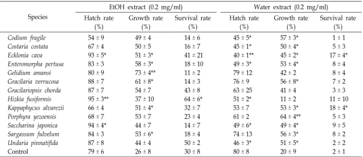
Table 1. Effects of seaweed extracts on hatching, growth, and survival rates in C. elegans
Species
EtOH extract (0.2 mg/ml) Water extract (0.2 mg/ml) Hatch rate
(%)
Growth rate (%)
Survival rate (%)
Hatch rate (%)
Growth rate (%)
Survival rate (%) Codium fragile
Costaria costata Ecklonia cava Enteromorpha pertusa Gelidium amansi Gracilaria verrucosa Gracilariopsis chorda Hizkia fusiformis Kappaphycus alvarezii Porphyra yezoensis Saccharina japonica Sargassum fulvelum Undaria pinnatifida Control
54 ± 9 67 ± 4 93 ± 5*
83 ± 3 80 ± 9 88 ± 7 87 ± 7 95 ± 3**
66 ± 4 68 ± 7 94 ± 4*
84 ± 3 87 ± 8 79 ± 6
49 ± 4 50 ± 5 51 ± 3*
58 ± 3*
73 ± 4**
61 ± 8*
54 ± 7 37 ± 10 51 ± 4*
53 ± 7 44 ± 7 53 ± 6*
44 ± 4 26 ± 8
14 ± 6 16 ± 7 41 ± 21 18 ± 10 11 ± 2 14 ± 3 43 ± 8 64 ± 6*
32 ± 7 23 ± 4 14 ± 7 18 ± 4 50 ± 2 30 ± 8
45 ± 5*
45 ± 1*
40 ± 1**
49 ± 3*
79 ± 12 76 ± 9 63 ± 25 51 ± 2*
53 ± 7 61 ± 2 49 ± 6*
74 ± 13 46 ± 3*
80 ± 8
57 ± 3*
50 ± 4*
45 ± 2*
53 ± 4*
42 ± 2 56 ± 8*
41 ± 4 11 ± 2 53 ± 3*
64 ± 4**
49 ± 4*
56 ± 3*
51 ± 5*
20 ± 9
1 ± 1 5 ± 3 17 ± 4*
8 ± 4 8 ± 4 7 ± 2 3 ± 3 11 ± 10 18 ± 4*
5 ± 3 9 ± 5 8 ± 2 2 ± 2 2 ± 1 Eggs (n≥100) or animals (n≥30) were cultured in 3 ml of NGM broth containing an ethanol or water extract of each seaweed (0.2 mg/ml) at 20℃. Hatch rate (%) is expressed as the number of hatched eggs at day 1 against the total eggs tested. Growth rate (%) is expressed as [(body length at day 2 - length at day 1) / length at day 1] x100. Survival rate (%) is expressed as the number of living animals at day 30 against the number of animals tested. Means ± SE (n=3). *p<0.05 and **p<0.01.
agar plate (20 g agar, 1 mM CaCl2, 1 mM MgSO4, 5 mM KPO4, pH 6, 1 l H2O) [5]. After adding 2.5 μl NaN3 (0.25 M) to immobilize animals within the target area, the number of animals in the area containing the attractant (10 μl of 1.25 M NH4Cl) or control were counted after 2 hr of incubation.
Chemotaxis index = (A–C) / T, where A is the number of animals at the attractant, C is the number of animals at the control, and T is total animals used. The brood size of progeny numbers per each adult was measured on NGM agar containing HFE (0.2 mg/ml). Six synchronized L1 ani- mals were transferred daily to fresh NGM containing extract and E. coli (killed) until no progenies were produced.
Hatched progenies were counted 2 days later, when the ani- mals reached the young adult stage. The total brood size was calculated by adding the numbers of progenies pro- duced during the animals’ lifetime. To examine effects of H.fusiformis extract on egg-laying time, approximately 30 an- imals at the L4 stage were placed on NGM agar, and egg-to- egg time was measured until the first egg was laid.
Analysis of chemical composition using GC-MS Chemical composition of the HFE was analyzed by gas chromatography–mass spectrometry (GC-MS) using a QP 5050A instrument (Shimadzu, Kyoto, Japan) equipped with a flame-ionization detector and compared with spectral data from the database. Analysis was performed on an HP-5 col-
umn (30 m × 0.25 mm, 0.25 μm; Agilent Technologies, Santa Clara, CA, USA). The temperature was initially held at 50°C for 2 min and raised to 150°C at 4°C/min and to 250°C at 7°C/min. Helium carrier gas was controlled at 0.6 ml/min with a split ratio of 1:50. The mass spectrometer was oper- ated in electron-ionization mode at 70 eV.
Statistical analysis
Statistical analysis was performed using one-way analysis of variance (ANOVA) followed by Duncan’s multiple range post hoc test and Student’s t-test. Values are presented as means ± standard error (SE) of at least three independent experiments. Mean values denoted by different letters were significantly different (p<0.05).
Results
To compare the effects of 13 common seaweed species (12 Korean + 1 Indonesisan K. alvarezii) on lifespan extension in C. elegans, we prepared ethanol and water extracts from each seaweed and measured their anti-aging potential as in- dicated by hatching, growth, and survival rates (Table 1).
When ethanol extracts were added to NGM broth to a final concentration of 0.05 mg/ml, H. fusiformis, Saccharina japon- ica, and Ecklonia cava showed significant positive effects on hatching rate (>90%) compared with the 5% Tween-80 con-
A B
C D
E
Fig. 1. Lifespan, chemotaxis, brood size, and egg-laying time in C. elegans. Lifespan of animals cultured on NGM agar and broth containing HFE was measured (A). Animals grown on NGM agar containing the extract (0.05 mg/
ml) for 3 or 7 days were tested with the chemotaxis in- dex (B). Brood size was calculated by number of prog- eny produced during the animals’ lifetime (C). Egg-lay- ing time was measured as the egg-to-egg time until the first egg was laid (D). Lifespan of animals cultured on NGM agar containing fucosterol was measured (E).
Different letters (a-b) indicate significant differences compared with control (p<0.05). Data represent means
± SE.
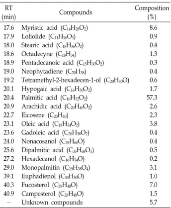
Table 2. Profile of the major compounds in the ethanolextract of H. fusiformis using GC-MS
RT
(min) Compounds Composition
(%) 17.6
17.9 18.0 18.6 18.9 19.0 19.2 20.1 20.4 20.9 22.7 23.1 23.6 24.0 25.6 27.2 29.0 39.1 40.3 40.9
-
Myristic acid (C14H28O2) Loliolide (C11H16O3) Stearic acid (C18H36O2) Octadecyne (C18H34)
Pentadecanoic acid (C15H30O2) Neophytadiene (C20H38)
Tetramethyl-2-hexadecen-1-ol (C20H40O) Hypogaic acid (C16H30O2)
Palmitic acid (C16H32O2) Arachidic acid (C20H40O2) Eicosene (C20H40) Oleic acid (C18H34O2) Gadoleic acid (C20H38O2) Nonacosanol (C29H60O) Dipalmitic acid (C35H68O5) Hexadecanol (C16H34O) Monopalmitin (C19H38O4) Euphadienol (C30H50O) Fucosterol (C29H48O) Campesterol (C28H48O) Unknown compounds
8.6 0.9 0.4 1.3 0.3 0.4 0.6 1.7 57.3 2.6 2.3 3.8 0.4 0.4 0.5 0.2 3.1 1.0 7.0 1.5 5.7 Composition values are percentages of the relative peak areas.
trol (79%). Most water extracts repressed hatching; espe- cially, E. cava water extract repressed the hatching rate down to 40%. Adding both ethanol and water extracts of H. fusi- formis (0.05 mg/ml) into NGM produced the least body length growth, presumably indicating an inverse relation- ship with longevity. Gelidium amansii ethanol extract and Porphyra yezoensis water extract enhanced body length.
Adding HFE significantly increased animal survival rate at d 30 to 64%, compared with the control (30%). Thus, we further investigated HFE for enhancement of C. elegans lifespan.
To evaluate the concentration dependency of the lon- gevity effects, HFE up to 1 mg/ml (as a final concentration in NGM) was added to NGM agar and broth (Fig. 1A). The lifespan of animals grown on NGM agar increased at 0.05 mg/ml (p<0.05). The mean lifespan at 0.05 mg/ml was 30.4
days, a significant 154% increase compared to the control (19.7 days). At higher concentrations, the lifespan gradually decreased. Therefore, in subsequent experiments, HFE was added to animal cultures at 0.05 mg/ml as the final concen- tration. In NGM broth, the lifespan peaked at 0.1 mg/ml with a 111% increase compared to the control, and then de- creased at higher doses.
Sensory function alterations could represent early in- dicators of life expectancy. By adding HFE (0.05 mg/ml), chemotaxis of the 3- and 7-day-old animals grown on NGM agar containing the extract increased slightly to 112% and 113%, respectively, compared to the control (Fig. 1B). This indicates that increasing trends in sensory functions with HFE appear to reflect lifespan extension, even though the values are not significantly different. Longevity has inverse correlations with fecundity and development; namely, long-lived animals have reduced brood size and delayed de- velopment time. With the addition of HFE (0.05 mg/ml), brood size of progeny per individual animal significantly decreased to 74% of the control (control: 396 progenies; ex- tract group: 294) (p<0.05) (Fig. 1C). This indicates that re- duced brood size with HFE demonstrates extension of lifespan. Developmental time or egg-laying time also sig-
nificantly decreased to 95% of the control (control: 66 hr;
extract group: 63 hr) (p<0.05) (Fig. 1D). The delayed devel- opmental time with HFE demonstrates extension of lifespan.
The chemical composition of the active HFE was analyzed by GC-MS. The major components by relative mass percent- age were palmitic acid (57.3%), myristic acid (8.6%), and fu- costerol (7.0%) (Table 2). Palmitic and myristic acids are common fatty acids in most organisms. Fucosterol is a phy- tosterol found in brown seaweed and has diverse biological activities to support nutraceutical applications. Therefore, we tentatively assumed that fucosterol was the main active constituent in HFE. Other minor sterol components such as campesterol and euphadienol and other compounds in HFE may also influence the effects of HFE on longevity. To eval- uate the effects of the main active compound fucosterol on lifespan, fucosterol was added to NGM agar at final concen- trations of up to 0.1 mg/ml (Fig. 1E). The lifespan of C. ele- gans significantly increased at 0.05 and 0.1 mg/ml fucosterol;
therefore, we determined the optimal effective dose to be 0.05 mg/ml. The lifespan at this dose was 14.3 days, sig- nificantly increased to 123% compared to the control (11.6 days) (p<0.05).
Discussion
Using C. elegans as a model, the edible H. fusiformis was the most effective seaweed in extending lifespan. H. fusi- formis has several health-promoting and beneficial nutrients, such as essential amino acids, n-3 polyunsaturated fatty acids (> 50% of total fatty acids), and dietary fiber (62% of biomass) [3]. It has also shown potent antioxiant [9], allelo- pathic [14], and proliferation activities on the osteosarcoma- derived cell line MG63 [8].
Deficits in sensory functions appear early in life, and sen- sory neuron functionality declines faster than locomotion during animal aging [16]. Wild-type 7-day-old C. elegans al- ready showed strong impairment of their sensory functions.
In these old animals, HFE showed a tendency to increase chemotaxis, albeit non-significantly. Preventing or retarding the loss of sensory functionality with HFE may accompany longevity of C. elegans. Moreover, we quantified the reduc- tion in broodsize and the delay in egg-laying time, parame- ters that inversely correlated with lifespan [13]. HFE sig- nificantly reduced fecundity and delayed development;
these changes might induce longevity by regulating over-ac- tivation of biological processes, such as development and
reproduction leading to aging in adulthood [6].
One of the major phytosterols in HFE was fucosterol (C29H48O; CAS number 17605-67-3), which isolated from brown algae mostly. Fucosterol elevates the enzyme activ- ities of free radical-scavenging superoxide dismutase, cata- lase, and glutathione peroxidase [11]. It also exhibits various biological activities, including antidiabetic, antioxidant, anti- photoaging, hepatoprotective, antihyperlipidemic, anti-in- flammatory, anticancer, antimicrobial, anti-obesity, anti-atop- ic, anticholinergic, anti-osteoporotic, and angiotensin-con- verting enzyme inhibitory properties [1]. Sterols are im- portant for mechanically strengthening membranes because they were competent in interactions with the phospholipid layers [2]. However, to date, no study has evaluated the ef- fects of HFE and fucosterol on longevity. Although the un- derlying mechanisms for extending the lifespan of C. elegans or other animals are not clear, we assume that they may involve antioxidant-related effects during the aging process that increase longevity. Additionally, when HFE and fucos- terol are compared, HFE shows generally higher longevity activities than fucosterol. This might be because of the pres- ence of various active compounds in HFE and which pro- vide synergistic effects. The practical synergic effects for life- span from HFE compounds have not been determined yet, but our data suggest that the edible and aquaculturable H.
fusiformis and/or fucosterol may have beneficial effects as a dietary supplement to support health care.
Acknowledgement
This work was supported by a research grant from Pukyong National University (Year 2019).
References
1. Abdul, Q. A., Choi, R. J., Jung, H. A. and Choi, J. S. 2016.
Health benefit of fucosterol from marine algae: a review.
J. Sci. Food Agric. 96, 1856-1866.
2. Dai, M. C., Chiche, H. B., Düzgünes, N., Ayanoglu, E. and Djerassi, C. 1991. Phospholipid studies of marine organisms:
26. Interactions of some marine sterols with 1-stearoyl02- oleoylphosphatidylcholine (SOPC) in model membranes.
Chem. Phys. Lipids 59, 245-253.
3. Dawczynski, C., Schubert, R. and Jahreis, G. 2007. Amino acids, fatty acids, and dietary fibre in edible seaweed prod- ucts. Food Chem. 103, 891-899.
4. Donguibogam Committee. 1999. Translated Donguibogam.
pp. 2198, Bubinmunwha Press, Seoul, Korea.
초록:갈조류 톳(
Hizikia fusiformis
)의 에탄올추출물 및 이의 활성성분 fucosterol에 의한 예쁜꼬마 선충의 수명 연장디야 파티마 옥타비아니1․배영석2․마리아 디아 누르 메이니타3․문일수4․홍용기1,5*
(1부경대학교 세계수산대학원, 2경북대학교 비케이21플러스그룹 생명과학부, 3인도네시아 젠데랄 소에디르만 대학
교 수산해양과학부, 4동국대학교 의과대학 해부학교실, 5부경대학교 생물공학과)
수명이 짧은 예쁜꼬마선충은 수명 연장 등 많은 연구의 모델생물으로서 사용되고있다. 해조류 추출물들이 포함 된 선충배양용 한천배지에서 선충(N2 야생형)을 키우면서 그 수명을 측정하였다. 13종의 흔한 해조류 중에서 갈 조류 톳의 에탄올추출물이 난 부화, 성장 및 생존율에서 가장 큰 효과를 보였다. 그 수명은 에탄올추출물(0.05 mg/ml) 및 주 활성성분인 fucosterol (0.05 mg/ml) 첨가에 의하여 1.54배 및 1.23배 정도로 유의미하게 증가되었 다. 또한 에탄올추출물에 의하여 chemotaxis는 1.13배 증가, 한 배에서의 새끼는 0.74배 감소, 첫 산란시기는 0.96 배 단축되었다. 이와 같은 결과들로 보아서 양식 가능한 해조류 톳은 건강에 이로운 건강보조 식품으로서의 좋은 재료가 될 수 있을 것이다.
5. Frøkjær-Jensen, C., Ailion, M. and Lockery, S. 2008. Ammo- nium-acetate is sensed by gustatory and olfactory neurons in Caenorhabditis elegans. Plos One 3, e2467.
6. Gems, D. and de la Guardia, Y. 2013. Alternative perspectives on aging in Caenorhabditis elegans: reactive oxygen species or hyperfunction? Antioxid. Redox. Signal. 19, 321-329.
7. Guiry, M. D. and Guiry, G. M. 2019. AlgaeBase: World-wide electronic publication, National University of Ireland, Galway.
http://www.algaebase.org; searched on 29 May 2019.
8. Huh, G. W., Lee, D. Y., In, S. J., Lee, D. G., Park, S. Y. and Yi, T. H. 2012. Fucosterols from Hizikia fusiformis and their proliferation activities on osteosarcoma-derived cell MG63.
J. Kor. Soc. Appl. Biol. Chem. 55, 551-555.
9. Karawita, R., Siriwardhana, N., Lee, K. W., Heo, M. S., Yeo, I. K., Lee, Y. D. and Jeon, Y. J. 2005. Reactive oxygen species scavenging, metal chelating, reducing power and lipid oxy- dation inhibition properties of different solvent fractions from Hizikia fusiformis. Eur. Food Res. Technol. 220, 363-371.
10. Kim, B., Suo, B. and Emmons, S. W. 2016. Gene function prediction based on developmental transcriptomes of the two sexes in C. elegans. Cell Rep. 17, 917-928.
11. Kim, S. K. and Ta, Q. V. 2011. Potential beneficial effects of marine algal sterols on human health. Adv. Food Nutr.
Res. 64, 191-198.
12. Korea Fisheries Association. 2017. Korean Fisheries Yearbook, pp. 541, Uno Design Press, Seoul, Korea.
13. Lee, Y., Hwang, W., Jung, J., Park, S., Cabatbat, J. J. T., Kim, P. J. and Lee, S. J. 2016. Inverse correlation between lon- gevity and developmental rate among wild C. elegans strains.
Aging 8, 986-999.
14. Ma, Z., Wu, M., Lin, L., Thring, R. W., Yu, H., Zhang, X.
and Zhao, M. 2017. Allelopathic interactions between the macroalga Hizikia fusiformis (Harvey) and the harmful blooms-forming dinoflagellate Karenia mikimotoi. Harmful Algae 65, 19-26.
15. Maeda, H., Hosokawa, M., Sashima, T., Funayama, K. and Miyashita, K. 2005. Fucoxanthin from edible seaweed, Unda- ria pinnatifida, shows antiobesity effect through UCP1 ex- pression in white adipose tissues. Biochem. Biophys. Res.
Commun. 332, 392-397.
16. Maglioni, S., Schiavi, A., Runci, A., Shaik, A. and Ventura, N. 2014. Mitochondrial stress extends lifespan in C. elegans through neuronal hormesis. Exp. Gerontol. 56, 89-98.
17. Pangestuti, R. and Kim, S. K. 2011. Neuroprotective effects of marine algae. Marine Drugs 9, 803-818.
18. Pant, A. and Pandey, R. 2015. Bioactive phytomolecules and ageing in Caenorhabditis elegans. Health Aging Res. 4, e19.
19. Strange, K. 2006. C. elegans Methods and Applications, pp. 287, Humana Press, New Jersey, USA.
20. Smit, A. J. 2004. Medicinal and pharmaceutical uses of sea- weed natural products: a review. J. Appl. Phycol. 16, 245-262.
21. Sundararajan, L., Stern, J. and Miller, D. M. 2019. Mechan- isms that regulate morphogenesis of a highly branched neu- ron in C. elegans. Develop. Biol. 451, 53-67.
22. The C. elegans sequencing consortium. 1998. Genome se- quence of the nematode C. elegans: a platform for investigat- ing biology. Science 282, 2012-2018.
23. Tseng, C. K. and Chang, C. F. 1984. Chinese seaweeds in herbal medicine. Hydrobiologia 116/117, 152-154.
24. Yagi, K. 1987. Lipid peroxides and human diseases. Chem.
Phys. Lipids 45, 337-351.