• 검색 결과가 없습니다.

Slow Coronary Flow is Related to Increased Carotid Intima-Media Thickness but Not Pulse Wave Velocity

N/A
N/A
Protected

Academic year: 2022

Share "Slow Coronary Flow is Related to Increased Carotid Intima-Media Thickness but Not Pulse Wave Velocity"

Copied!
5
0
0

로드 중.... (전체 텍스트 보기)

전체 글

(1)

666

Open Access

Slow Coronary Flow is Related to Increased Carotid Intima-Media Thickness but Not Pulse Wave Velocity

Bum Sung Kim, MD, Hyun-Joong Kim, MD, Seong Woo Han, MD, Sung Hea Kim, MD, Soon Yong Suh, MD, Sang Man Chung, MD, and Kyu Hyung Ryu, MD

Department of Cardiovascular Medicine, Konkuk University School of Medicine, Seoul, Korea ABSTRACT

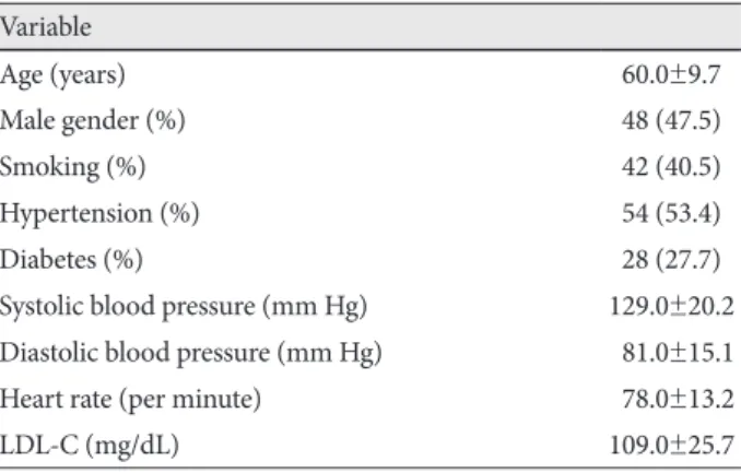
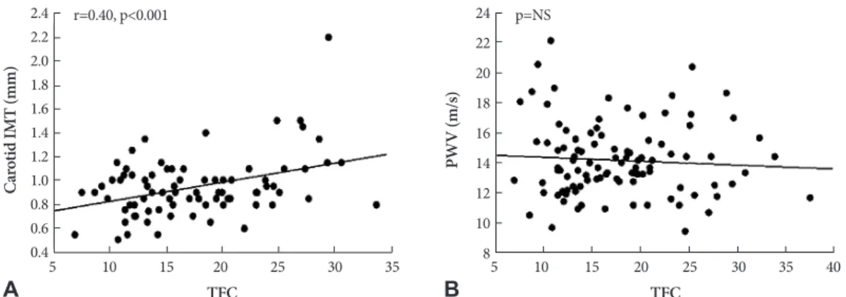
Background and Objectives: Slow coronary flow (SCF) is characterized by delayed contrast dye opacification without sig- nificant stenosis of epicardial coronary arteries. However, the pathophysiology and clinical implications of SCF are not fully understood. Some reports have suggested that SCF might be caused by atherosclerosis in the coronary artery microvascula- ture. Measuring carotid intima-media thickness (IMT) and pulse wave velocity (PWV), which are non-invasive and simple diagnostic tools, was developed to detect subclinical atherosclerosis. Thus, we determined IMT and PWV, and their possible relationship in a SCF group and a normal coronary flow (NCF) group of patients. Subjects and Methods: We included 101 patients who complained of chest pain but had a normal coronary angiogram. Thrombolysis in Myocardial Infarction frame count (TIMI frame count, TFC) was evaluated in the left and right coronary arteries. We defined SCF as a TFC of more than 25. Carotid IMT was measured by ultrasonography in both common carotid arteries. PWV was calculated from pulse tran- sit time between the brachial and ankle arteries. Results: Fifteen patients were included in the SCF group and 86 patients in the NCF group. Male patients (n=11, 73.3%) were significantly more common in the SCF group than in the NCF group (n=37, 43.0%, p<0.05). The TFC of the SCF and NCF groups were 28.8±3.5 and 15.7±4.5, respectively. The carotid IMT in the SCF group increased significantly compared to that in the NCF group (1.2±0.3 mm vs. 0.8±0.1 mm, p<0.01). However, no signifi- cant difference in PWV was observed between the two groups. Conclusion: SCF may reflect early atherosclerotic changes in the coronary artery microvasculature. (Korean Circ J 2011;41:666-670)

KEY WORDS: Microcirculation; Slow coronary flow; Carotid intima-media thickness.

Received: October 4, 2010 Revision Received: February 9, 2011 Accepted: April 19, 2011

Correspondence: Kyu Hyung Ryu, MD, Department of Cardiovascular Medicine, Konkuk University School of Medicine, 4-12 Hwayang-dong, Gwangjin-gu, Seoul 143-729, Korea

Tel: 82-2-2030-7512, Fax: 82-2-2030-6069 E-mail: [email protected]

• The authors have no financial conflicts of interest.

cc

This is an Open Access article distributed under the terms of the Cre- ative Commons Attribution Non-Commercial License (http://creativecom- mons.org/licenses/by-nc/3.0) which permits unrestricted non-commer- cial use, distribution, and reproduction in any medium, provided the origi- nal work is properly cited.

Introduction

Slow coronary flow (SCF) is characterized by delayed con- trast dye opacification without significant stenosis of epicar- dial coronary arteries. Most patients with SCF are debilitated with recurrent chest pain, commonly resulting in repeated hos- pital admissions.

1)

However, the pathophysiology and clini-

cal implications of SCF are not fully understood. It is likely that endothelial inflammation and microvascular vasomotor dys- function are associated with SCF pathophysiology.

2-8)

Some reports have suggested that SCF might be caused by athero- sclerotic changes in the coronary artery microvasculature and could be an early marker of subclinical atherosclerosis.

9)

Non-invasive and simple diagnostic tools have been deve-

loped to detect subclinical atherosclerosis. Intima-media th-

ickness (IMT) of the carotid artery reflects atherosclerotic

changes and strongly correlates with the presence and extent

of coronary artery disease.

10)11)

Arterial stiffness is also an im-

portant aspect of atherosclerosis related to vessel function that

can be evaluated noninvasively by measuring pulse wave ve-

locity (PWV), which is reportedly associated with the severi-

ty of vascular damage.

12)

Many studies in different populations

have shown that these are good parameters that indicate the

degree of vascular atherosclerosis.

11)12)

We hypothesized that

SCF is related to coronary artery atherosclerosis, so we com-

(2)

pared carotid IMT and PWV between an SCF and normal co- ronary flow (NCF) patient groups.

Subjects and Methods

Study population

The study was conducted from August 2005 to September 2006. We included 101 consecutive patients who complain- ed of chest pain but showed no significant coronary stenosis on coronary angiography. A complete history, as well as phy- sical and laboratory examinations, were obtained from all pa- tients before performing the coronary angiography. Smokers were defined as subjects who had smoked regularly for more than 1 year. Hypertension was defined as systolic blood pres- sure ≥140 mm Hg or diastolic blood pressure ≥90 mm Hg or current use of antihypertensive agents. Diabetes was defined as the use of insulin, oral hypoglycemic agents, or a fasting pla- sma glucose level of ≥126 mg/dL. After the coronary angio- gram, we confirmed the clinical diagnosis and categorized the patient as follows: acute myocardial infarction, unstable angina, microvascular angina, non-cardiac chest pain, or con- gestive heart failure. The definition of acute myocardial infarct was a typical rise and/or gradual fall (troponin) or a more rapid rise and fall (creatine kinase-MB fraction) of biochem- ical markers of myocardial necrosis, with at least one isch- emic symptom (chest pain) or electrocardiogram (ECG) ch- anges indicative of ischemia. Unstable angina was consider- ed as ischemic symptoms (chest pain) and no elevation in bio- chemical markers with ECG changes (new 1 mm or greater ST-segment depression or T-wave inversion in multiple pre- cordial leads). The definition of microvascular angina re- quired ischemic symptoms (chest pain) but no elevation in biochemical markers with the following ECG changes: ST- segment depression of 0.5-1 mm, T-wave flattening, or inver- sion <1 mm in the lead with a dominant R wave. The non- cardiac chest pain group complained of ischemic symptoms (chest pain) but no elevation in biochemical markers and a normal or unchanged ECG. Congestive heart failure was di- agnosed clinically using the Framingham criteria.

Coronary angiography and thrombolysis in myocardial infarction frame count

Coronary angiography was performed with the radial or femoral approach using the standard Judkins technique. Co- ronary arteries were shown in the left and right oblique planes and in cranial and caudal angulations. All angiograms were filmed at 15 frames per second. The Thrombolysis in Myo- cardial Infarction frame count (TIMI frame count, TFC) was obtained by the method of Gibson et al.

13)

The first frame used for counting was the first frame in which dye fully entered the artery. The last frame counted was the one in which dye first entered the end point branch off the target artery. The follow-

ing distal landmark branches of the target artery were used for the analysis: the distal bifurcation in the left anterior descend- ing artery (LAD), the distal bifurcation of the segment with the longest total distance in the left circumflex artery (LCX), and the first branch of the posterolateral artery in the right coronary artery (RCA). Because the length of the LAD is ana- tomically longer than that of the LCX and RCA, the LAD fra- me counts were corrected by dividing by 1.7 to obtain a cor- rected TFC, as previously reported.

13)

We calculated the mean TFC for the LAD, LCX, and RCA. All participants with a mean TFC greater than two standard deviations from mean TFC in the noncardiac chest pain subgroup were considered to have SCF, whereas those with a value below this threshold were considered to have NCF. The threshold TFC value was 25 frames.

Intima-media thickness and pulse wave velocity The carotid IMT was calculated by ultrasound equipment with a linear transducer (12-MHz, Vivid 7 dimension, GE He- althcare, Piscataway, NJ, USA). The right and left common carotid arteries were scanned in a longitudinal plane with loss of the parallel configuration of the near and far walls of the common carotid artery. The reference point for measuring IMT was the beginning of the dilatation of the carotid bulb in the bifurcation area. The IMT was measured manually as the thickest point of the transition zone between the lumen- intima and media-adventitia, 1 cm proximal to the reference point along at least 1 cm of axial length.

14)

The values for each carotid artery were averaged to obtain the mean carotid IMT.

Carotid IMT was assessed by two cardiologists blinded to other clinical data. Disagreements were resolved by a third observer.

We measured PWV on the left and right side using a Vase- ra VS-1000 instrument (Fukuda Denshi Co. Ltd, Tokyo, Japan), which simultaneously records PWV, blood pressure, ECG and heart sounds. The PWV is theoretically calculated as the distance from two sites over the pulse transit time. The time interval between the brachium and the ankle (ΔTba; pulse wave transit time) was defined as the time interval between the wave front of the brachial waveform and that of the ankle wave form. The distance between sampling points was calculated automatically according to subject height. The path length from the heart to the brachium (Lb) was expressed using the following equation: Lb=0.2195×height of the patient (cm)- 2.0734.

15)

The path length from the heart to the ankle (La) was expressed using the following equation: La=0.8129×height of the patient (cm)+12.328.

15)

Finally, the PWV was calculated from the following equation: PWV=(La-Lb)/ΔTba.

15)

The mean PWV value was calculated as the average of both PWV values.

Statistics

The statistical analysis was performed using the general

(3)

linear model in JMP7.0 software (SAS Inc., Cary, NC, USA).

The categorical variables were expressed as counts and per- cents. The categorical variables were compared with Pearson’s chi-square test and an analysis of variance. Continuous vari- ables are expressed as the mean±standard deviation (SD), and the independent t-test was used to compare the two gr- oups. The Pearson’s correlation test was used to determine the correlation between the TFC and the carotid IMT.

Results

Baseline characteristics

The clinical characteristics of the 101 patients in this study are summarized in Table 1. Overall, 54% of the subjects had hy- pertension, 28% had diabetes mellitus (DM), and 42% were smokers. Eighty-six patients were categorized into the NCF group, and 15 patients in the SCF group. Male patients (n=

11, 73.3%) were significantly more common in the SCF gr- oup than those in the NCF group (n=37, 43.0%, p<0.05). How- ever, age, presence of DM, hypertension, and low-density li- poprotein levels did not differ between the NCF and SCF gr- oups. Smoking tended to be more common in the SCF group, but the difference was not statistically significant (Table 2).

Clinical diagnosis and thrombolysis in myocardial infarction frame count

A significant difference was observed in the clinical diagno- ses between the SCF and NCF groups. Microvascular angina was the most common clinical diagnosis (n=11, 73.3%) in the SCF group, whereas non-cardiac chest pain was the most common diagnosis in the NCF group (n=41, 47.7%). The TFC was 28.8±3.5 frames in the SCF group and 15.7±4.5 frames in the NCF group (Table 2). In particular, the TFC increased significantly in a subgroup with microvascular angina (21.8±

7.8 frame count), compared to that in the other clinical sub- groups (Fig. 1). We also compared the TFC according to risk factors. The TFC in male patients tended to be higher than that of female patients (18.9±7.0 vs. 16.6±5.5, respectively),

but the result was not statistically significant. The TFC was not different with the presence of other risk factors, such as DM, hypertension, or smoking.

Carotid intima-media thickness and pulse wave velocity

Carotid IMT was positively correlated with TFC (r=0.40, p<0.001), but no significant correlation was found between PWV and TFC (Fig. 2). Carotid IMT increased significantly in the SCF group compared to that in the NCF group (1.2±

0.3 mm and 0.8±0.1 mm, respectively, p<0.01). However, no difference was found in the PWV between the SCF and NCF groups (SCF; 14.1±2.4 m/s, NCF; 14.5±2.8 m/s) (Fig. 3).

Table 1. Clinical characteristics of the study population (n=101) Variable

Age (years) 60.0±9.7

Male gender (%) 48 (47.5)

Smoking (%) 42 (40.5)

Hypertension (%) 54 (53.4)

Diabetes (%) 28 (27.7)

Systolic blood pressure (mm Hg) 129.0±20.2 Diastolic blood pressure (mm Hg) 081.0±15.1

Heart rate (per minute) 078.0±13.2

LDL-C (mg/dL) 109.0±25.7

LDL-C: low density lipoprotein-cholesterol

Table 2. Baseline clinical characteristics of the SCF and NCF groups

SCF (n=15) NCF (n=86) p Gender (%)

Male 11 (73.3) 37 (43.0)

p<0.05

Female 04 (26.7) 49 (57.0)

Age (years) 60.9±9.3 59.3±9.7 p=NS

DM (%) 05 (33.3) 23 (26.7) p=NS

Hypertension (%) 06 (40.0). 48 (55.8) p=NS

Smoking (%) 09 (60.0). 32 (37.2) p=NS

LDL-C (mg/dL) 114.4±28.2 103.9±28.4 p=NS

TFC (frame) 28.8±3.5 15.7±4.5 p <0.01

Clinical diagnosis (%)

Non-cardiac chest pain 02 (13.3) 41 (47.7) p<0.01

MA 11 (73.3) 18 (20.9) p<0.01

UA 000 (0.0).0 09 (10.5) p=NS

AMI 1 (6.7) 8 (9.3) p=NS

CHF 1 (6.7) 6 (7.0)0. p=NS

AMI: acute myocardial infarction, CHF: congestive heart failure, MA: microvascular angina, NCF: normal coronary flow, NS: not sig- nificant, SCF: slow coronary flow, TFC: Thrombolysis in Myocardial Infarction frame count, UA: unstable angina, DM: diabetes melli- tus, LDL-C: low density lipoprotein-cholesterol

Fig. 1. TFC for each clinical diagnosis. TFC increased significant- ly in the microvascular angina group compared to other clinical diagnoses (p<0.05 by analysis of variance). AMI: acute myocardi- al infarction, CHF: chronic heart failure, MA: microvascular angi- na, NC: non-cardiac chest pain, TFC: Thrombolysis in Myocardial Infarction frame count, UA: unstable angina.

30 25 20 15 10 5 0

p<0.05

TF C

NC UA AMI CHF MA Others

(4)

Discussion

After the first description of the SCF phenomenon in six pa- tients with typical angina pectoris by Tambe et al.

2)

SCF has been widely recognized during routine coronary angiogra- phy. SCF reveals various clinical presentations such as uns- table angina, acute myocardial infarction, ventricular tachy- cardia, hypertrophic cardiomyopathy, and microvascular angina.

1)16)17)

Camsari et al.

9)

used intravascular ultrasound to show that diffuse intimal thickening of the coronary arteries and ca- rotid IMT are correlated with TFC in patients with SCF.

18)

Some reports have suggested that homocysteine, a well-kn- own factor associated with generalized atherosclerosis, in- creases in patients with SCF.

14)

In other reports, simvastatin restores endothelial function and improves myocardial per- fusion in patients with SCF.

19)

These findings suggest that dif- fuse atherosclerosis may play a role in the pathophysiology of SCF. Similar to previous studies, our findings showed that TFC was positively correlated with the degree of carotid IMT, and that carotid IMT increased more significantly in the SCF group than that in the NCF group. According to these find- ings, it seems that the degree of atherosclerosis is a determin- ing factor of SCF.

In our study, SCF was defined as a TFC greater than 25 fr- ames (filmed at 15 frames per second). This is extreme SCF compared to previous studies.

9)13)14)

IMT of the carotid arteries in the SCF group increased greater in our results than those of other reports.

13)14)20)

These findings indicate that our subjects were more severe cases of SCF.

The results also showed no difference in the PWV between the SCF and NCF groups. PWV is considered a good indica- tor of the degree of atherosclerosis; however; compared to IMT, it seems to be less sensitive for coronary artery athero- sclerosis. Similar to the present study, research on the rela- tionship between carotid IMT, brachial-ankle PWV (baPWV), and the severity of coronary artery atherosclerosis, showed a relatively good correlation between carotid IMT and the Gen- sini score, which is a measure of the coronary artery disease severity, whereas baPWV correlated only weakly with this sc- ore.

21)

That is, carotid IMT reveals the degree of atherosclero- sis in coronary arteries, and it may be more suitable for evaluat- ing coronary microcirculation than baPWV.

21-23)

However, the reason for the different correlation in carotid IMT and baPWV is unclear. Therefore, further study is needed for confirmation.

Because SCF is associated with coronary atherosclerosis, it can be influenced by various cardiovascular risk factors such as gender, age, DM, hypertension, hyperlipidemia, and smok- ing. However, no significant differences were observed for age, gender, hypertension, DM, smoking, and total choleste- rol in a series of 50 patients with SCF.

9)

In another report, 47 patients with SCF also showed no significant differences in risk factors from the control group.

1)

Furthermore, we found no significant difference in the risk factors between the SCF and the NCF groups, except for gender. Although conven- tional cardiovascular risk factors reflect an advanced stage of atherosclerosis, we found no significant difference in conven- tional cardiovascular risk factors between the SCF and NCF groups. Thus, it is possible that risk factors other than conven- tional cardiovascular risk factors are present in patients with SCF. This should be explored further in a larger study.

There were some limitations to our study. First, we showed Fig. 2. A: a positive correlation was observed between carotid intima-media thickness (IMT) and the Thrombolysis in Myocardial Infarction frame count (TFC) (r=0.40, p<0.001). B: no significant correlation was found between pulse wave velocity (PWV) and the TFC. NS: not sig- nificant.

2.4 2.2 2.0 1.8 1.6 1.4 1.2 1.0 0.8 0.6 0.4

24 22 20 18 16 14 12 10 8

r=0.40, p<0.001 p=NS

Ca ro tid IM T (m m ) PW V (m /s)

TFC TFC

5 10 15 20 25 30 35 5 10 15 20 25 30 35 40

A B

Fig. 3. Comparison of the carotid intima-media thickness (IMT) and pulse wave velocity (PWV) between the normal coronary flow (NCF) and slow coronary flow (SCF) groups. A: carotid IMT increased significantly in the SCF group compared to that in the NCF group. B: no difference in the PWV between the SCF and NCF groups. NS: not significant.

p<0.01 p=NS

Ca ro tid IM T (m m ) PW V (m /s)

NCF SCF NCF SCF

0.8 14.1

1.2

14.5

A B

(5)

a correlation between SCF and carotid IMT and the possibi- lity of atherosclerosis in coronary microvasculature, but we did not directly assay the myocardial microvasculature itself to evaluate the degree of atherosclerosis. A pathological com- parison in patients with SCF will help in the understanding of the SCF mechanism. Second, because we included a small number of patients, some difficulties were encountered when identifying the characteristics of the cardiovascular risk fac- tors. A large multi-center based clinical trial is needed to iden- tify clinical characteristics and to manage patients with cor- onary microvascular dysfunction.

Conclusion

The results showed that SCF in patients with a normal an- giogram may reflect early atherosclerotic changes in the cor- onary microcirculation before the development of epicardial coronary artery atherosclerosis.

REFERENCES

1) Beltrame JF, Limaye SB, Horowitz JD. The coronary slow flow phe- nomenon: a new coronary microvascular disorder. Cardiology 2002;

97:197-202.

2) Tambe AA, Demany MA, Zimmerman HA, Mascarenhas E. Angina pectoris and slow flow velocity of dye in coronary arteries: a new angiographic finding. Am Heart J 1972;84:66-71.

3) Vrints C, Herman AG. Role of the endothelium in the regulation of coronary artery tone. Acta Cardiol 1991;46:399-418.

4) Kurtoglu N, Akcay A, Dindar I. Usefulness of oral dipyridamole thera- py for angiographic slow coronary artery flow. Am J Cardiol 2001;87:

777-9.

5) Motz W, Vogt M, Rabenau O, Scheler S, Lückhoff A, Strauer BE.

Evidence of endothelial dysfunction in coronary resistance vessels, in patients with angina pectoris and normal coronary angiograms. Am J Cardiol 1991;68:996-1003.

6) Quyyumi AA, Cannon RO 3rd, Panza JA, Diodati JG, Epstein SE.

Endothelial dysfunction in patients with chest pain and normal coro- nary arteries. Circulation 1992;86:1864-71.

7) Egashira K, Inou T, Hirooka Y, Yamada A, Urabe Y, Takeshita A. Ev- idence of impaired endothelium dependent coronary vasodilatation in patients with angina pectoris and normal coronary angiograms. N Engl J Med 1993;328:1659-64.

8) Zeiher AM, Krause T, Schächinger V, Minners J, Moser E. Impaired endothelium: dependent vasodilation of the coronary resistance ves- sels is associated with exercise-induced myocardial ischemia. Circu-

lation 1995;91:2345-52.

9) Camsari A, Ozcan T, Ozer C, Akcay B. Carotid artery intima-media thickness correlates with intravascular ultrasound parameters in pa- tients with slow coronary flow. Atherosclerosis 2008;200:310-4.

10) Wofford JL, Kahl FR, Howard GR, McKinney WM, Toole JF, Crouse JR 3rd. Relation of extent of extracranial carotid artery atherosclero- sis as measured by B-mode ultrasound to the extent of coronary ath- erosclerosis. Arterioscler Thromb 1991;11:1786-94.

11) Craven TE, Ryu JE, Espeland MA, et al. Evaluation of the associa- tions between carotid artery atherosclerosis and coronary artery ste- nosis: a case-control study. Circulation 1990;82:1230-42.

12) van Popele NM, Grobbee DE, Bots ML, et al. Association between arterial stiffness and atherosclerosis: the Rotterdam Study. Stroke 2001;

32:454-60.

13) Gibson CM, Cannon CP, Daley WL, et al. TIMI frame count: a quan- titative method of assessing coronary artery flow. Circulation 1996;

93:879-88.

14) Tanriverdi H, Evrengul H, Tanriverdi S, et al. Carotid intima-media thickness in coronary slow flow: relationship with plasma homocyste- ine levels. Coron Artery Dis 2006;17:331-7.

15) Yamashina A, Tomiyama H, Takeda K, et al. Validity, reproducibility, and clinical significance of noninvasive brachial-ankle pulse wave velocity measurement. Hypertens Res 2002;25:359-64.

16) Saya S, Hennebry TA, Lozano P, Lazzara R, Schechter E. Coronary slow flow phenomenon and risk for sudden cardiac death due to ventri- cular arrhythmias: a case report and review of literature. Clin Cardiol 2008;31:352-5.

17) Olivotto I, Cecchi F, Gistri R, et al. Relevance of coronary microvas- cular flow impairment to long-term remodeling and systolic dysfunc- tion in hypertrophic cardiomyopathy. J Am Coll Cardiol 2006;47:

1043-8.

18) Cin VG, Pekdemir H, Camsar A, et al. Diffuse intimal thickening of coronary arteries in slow coronary flow. Jpn Heart J 2003;44:907-19.

19) Cakmak M, Tanriverdi H, Cakmak N, Evrengul H, Cetemen S, Kuru O. Simvastatin may improve myocardial perfusion abnormality in slow coronary flow. Cardiology 2008;110:39-44.

20) Yigit F, Sezgin AT, Demircan S, Tekin G, Erol T, Muderrisoglu H.

Slow coronary flow is associated with carotid artery dilatation. Tohoku J Exp Med 2006;209:41-8.

21) Matsushima Y, Kawano H, Koide Y, et al. Relationship of carotid inti- ma-media thickness, pulse wave velocity, and ankle brachial index to the severity of coronary artery atherosclerosis. Clin Cardiol 2004;27:

629-34.

22) Megnien JL, Simon A, Denarie N, et al. Aortic stiffening does not predict coronary and extracoronary atherosclerosis in asymptomatic men at risk for cardiovascular disease. Am J Hypertens 1998;11:

293-301.

23) Seo WW, Chang HJ, Cho I, et al. The value of brachial-ankle pulse

wave velocity as a predictor of coronary artery disease in high-risk pa-

tients. Korean Circ J 2010;40:224-9, doi: 10.4070/kcj.2010.40.5.224.

수치

Fig. 1. TFC for each clinical diagnosis. TFC increased significant- significant-ly in the microvascular angina group compared to other clinical  diagnoses (p&lt;0.05 by analysis of variance)
Fig. 3. Comparison of the carotid intima-media thickness (IMT)  and pulse wave velocity (PWV) between the normal coronary  flow (NCF) and slow coronary flow (SCF) groups

참조

관련 문서

This study was conducted to define the related risk factors with carotid intima-media thickness (MT) of the rural elderly using B mode ultrasonography.. The

Correlations between aortic stiffness index β and intima-media thickness (IMT) of carotid and descending thoracic aorta.. cal reduction of perfusion that impairs neurological function

Linear regression analysis among the mean carotid intima-media thickness (IMT) (A), total plaque area (TPA) (B), total plaque volume (TPV) (C), and the severity of the coronary

경동맥 내중막 두께 (Intima-media thickness, IMT) 측정 본 연구에서는 원위 총 경동맥(Distal common carotid artery) 의 1 cm 분기점 이후의 원 벽(Far wall)에서

Keywords : permanent magnet, Hall device, atherosclerosis, pulse wave velocity (PWV), pulse transit time (PTT), pulse wave of carotid artery, pulse wave of radial

Association of Carotid Artery Intima-Media Thickness and Cardiovascular Risk Factors in Adult. Mi-Young Kim ․ Hwa-Sun Kim 1) ․ Shin-Young Kim 2) Dept. of Diagnostic

초음파를 이용한 경동맥 내-중막 두께와 대사증후군 진단지표의 연관성 ― Relationship Between Carotid Intima-Media Thickness Using Ultrasonography and Diagnostic

CVD, cardiovascu- lar disease; RA, rheumatoid arthritis; CIMT, carotid intima media thickness; T-Chol, total cholesterol; TG, triglyceride; HDL, high density lipoprotein; LDL,