Endothelial Dysfunction and Increased Carotid Intima-Media Thickness in the Patients with Slow Coronary Flow
Flow mediated brachial dilatation (FMD) and carotid intima-media thickness (IMT) have been a surrogate for early atherosclerosis. Slow coronary flow in a normal coronary angiogram is not a rare condition, but its pathogenesis remains unclear. A total of 85 patients with angina were evaluated of their brachial artery FMD, carotid IMT and conventional coronary angiography. Coronary flow was quantified using the corrected thrombosis in myocardial infarction (TIMI) frame count method. Group I was a control with normal coronary angiography (n = 41, 56.1 ± 8.0 yr) and group II was no significant coronary stenosis with slow flow (n = 44, 56.3 ± 10.0 yr). Diabetes was rare but
dyslipidemia and family history were frequent in group II. Heart rate was higher in group II than in group I. White blood cells, especially monocytes and homocysteine were higher in group II. The FMD was significantly lower in group II than in group I. Elevated heart rate, dyslipidemia and low FMD were independently related with slow coronary flow in regression analysis. Therefore, endothelial dysfunction may be an earlier vascular phenomenon than increased carotid IMT in the patients with slow coronary flow.
Key Words: Endothelium; Coronary Artery; Carotid Intima Hyun Ju Yoon1, Myung Ho Jeong1,
Sook Hee Cho2, Kye Hun Kim1, Min Goo Lee1, Keun Ho Park1, Doo Sun Sim1, Nam Sik Yoon1, Young Joon Hong1, Ju Han Kim1, Youngkeun Ahn1, Jeong Gwan Cho1, Jong Chun Park1, and Jung Chaee Kang1
1The Heart Center of Chonnam National University Hospital and 2Department of Nursing, Nambu University, Gwangju, Korea
Received: 8 November 2011 Accepted: 13 March 2012 Address for Correspondence:
Myung Ho Jeong, MD
Director of Heart Research Center Nominated by Korea Ministry of Health and Welfare, Chonnam National University Hospital, 167 Jaebong-ro, Dong-gu, Gwangju 501-757, Korea Tel: +82.62-220-6243, Fax: +82.62-228-7174 E-mail: [email protected]
This study was supported by a grant from the Korea Healthcare Technology R&D Project, Ministry for Health, Welfare & Family Affairs, Republic of Korea Government (A084869).
http://dx.doi.org/10.3346/jkms.2012.27.6.614 • J Korean Med Sci 2012; 27: 614-618
INTRODUCTION
Coronary slow flow is not a rare finding which characterized by angiographically normal or near-normal coronary arteries with delayed progression of the contrast agent into distal vasculature.
It is characterized by slow velocity flow of dye through the coro- nary artery in the absence of any evident obstructive lesion in it grossly. Since first description of this phenomenon in 1972, the mechanism of this phenomenon has not been extensively stud- ied so far (1). On the basis of myocardial biopsy studies, it can be suggested that a combination of structural and functional obstruction exists in the coronary microcirculation (2, 3).
Previous studies have shown that impaired endothelial func- tion plays an important role in this abnormal coronary vasore- activity (4). Several surveys have investigated the relationship between coronary slow flow phenomenon and endothelial dys- function as a probable etiology.
Among several atherosclerotic surrogate, increased carotid intima-media thickness (IMT) was positively related with coro- nary artery severity and cardiovascular event; therefore it regard- ed as an early indicator of overall atherosclerosis (5). In the pres-
ent studies, the patients with obesity or ischemic heart disease proven coronary angiography were found to have increased IMT and impaired FMD compared to the healthy controls (6).
However, the situation might not be same in patients with coro- nary slow flow. Therefore, we aimed to determine the relation- ship among the vascular risk factors, including endothelial dys- function and carotid IMT in the patients with slow coronary flow.
MATERIALS AND METHODS Study populations
The study population consisted of 150 patients with newly diag- nosed stable angina. All of them were evaluated conventional coronary angiography, brachial artery FMD, and carotid IMT.
We excluded subjects with significant narrowed coronary artery from coronary angiography (n = 65, 59.5 ± 8 yr). Eighty-five pa- tients without significant coronary stenosis were divided into two groups according to the presence of coronary slow flow:
Group I included patients without slow flow (n = 41, 56.1 ± 8.0 yr) and group II patients with slow flow (n = 44, 56.3 ± 10.0 yr).
We evaluated the relationship of the vascular risk factors, includ-
ing endothelial dysfunction and carotid IMT in the patients with slow coronary flow.
Definition of slow coronary flow
Determination of frame counts was carried out by the method described previously by Gibson et al. (7). According to correct- ed thrombolysis in myocardial infarction (TIMI) frame count, slow flow was defined as more than 2 standard deviations of frame count (TIMI 2) from the normal published range for that particular vessel (7). We corrected the TIMI frame count for the left anterior descending (LAD) to take account of the longer dis- tance to the TIMI landmark. This ratio was obtained by dividing the mean TIMI frame count of the LAD by the mean TIMI frame count of the circumflex and the right coronary artery. More than two experienced cardiologists reviewed all the patients’ angiog- raphy and calculated the frame count. Interobserver and intrao- bserver agreement (κ-value) were 0.73 and 0.78 for TIMI flow grade respectively. Interobserver and intraobserver coefficients of variation were 9.72% and 5.86% for corrected TIMI frame count. Normal coronary artery was defined as no significant stenosis even at < 50% of the diameter of the coronary artery.
Measurement of endothelial and vascular smooth muscle dysfunction
A Sequoia 512 ultrasound system (Siemens Corp, Upplands- Väsby, Sweden) was used with a 15L8 transducer for brachial artery and a 8L5 transducer for carotid artery studies. All investi- gations were digitally stored for analyses, which were performed by a single observer (EL, intraobserver variability r = 0.988) on a Sequoia 512. R-wave triggered end diastolic right brachial ar- tery longitudinal images proximal to the antecubital fossa were recorded at baseline after 15 min of supine rest. Transducer po- sition was carefully noted for subsequent investigations. For flow mediated dilatation (FMD), R-wave triggered images were stored for 90 sec following ischemia. The ischemia was induced using a cuff on the forearm inflated 20 mmHg above the systolic blood pressure for five minutes with additional lower arm muscular work by repetitively squeezing a small ball during the last min- ute of ischemia. Maximal brachial artery diameter was calcu- lated for both conditions as a mean of three measurements (8).
After 15 min of recovery to the baseline diameter, 400 μg sublin- gual nitroglycerine was administered and nitrogen mediated diameter (NMD) was assessed. FMD and NMD values were ex- pressed as percentage change from baseline value. The repro- ducibility of this method was reliable level in this study (r = 0.997, P < 0.001).
Measurement of carotid artery intima-media thickness Carotid artery studies were performed with the individual in the supine position with the neck extended and the chin turned away from the side being examined. The right and left common
carotid artery proximal to the bulb was imaged in multiple lon- gitudinal planes for the best resolution of the IMT of the far wall.
The mean IMT was obtained manually tracing the intima-me- dia in the far wall of the artery for a distance of approximately 10 mm (9). Measurements were performed on three end dia- stolic images and averaged.
Statistical analysis
All eligible patients enrolled in this study were included in the analysis. Standard statistics were used to describe the baseline clinical characteristics. Continuous variables are presented as mean ± standard deviations. Comparisons of means between the groups were done using a Student’s t-test and analysis of variance (ANOVA) as appropriate. A P value < 0.05 was con- sidered statistically significant. To find out optimal cutoff val- ues, receiver operating characteristic (ROC) analysis was per- formed. Sensitivity, specificity were calculated using typical for- mulas. No missing value imputation was performed. All statisti- cal analysis were performed with SPSS 15.0 for Windows (SPSS Inc., Chicago, IL, USA).
Ethics statement
The study protocol was reviewed and approved by the institu- tional review board of Chonnam National University Hospital (No. 2010-05-092). All subjects provided their informed, written consent before participation.
RESULTS
Baseline characteristics
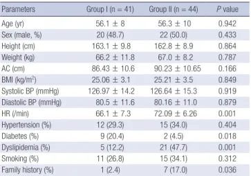
As shown in Table 1, there were no significant differences in age and gender between two groups. Baseline heart rate was higher in group II than in group I. Dyslipidemia and family history of premature cardiovascular disease were significantly frequent in
Table 1. Baseline clinical characteristics
Parameters Group I (n = 41) Group II (n = 44) P value
Age (yr) 56.1 ± 8 56.3 ± 10 0.942
Sex (male, %) 20 (48.7) 22 (50.0) 0.433
Height (cm) 163.1 ± 9.8 162.8 ± 8.9 0.864
Weight (kg) 66.2 ± 11.8 67.0 ± 8.2 0.787 AC (cm) 86.43 ± 10.6 90.23 ± 10.65 0.166
BMI (kg/m2) 25.06 ± 3.1 25.21 ± 3.5 0.849
Systolic BP (mmHg) 126.97 ± 14.2 126.64 ± 15.3 0.919 Diastolic BP (mmHg) 80.5 ± 11.6 80.16 ± 11.0 0.879
HR (/min) 66.1 ± 7.3 72.09 ± 6.26 0.001
Hypertension (%) 12 (29.3) 15 (34.0) 0.404
Diabetes (%) 9 (20.4) 2 (4.5) 0.018
Dyslipidemia (%) 5 (12.2) 21 (47.7) 0.001
Smoking (%) 11 (26.8) 15 (34.1) 0.312
Family history (%) 1 (2.4) 7 (17.0) 0.036
Data are given as mean ± SD or No. (%). AC, abdominal circumference; BMI, body mass index; BP, blood pressure; HR, heart rate.
group II. The prevalence of diabetes was lower in groups II than in group I. The percentage of hypertension and smoking were not different between groups.
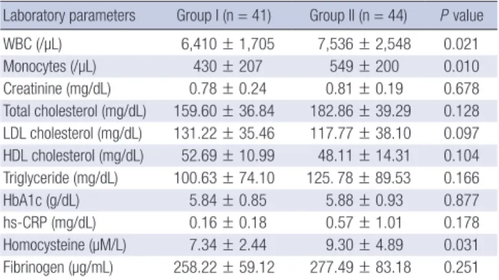
Laboratory findings in the patients with coronary slow flow
White blood cell count, especially monocytes was higher in group II than in group I. In spite of dyslipidemia was more fre- quent in group II, the level of lipid profile contained total cho- lesterol, low density lipoprotein (LDL) cholesterol, high density lipoprotein (HDL) and triglyceride was not significant different between groups. The level of homocysteine was higher in group II than in group I significantly. Other inflammatory marker such as high sensitivity C-reactive protein and fibrinogen were not significantly different between groups (Table 2).
The difference of endothelial and vascular smooth muscle function
The FMD showed normal distribution pattern; mean FMD was 7.22 ± 3.62 (minimum 1.79% to maximum 21.43%). The FMD was significantly lower in group II than in group I (5.52 ± 2.18 vs
9.03 ± 3.98, P < 0.001). NMD was not significantly different be- tween two groups (Fig. 1). Avarage carotid IMT was 0.61 ± 0.15 in the patients of this study. Carotid IMT was tended to be high- er in group II, without no statistical significance (Table 3).
The independent risk factors of slow coronary flow In regression analysis, elevated heart rate, dyslipidemia and low FMD were independently related with slow coronary flow in regression analysis (Table 4). The cut off value of FMD for pre-
Table 2. Laboratory findings in the patients with slow coronary flow
Laboratory parameters Group I (n = 41) Group II (n = 44) P value WBC (/µL) 6,410 ± 1,705 7,536 ± 2,548 0.021
Monocytes (/µL) 430 ± 207 549 ± 200 0.010
Creatinine (mg/dL) 0.78 ± 0.24 0.81 ± 0.19 0.678 Total cholesterol (mg/dL) 159.60 ± 36.84 182.86 ± 39.29 0.128 LDL cholesterol (mg/dL) 131.22 ± 35.46 117.77 ± 38.10 0.097 HDL cholesterol (mg/dL) 52.69 ± 10.99 48.11 ± 14.31 0.104 Triglyceride (mg/dL) 100.63 ± 74.10 125. 78 ± 89.53 0.166
HbA1c (g/dL) 5.84 ± 0.85 5.88 ± 0.93 0.877
hs-CRP (mg/dL) 0.16 ± 0.18 0.57 ± 1.01 0.178 Homocysteine (µM/L) 7.34 ± 2.44 9.30 ± 4.89 0.031 Fibrinogen (µg/mL) 258.22 ± 59.12 277.49 ± 83.18 0.251 Data are given as mean ± SD. HDL, high density lipoprotein; hs-CRP, high-sensitivity C-reactive protein; WBC, white blood cells.
%
P = 0.001
P = NS
FMD NMD
20
15
10
5
0
Control Slow flow
Fig. 1. Differences of endothelial and vascular smooth muscular function. FMD, flow mediated dilatation; NMD, nitrogen mediated dilatation.
Fig. 2. Receiver operator characteristics curves of slow coronary flow. Arrow means cut off value of FMD (Area under the curve = 0.787, sensitivity = 0.773, specificity = 0.732).
Sensitivity
1.0
0.8
0.6
0.4
0.2
0.0
1-Specificity
FMD = 6.9767
0.0 0.2 0.4 0.6 0.8 1.0
Table 3. The difference of atherosclerotic surrogate in the patients with slow coro- nary flow
Parameters Group I (n = 41) Group II (n = 44) P value Pre FMD diameter (mm) 3.93 ± 0.67 4.09 ± 0.8 0.327 Post FMD diameter (mm) 4.27 ± 0.65 4.31 ± 0.81 0.783
FMD (%) 9.03 ± 3.93 5.52 ± 2.18 0.001
Pre NMD diameter (mm) 4.53 ± 0.27 4.12 ± 0.814 0.444 Post NMD diameter (mm) 4.69 ± 0.68 4.76 ± 0.81 0.660
NMD (%) 16.87 ± 18.4 16.37 ± 8.4 0.871
Left carotid IMT (mm) 0.55 ± 0.14 0.62 ± 0.17 0.095 Right carotid IMT (mm) 0.56 ± 0.13 0.60 ± 0.24 0.529 Data are given as mean ± SD. IMT, intima-media thickness; FMD, flow mediated dilatation; NMD, nitrogen mediated dilatation.
Table 4. Independent predictors of slow coronary flow
Predictors Hazard ratio Confidence interval P value Heart rate ( > 70/min) 4.971 1.264-19.552 0.022
Dyslipidemia 13.050 1.739-97.910 0.012
Family history 5.229 0.091-302.080 0.424
WBCs 2.642 0.642-10.877 0.178
Monocytes 0.862 0.200-3.707 0.841
Homocysteine 1.468 0.370-5.830 0.585
FMD 17.228 3.006-98.774 0.001
CCA IMT, common carotid artery intima-media thickness; FMD, flow mediated dilata- tion.
diction of slow flow was 6.97% (area under the curve = 0.787, sensitivity = 77.3%, specificity = 73.2%) (Fig. 2).
DISCUSSION
Slow coronary flow means the slow dye progression into distal vasculature of coronary arteries during coronary angiography.
It was reported in approximately 1% of patients undergoing con- ventional coronary angiography (10). The exact mechanism was not clear until now; small vessel dysfunction may be involved in this phenomenon. Increased flow resistance, increased mi- crovascular tone, platelet dysfunction (11), early demonstration of diffuse atherosclerosis (12), inflammation (13, 14), and an imbalance of vasoactive substances (15) have been suggested as underlying mechanisms.
In our study, baseline heart rate was higher in slow flow group and heart rate more than 70/min was the independent factor of slow flow. Increased sympathetic tone with elevated catechol- amine levels may have direct effects on vessel or it may affect other factors promoting the progression of atherosclerosis. If we could measure the baseline catecholamine level, it would be more supportive presenting this phenomenon. This cate- cholamine may be a role for the difference of family history in this study due to emotional stress. In spite of insignificance of the level of lipid profile, dyslipidemia was also significantly fre- quent in slow flow group and an independent predictor of slow flow in this study. Fortunately, the patients with previous medi- cated statin were excluded in this study, because they contained the group of significant coronary stenosis. Hyperlipidemia has been associated with an increased blood viscosity, which causes an increase in capillary resistance (16). Although diabetes was frequent risk factor of cardiovascular disease increasing inflam- mation and vascular damage, diabetes was significantly more common in the control group than in slow flow group in this study. Because we analyzed the control and slow flow group after excluded the significant coronary artery lesion, it may be a result from analysis of the covariance for omission of the con- founding factor. Vascular inflammatory marker was variety. In this study, we checked white blood cells with differential count, fibrinogen, hs-CRP and homocysteine. White blood cells, espe- cially monocytes and homocysteine were elevated in the pa- tient with slow flow. In a previous study, homocysteine levels increased but folate levels decreased in patients with slow coro- nary flow (17). Even though we did not check the folate levels, the possible disturbance in the metabolism of homocysteine in patients with coronary slow flow may have a role in the patho- genesis of this phenomenon associated with inflammation by causing generalized atherosclerosis.
Different theories have been postulated about the cause of small-vessel dysfunction, including microvascular tone altera- tion, small-vessel wall thickening (18), patchy fibrosis and im-
paired endothelial release of nitric oxide (NO) (19, 20). The en- dothelium plays a crucial role for initiation of atherosclerosis in early stage. Endothelial dysfunction was considered as early marker of atherosclerosis (21, 22). In this study, the FMD was significantly lower in slow flow group than in control and low FMD were independently related with slow coronary flow in regression analysis. The cut off value of FMD for prediction of slow flow was 6.97% from ROC curve. Vascular smooth muscle dysfunction from NMD was not affected in coronary slow flow.
Collectively, it can be hypothesized that the impaired peripheral endothelial function (assessed by FMD of the brachial artery), not vascular smooth muscle, may also suggest the involvement of epicardial coronary arteries with slow flow. Another surro- gate of early atherosclerosis is carotid IMT. Carotid arterial IMT correlates well with most cardiovascular risk factors and, in pop- ulation studies, increasing carotid arterial wall thickness (IMT and plaques) has been associated with worsening coronary ar- tery disease. Several studies have now demonstrated that carot- id IMT is significantly increased in patients with slow coronary flow (23). However, our data showed carotid IMT was not sig- nificantly different between normal and slow coronary flow pa- tients. Since FMD constitutes a physiologic assessment of en- dothelial dysfunction and carotid IMT is an anatomic structural measure of subclinical atherosclerosis, endothelial dysfunction is an earlier phenomenon than increased carotid IMT on the cas- cade of atherosclerosis in the patients with slow coronary artery.
This study had several limitations. There was a patient selec- tion bias. Because this study was cross-sectional, baseline char- acteristics of this study could not be controlled, especially risk factors. Also, we did not consider about previous medication history. Although we measured FMD and carotid IMT in the state of holding drug during one day, previous duration of anti- anginal or antiplatelets might affect the results of frame count in coronary angiography cession. There had insufficient impact for endothelial dysfunction due to absence of laboratory mark- ers. Finally, our study population was relatively small, and fur- ther large-scale clinical and experimental studies are warranted to detect precise pathogenesis and effective therapy for this sys- temic disorder.
Notwithstanding all these limitations, the data suggest that the slow coronary flow is not a simple, isolated change local- ized in epicardial coronary arteries, but may be a part of sys- temic vascular disturbance.
REFERENCES
1. Tambe AA, Demany MA, Zimmerman HA, Mascarenhas E. Angina pectoris and slow flow velocity of dye in coronary arteries: a new angio- graphic finding. Am Heart J 1972; 84: 66-71.
2. Mangieri E, Macchiarelli G, Ciavolella M, Barillà F, Avella A, Martinotti A, Dell’ltalia LJ, Scibilia G, Motta P, Campa PP. Slow coronary flow: clini-
cal and histopathological features in patients with otherwise normal epicardial coronary arteries. Cathet Cardiovasc Diagn 1996; 37: 375-81.
3. Mosseri M, Yarom R, Gotsman MS, Hasin Y. Histologic evidence for small- vessel coronary artery disease in patients with angina pectoris and pat- ent large coronary arteries. Circulation 1986; 74: 964-72.
4. Singh S, Kothari SS, Bahl VK. Coronary slow flow phenomenon: an an- giographic curiosity. Indian Heart J 2004; 56: 613-7.
5. Simon A, Gariepy J, Chironi G, Megnien JL, Levenson J. Intima-media thickness: a new tool for diagnosis and treatment of cardiovascular risk.
J Hypertens 2002; 20: 159-69.
6. Roh EJ, Lim JW, Ko KO, Cheon EJ. A useful predictor of early atheroscle- rosis in obese children: serum high-sensitivity C-reactive protein. J Korean Med Sci 2007; 22: 192-7.
7. Gibson CM, Cannon CP, Daley WL, Dodge JT Jr, Alexander B Jr, Marble SJ, McCabe CH, Raymond L, Fortin T, Poole WK, et al. TIMI frame count:
a quantitative method of assessing coronary artery flow. Circulation 1996; 93: 879-88.
8. Corretti MC, Anderson TJ, Benjamin EJ, CelermajerD, Charbonneau F, Creager MA, Deanfield J, Drexler H, Gerhard-Herman M, Herrington D, et al. Guidelines for the ultrasound assessment of endothelial-depen- dent flow-mediated vasodilation of the brachial artery: a report of the International Brachial Artery Reactivity Task Force. J Am Coll Cardiol 2002; 39: 257-65.
9. Adams MR, Nakagomi A, Keech A, Robinson J, McCredie R, Bailey BP, Freedman SB, Celermajer DS. Carotid intima-media thickness is only weakly correlated with the extent and severity of coronary artery disease.
Circulation 1995; 92: 2127-34.
10. Beltrame JF, Limaye SB, Wuttke RD, Horowitz JD. Coronary hemody- namic and metabolic studies of the coronary slow flow phenomenon.
Am Heart J 2003; 146: 84-90.
11. Gökçe M, Kaplan S, Tekelioğlu Y, Erdoğan T, Küçükosmanoğlu M. Plate- let function disorder in patients with coronary slow flow. Clin Cardiol 2005; 28: 145-8.
12. Cin VG, Pekdemir H, Camsar A, Ciçek D, Akkus MN, Parmaksýz T, Katýrcybaý T, Döven O. Diffuse intimal thickening of coronary arteries
in slow coronary flow. Jpn Heart J 2003; 44: 907-19.
13. Li JJ, Xu B, Li ZC, Qian J, Wei BQ. Is slow coronary flow associated with inflammation? Med Hypotheses 2006; 66: 504-8.
14. Jin SM, Noh CI, Yang SW, Bae EJ, Shin CH, Chung HR, Kim YY, Yun YS.
Endothelial dysfunction and microvascular complications in type 1 dia- betes mellitus. J Korean Med Sci 2008; 23: 77-82.
15. Sezgin N, Barutcu I, Sezgin AT, Gullu H, Turkmen M, Esen AM, Karaka- ya O. Plasma nitric oxide level and its role in slow coronary flow phe- nomenon. Int Heart J 2005; 46: 373-82.
16. Rim SJ, Leong-Poi H, Lindner JR, Wei K, Fisher NG, Kaul S. Decrease in coronary blood flow reserve during hyperlipidemia is secondary to an increase in blood viscosity. Circulation 2001; 104: 2704-9.
17. Riza Erbay A, Turhan H, Yasar AS, Ayaz S, Sahin O, Senen K, Sasmaz H, Yetkin E. Elevated level of plasma homocysteine in patients with slow coronary flow. Int J Cardiol 2005; 102: 419-23.
18. Fineschi M, Bravi A, Gori T. The “slow coronary flow” phenomenon: evi- dence of preserved coronary flow reserve despite increased resting micro- vascular resistances. Int J Cardiol 2008; 127: 358-61.
19. Sezgin AT, Sigirci A, Barutcu I, Topal E, Sezgin N, Ozdemir R, Yetkin E, Tandogan I, Kosar F, Ermis N, et al. Vascular endothelial function in pa- tients with slow coronary flow. Coron Artery Dis 2003; 14: 155-61.
20. Wu WC, Sharma SC, Choudhary G, Coulter L, Coccio E, Eaton CB. Flow- mediated vasodilation predicts the presence and extent of coronary ar- tery disease assessed by stress thallium imaging. J Nucl Cardiol 2005; 12:
538-44.
21. Fineschi M, Gori T. Coronary slow flow: description of a new “cardiac Y”
syndrome. Int J Cardiol 2009; 137: 308-10.
22. Tanriverdi H, Evrengul H, Mergen H, Acar C, Seleci D, Kuru O, Tanriverdi S, Kaftan A. Early sign of atherosclerosis in slow coronary flow and rela- tionship with angiotensin-converting enzyme I/D polymorphism. Heart Vessels 2007; 22: 1-8.
23. Tanriverdi H, Evrengul H, Tanriverdi S, Kuru O, Seleci D, Enli Y, Kaftan A, Kilic M. Carotid intima-media thickness in coronary slow flow: relation- ship with plasma homocysteine levels. Coron Artery Dis 2006; 17: 331-7.