Effects of Methotrexate on Carotid Intima-media Thickness in Patients with Rheumatoid Arthritis
The purpose of this study was to evaluate the effects of rheumatoid arthritis (RA) and anti- rheumatic drugs on atherosclerosis by comparing carotid intima-media thickness (CIMT) as an indicator for cardiovascular diseases (CVD). This study included 44 female RA patients who met the 2010 ACR/EULAR criteria and age-matched 22 healthy females. CIMT was measured on both carotid arteries using a B-mode ultrasound scan. The mean value of both sides was taken as the CIMT of the subject. The CIMT was evaluated according to the use of drugs, disease activity and CVD risk factors in RA patients as a case-control study.
Higher CIMT was observed in RA patients as compared with healthy subjects
(0.705 ± 0.198 mm, 0.611 ± 0.093 mm, respectively, P < 0.05). With adjustment for the CVD risk factors, disease activity and the use of anti-rheumatic drugs, methotrexate (MTX) only showed a favorable effect on CIMT in RA. A significantly lower CIMT was observed in RA with MTX as compared with RA without MTX (0.644 ± 0.136 mm, 0.767 ± 0.233 mm, respectively, P < 0.05). The effects were correlated with MTX dosage (β = -0.029, P < 0.01). The use of MTX should be considered in high priority not only to control arthritis but also to reduce the RA-related CVD risk to mortality.
Keywords: Arthritis, Rheumatoid; Carotid Intima Media Thickness (CIMT); Methotrexate;
Atherosclerosis Hyun-Je Kim, Min-Jung Kim,
Choong-Ki Lee, and Young-Hoon Hong Department of Internal Medicine, School of Medicine, Yeungnam University, Daegu, Korea Received: 22 April 2015
Accepted: 14 July 2015 Address for Correspondence:
Young-Hoon Hong, MD
Department of Internal Medicine, School of Medicine, Yeungnam University, 170 Hyeonchung-ro, Nam-gu, Daegu 42415, Korea
Tel: +82.53-620-3820, Fax: +82.53-654-8386 E-mail: [email protected]
Funding: This study was supported by a grant from the Chunma medical research foundation, Korea, 2014 to Choong-Ki Lee.
http://dx.doi.org/10.3346/jkms.2015.30.11.1589 • J Korean Med Sci 2015; 30: 1589-1596
INTRODUCTION
Rheumatoid arthritis (RA) is a systemic inflammatory disease of unknown cause. RA patients have higher all-cause mortality compared to general population of the same age and gender without RA. The most common cause of death is due to accel- erated atherosclerosis, especially coronary artery disease; the incidence of myocardial infarction is 30%-60% higher (1), and its onset is approximately 10 yr earlier in RA (2,3). The age- and sex-adjusted mortality rate from cardiovascular disease (CVD) in RA patients is 50%-100% higher than that in the general pop- ulation (4). Increased prevalence of diabetes, hypertension, hy- percholesterolemia, and metabolic syndrome in RA can explain the higher mortality, however, RA itself is also responsible for the excess of cardiovascular morbidity and mortality (5). The traditional atherosclerotic risk factors alone cannot explain the accelerated atherosclerosis in RA patients. Some studies have reported that chronic inflammation might play a major role in accelerated atherosclerosis (6). Inflammation can cause not only abnormal lipid metabolism, but also transformation of fat- ty streak to unstable plaque. In addition, there is growing evi- dence that atherosclerosis is an inflammatory disease (7).
Regarding the CVD risk, Framingham risk score (FRS), the 10-yr risk of CVD in age between 20 and 79, can be used - scor-
ing age, smoking history, systolic blood pressure, total choles- terol, high density lipoprotein, and fasting glucose according to sex (8). Atherosclerosis is a chronic disease that remains as- ymptomatic for decades, and complicates CVD such as stroke or heart attack. Flow mediated dilation (FMD), pulse wave ve- locity (PWV), augmentation index (AIx), central blood pressure, and carotid intima media thickness (CIMT) are available as non-invasive measurements for evaluation of atherosclerosis (9). CIMT is one of the most widely used non-invasive tests of atherosclerosis, and employed by clinicians and clinical inves- tigators to assess the risk of cardiovascular disease. The carotid artery is a good candidate for study using high-resolution ultra- sound devices because it is located superficially, relatively sta- tionary, and runs parallel to the surface of the neck at least to the level of the carotid bifurcation. In 1986, Pignoli et al. report- ed CIMT as a non-invasive method for measurement of athero- sclerosis (10), and a strong predictor of future vascular events.
In asymptomatic persons older than 45 yr, CIMT measurement can add incremental information to the traditional risk factor assessment for primary prevention. Furthermore, increases in CIMT were directly associated with the risk of stroke and coro- nary artery diseases such as acute myocardial infarction (AMI) in older adults (11).
Regarding increased prevalence of coronary artery disease
and high CVD mortality rate, chronic systemic inflammation and enhanced instability of atherosclerotic plaques are suggest- ed as other factors in RA in addition to the traditional CVD risk factors (12). RA patients treated with disease-modifying anti- rheumatic drugs (DMARDs) showed a reduced risk for CVD compared to those without DMARDs (13). Suissa et al. reported on a case control study that RA patients with any DMARDs show- ed decreased prevalence of AMI (14). The purpose of this study was to evaluate the effects of RA and anti-rheumatic drugs on atherosclerosis by comparing CIMT as an indicator of the risk of CVD in RA patients.
MATERIALS AND METHODS Procurement of subjects
This study was designed as a case-control study. With informed consent, age-, sex-matched control subjects and RA patients were recruited evenly according to age group. Of patients diag- nosed as RA at the Yeungnam University Hospital, a total of 44 female patients from each age group were included in this study (5 RA patients aged ≤ 40 yr; 17 of 41-50 yr; 15 of 51-60 yr; 7 of
≥ 61 yr). All the patients met the 2010 ACR/EULAR criteria for RA (15). Of persons who consented to routine medical exami- nations and CIMT measurement at the Yeungnam University Hospital Health Promotion Center, 22 age-matched healthy fe- males (2 aged ≤ 40 yr; 10 of 41-50 yr; 8 of 51-60 yr; 2 of ≥ 61 yr) without abnormal laboratory findings or past medical history were enrolled as control subjects. Those with a body mass in- dex (BMI) > 25 kg/m2, hypertension, history of smoking, car- diovascular diseases; coronary artery disease, heart failure, sym- ptomatic carotid artery disease (CAD), peripheral artery dis- ease, abdominal aortic aneurysm, diabetes, malignancy, active infection, other metabolic disorders, and currently taking lipid lowing drugs were excluded from this study.
Evaluation of subjects and measurement of CIMT
Blood pressure was measured twice using an electrical sphyg- momanometer. BMI was determined as the individual’s body mass in kilogram divided by the square of their height in meter.
Persons with normal blood pressure in the range from 120 over 80 (120/80 mmHg) to 140 over 90 (140/90 mmHg), and BMI within normal ranges (18.5 kg/m2 ≤ BMI ≤ 25 kg/m2, respec- tively) were included in this study. Serum C-reactive protein (CRP), erythrocyte sedimentation rate (ESR), white blood cell, platelet counts, hemoglobin, total cholesterol, high dense lipo- protein (HDL), triglyceride (TG), and low density lipoprotein (LDL) levels were tested. CIMT was measured on both carotid arteries using a B-mode ultrasound (Medison, KoreaTM EKO7, 12 MHz linear transducer, Samsung Medison Co., Seoul, Korea).
Images were obtained on the views parallel to the common ca- rotid artery from the claviculo-sternal junction to the carotid bi-
furcation. Double echo pattern representing the combined width of the intima-media complex could be readily and reproducibly visualized in all subjects. At three different points on each side where a double-line pattern is observed without plaque, CIMTs were measured, and the mean value of both sides was taken as the CIMT of the subject.
Assessment of RA disease activity
For assessment of RA activity, disease activity score 28 (DAS 28) was determined. A rheumatologist evaluated 28 joints for swell- ing and tenderness in RA patients. The 28 joints examined were, both metacarpo-phalangeal joints, both second to fifth proximal interphalangeal joints, both first interphalangeal joints, both el- bow, both shoulder and both knee joints. With the counts of tender and swollen joints, ESR and the patient’s subjective as- sessment (SA) score, DAS 28 was calculated using the following numeric formula.
DAS28 = 0.56 × TEN28 + 0.28 × SW28 + 0.70 × In(ESR) + 0.014
× SA,
TEN28, tender joint counts 28;
SW28, swollen joint counts;
ESR, erythrocyte sedimentation rate (mm/hr);
SA, subjective assessment of disease activity during the preced- ing 7 days on a scale between 0 and 100
In RA patients, the uses of steroids, non-steroidal anti-inflam- matory drugs (NSAIDs), and DMARDs were assessed as well as the traditional CVD risk factors. Accumulated dose of steroids was calculated equivalently to that of prednisolone. Hydroxy- chloroquine (HCQ), leflunomide (LFM), and MTX were includ- ed as DMARDs, and CIMT was evaluated in RA patients accord- ing to the CVD risk factors, disease activity and the use of anti- rheumatic drugs.
Statistical analysis
Statistical analysis was performed using SPSS version 18. Paired t-test, Student t-test, two-way ANOVA for parametric analysis and Wilcoxon singled rank test, Mann-Whitney test, Wilcoxon signed rank test, Spearman’s rank correlation test and Friedman test were used for non-parametric analysis. Analysis of correla- tion between the determinants was performed using Spearman’s rho test and adjusted using multivariate regression tests. Distri- bution of all parameters was checked by Kolmogorov-Smirnov test. The values were statistically significant with a confidence interval of 95% and a P value < 0.05.
Ethics statement
The study protocol was approved by institutional review board of the Yeungnam University Hospital (IRB No. YUH-2014-01-
518). This study was conducted according to the code of ethics of the World Medical Association (Declaration of Helsinki).
RESULTS
As the CVD risk factors, age, the levels of BMI and cholesterol showed no difference between healthy control subjects and RA patients. However, CIMT in RA patients was significantly thick- er than that of healthy control subjects (0.705 ± 0.198 mm, 0.611
± 0.093 mm, respectively, P < 0.05). Significantly higher levels of ESR and serum CRP were observed in RA patients compared to those in healthy control subjects (26.93 ± 23.46 mm/hr, 16.14
± 8.18 mm/hr, respectively, for ESR, P < 0.01; 0.71 ± 2.09 mg/dL, 0.05 ± 0.05 mg/dL, respectively, for CRP, P < 0.05) (Table 1).
With adjustment for the conventional CVD risk factors, the effects of RA and anti-rheumatic drugs on atherosclerosis were analyzed by comparison of CIMT between RA patients and heal- thy control subjects. Adequate control of inflammation and dis- ease activity is the target of treatment in RA. Glucocorticoids (GC), NSAIDs and DMARDs are used to attain low disease ac- tivity or remission. The accumulated dose of GC was calculated as the sum of the equivalent dose of prednisolone. Blood total cholesterol, TG, HDL-, LDL-cholesterol levels, disease duration, BMI, DAS 28 and CIMT did not differ significantly between the
Table 1. Comparison of the CVD risk factors between RA and healthy control
Parameters Healthy control (n = 22) RA (n = 44)
Mean Median (IQR) Mean Median (IQR)
CIMT (mm) 0.611 ± 0.093 0.635 (0.555-0.654) 0.705 ± 0.198 0.759 (0.550-0.830)*
Age (yr) 57.95 ± 6.87 59 (53-63) 59.98 ± 9.34 60.5 (53.25-66.75)
BMI (kg/m2) 23.00 ± 2.03 23.25 (21.51-24.26) 22.38 ± 3.18 21.91 (20.35-23.32)
ESR (mm/hr) 16.14 ± 8.18 13.00 (9.75-21.25) 26.93 ± 23.46 18.50 (12-37.5)*
CRP (mg/dL) 0.05 ± 0.05 0.032 (0.021-0.075) 0.71 ± 2.09 0.083 (0.043-0.672)†
T-Chol (mg/dL) 197.36 ± 33.80 196.00 (171.00-222.00) 195.95 ± 36.73 192.00 (165-223)
TG (mg/dL) 124.32 ± 72.06 92.00 (74.25-157.00) 131.20 ± 63.10 126.00 (83-152)
HDL (mg/dL) 59.41 ± 16.36 57.90 (43.50-75.13) 65.21 ± 17.93 60.75 (51.7-77.1)
LDL (mg/dL) 113.45 ± 28.70 117.65 (92.75-125.08) 104.51 ± 28.08 102.85 (84.63-120.28)
Values show as Mean ± SD and Median[Interquartile range], *P < 0.05, †P < 0.01. CVD, cardiovascular disease; RA, rheumatoid arthritis; CIMT, carotid intima media thick- ness; T-Chol, total cholesterol; TG, triglyceride; HDL, high density lipoprotein; LDL, low density lipoprotein; BMI, body mass index.
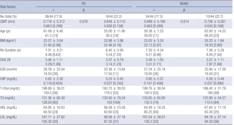
Table 2. Comparison of CVD risk factors according to the use of glucocorticoid and NSAID in RA
Risk factors PD NSAID
+ β - + β -
No./total (%) 36/44 (77.8) 8/44 (22.2) 34/44 (77.3) 10/44 (22.7)
CIMT (mm) 0.718 ± 0.212
0.663 [0.298] 0.076 0.648 ± 0.110
0.636 [0.158] 0.699 ± 0.180
0.663 [0.288] -0.014 0.726 ± 0.261
0.643 [0.168]
Age (yr) 61.08 ± 8.48
61.5 [14] 55.00 ± 11.90
56.5 [18] 58.38 ± 7.53
59.00 [11] 62.00 ± 14.25
68.50 [23]
BMI (kg/m2) 22.27 ± 3.04
21.85 [2.98]
22.88 ± 3.96 22.46 [4.16]
23.02 ± 3.24 22.13 [2.97]
20.23 ± 1.84 19.30 [2.80]† RA Duration (yr) 7.51 ± 4.21
9.46 [8.62] 6.40 ± 5.90
5.54 [7.23] 7.30 ± 4.34
9.21 [8.98] 7.36 ± 5.29
8.04 [7.00]
DAS 28 3.46 ± 1.11
3.29 [1.89] 3.37 ± 0.79
3.14 [1.23] 3.49 ± 1.05
3.51 [1.71] 3.27 ± 1.11
2.97 [1.89]
ESR (mm/hr) 28.39 ± 25.04
18.50 [30]
20.38 ± 13.64 17.50 [11]
27.24 ± 25.18 19.00 [26]
25.90 ± 17.39 18.00 [31]
CRP (mg/dL) 0.82 ± 2.30
0.116 [0.654] 0.23 ± 0.42
0.037 [0.335] 0.80 ± 2.37
0.101 [0.458] 0.38 ± 0.49
0.037 [0.890]
T-Chol (mg/dL) 198.89 ± 36.01
193.5 [56] 182.75 ± 39.53
178.5 [55] 198.76 ± 38.04
193.5 [55] 186.40 ± 31.79
180 [49]
TG (mg/dL) 131.36 ± 60.30
128.50 [60]
130.50 ± 79.24 102 [164]
133.65 ± 65.95 132.5 [74]
122.90 ± 54.57 110.5 [64]
HDL (mg/dL) 64.90 ± 16.83
60.50 [24] 66.58 ± 23.58
62.60 [33] 64.49 ± 18.33
60.25 [84] 67.64 ± 17.19
65.35 [25]
LDL (mg/dL) 107.71 ± 27.62
105.30 [30] 90.08 ± 27.16
87.25 [31] 107.54 ± 28.01
105.3 [32] 94.18 ± 27.14
94.50 [36]
Values show as Mean ± SD and Median [Interquartile range]. *P < 0.05, †P < 0.01. CVD, cardiovascular disease; RA, rheumatoid arthritis; NSAID, non-steroidal anti-inflam- matory drugs; CIMT, carotid intima media thickness; T-Chol, total cholesterol; TG, triglyceride; HDL, high density lipoprotein; LDL, low density lipoprotein; BMI, body mass index;
PD, prednisolone.
subgroups according to the use of GC. Age and ESR level were higher in RA patients treated with GC, as compared to those in RA patients without GC (61.08 ± 8.48 yr, 55.0 ± 11.90 yr for age, respectively, P < 0.05; 28.39 ± 25.04 mm/hr, 20.38 ± 13.64 mm/
hr for ESR, respectively, P < 0.01). Blood total cholesterol, TG, HDL-, LDL-cholesterol levels, disease duration, DAS 28 and CI- MT were not different between the subgroups by the use of NS- AIDs. Higher BMI was observed in RA subgroups with NSAIDs than in RA without NSAIDs (23.02 ± 3.24, 20.23 ± 1.84, P < 0.05, respectively) (Table 2).
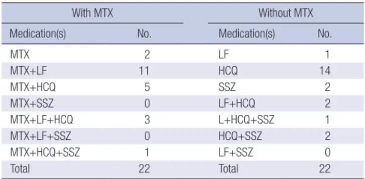
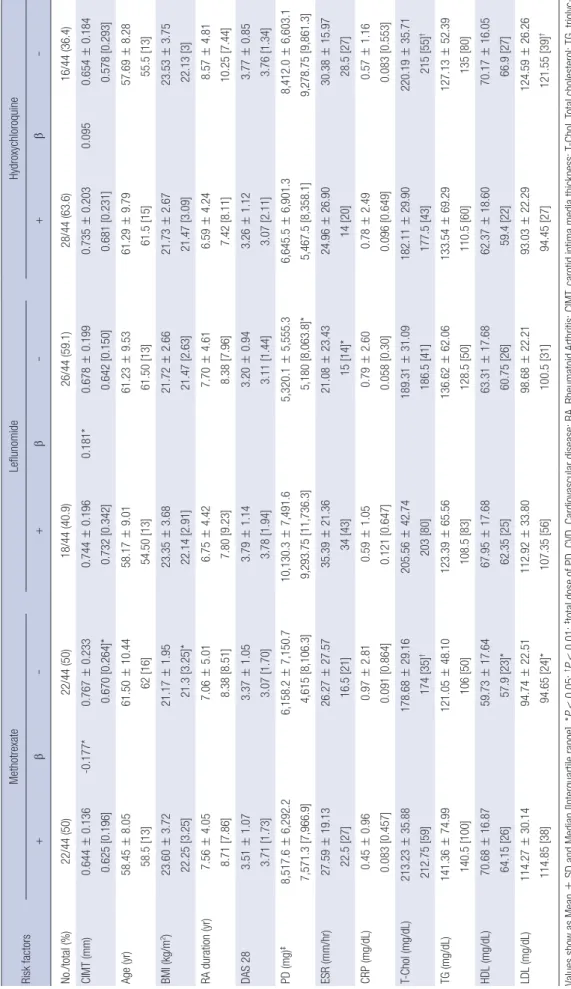
The most commonly used DMARDs in RA patients were MTX, LFM, HCQ, and sulfasalazine (SSZ). The DMARDs were used either singly or in combination (Table 3). In a dummy-variable regression analysis, among the DMARDs, a decrease of CIMT was observed only in RA patients using MTX (P < 0.01). The GC dose, DAS28, and disease duration did not differ between the age-, sex-matched RA subgroups with or without MTX. CIMT in RA without MTX was thicker than that with MTX (0.767 ± 0.233 mm, 0.644 ± 0.136 mm, respectively, P < 0.05) even with significantly lower levels of total cholesterol, LDL cholesterol, HDL cholesterol, and BMI. CIMT in RA showed no difference according to the use of LFM. However, higher ESR, BMI, GC dose, and LDL cholesterol level were observed in RA patients with LFM than in those without LFM. No difference in CIMT was observed in RA according to the use of HCQ either, even with significantly lower total cholesterol and LDL cholesterol levels in RA with HCQ. CIMT of RA patients and RA patients without MTX was thicker than that of healthy control subjects.
However, in RA patients treated with MTX, CIMT did not differ from that of healthy control subjects. In contrast to MTX, the use of LFM or HCQ yielded no effect on CIMT in RA patients.
The current study showed that among the DMARDs, MTX may exert favorable effects on CIMT independently to the disease activity of RA (Table 4).
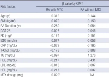
Additionally, in analyses of CIMT according to the clinical factors and the use of DMARDs in RA, a correlation between CIMT and age was observed in RA without MTX (r = 0.338, P <
0.05), but not in RA with MTX (Table 5). In a multivariate regres-
sion analysis among the drugs, the use of MTX showed signifi- cant correlation with CIMT (β = -0.177, P < 0.05). When adjust- ed for the conventional CVD risk factors, age, disease duration, DAS28, total GC dose, ESR and CRP levels, the maintenance dose of MTX in RA patients showed significant negative corre- lation with CIMT. For each 1 mg increase in MTX dose, there was a 0.029 mm decrease in CIMT (P < 0.01) (Table 6).
DISCUSSION
In RA patients, thicker CIMT and more frequent plaques of the carotid artery are observed (16); and the findings are correlated with the degree of articular damage (17). Coronary artery ath- erosclerosis detected as calcification was more severe and more prevalent in patients with established RA than in patients with early disease and control subjects (18,19). Ten-year cardiovas- cular mortality could be predicted in the group of patients with rheumatoid factor (RF)-positive inflammatory arthritis once the baseline CRP was elevated (20). The current study showed a higher CIMT in RA patients as compared to healthy subjects of the same age, sex, BMI, and cholesterol levels without history of smoking or hypertension.
Steroids can be associated with atherosclerosis, which can damage the vessel wall, and then increase the cardiovascular disease morbidity (21,22). Blood pressure can be elevated. And disturbance of glucose and lipid metabolism, hypercoagulabili- ty, platelet activation and endothelial cell damage can occur in subjects taking steroids. Davis et al. reported that RF-positive RA patients were at increased risk of CV events, particularly with higher cumulative exposure, higher average daily dosage (> 7.5 mg/day), and recent use of GC (23). Wei et al. reported that pa- tients with DMARDs and high dose steroid had higher risk of CVD in inflammatory arthritis (24). In addition to the steroids, excessive risk of CVD has been reported for some NSAIDs (25, 26). Lindhardsen et al. reported an association of NSAID expo- sure with a 22% increase in risk of cardiovascular events among RA patients; and this was significantly lower than the NSAID- associated risk increase (51%) observed in control subjects (27).
On the contrary, a small beneficial effect of NSAIDs on the risk of cardiovascular mortality was also reported in a cohort of in- flammatory polyarthritis patients (25). In this study, the use and accumulated dose of GC and NSAIDs had no effect on CIMT in RA. The effects of GC and NSAIDs on CIMT of RA in the current study may be a consequence of shared inflammatory mecha- nisms between RA and atherosclerosis.
Suissa et al. reported an association of the use of DMARDs with a decreased rate of myocardial infarction (14). However, Solomon et al. reported a higher risk for CV events in patients who had received cyclosporine, azathioprine, or LFM (22). Use of MTX in RA patients showed an association with a reduced risk of CV events compared to RA patients without MTX (28).
Table 3. The use of DMARDs in RA patients
With MTX Without MTX
Medication(s) No. Medication(s) No.
MTX 2 LF 1
MTX+LF 11 HCQ 14
MTX+HCQ 5 SSZ 2
MTX+SSZ 0 LF+HCQ 2
MTX+LF+HCQ 3 L+HCQ+SSZ 1
MTX+LF+SSZ 0 HCQ+SSZ 2
MTX+HCQ+SSZ 1 LF+SSZ 0
Total 22 Total 22
DMARDs, disease modifying anti-rheumatic drugs; MTX, methotrexate; LF, lefluno- mide; HCQ, hydroxychloroquine; SSZ, sulfasalazine.
Table 4. Comparison of the CVD risk factors according to the DMARDs used in RA Risk factorsMethotrexateLeflunomideHydroxychloroquine +β-+β-+β- No./total (%)22/44 (50)22/44 (50)18/44 (40.9)26/44 (59.1)28/44 (63.6)16/44 (36.4) CIMT (mm)0.644±0.136 0.625 [0.196]-0.177*0.767±0.233 0.670 [0.264]*0.744±0.196 0.732 [0.342]0.181*0.678±0.199 0.642 [0.150]0.735±0.203 0.681 [0.231]0.0950.654±0.184 0.578 [0.293] Age (yr)58.45±8.05 58.5 [13]61.50±10.44 62 [16]58.17±9.01 54.50 [13]61.23±9.53 61.50 [13]61.29±9.79 61.5 [15]57.69±8.28 55.5 [13] BMI (kg/m2)23.60±3.72 22.25 [3.25]21.17±1.95 21.3 [3.25]*23.35±3.68 22.14 [2.91]21.72±2.66 21.47 [2.63]21.73±2.67 21.47 [3.09]23.53±3.75 22.13 [3] RA duration (yr)7.56±4.05 8.71 [7.86]7.06±5.01 8.38 [8.51]6.75±4.42 7.80 [9.23]7.70±4.61 8.38 [7.96]6.59±4.24 7.42 [8.11]8.57±4.81 10.25 [7.44] DAS 283.51±1.07 3.71 [1.73]3.37±1.05 3.07 [1.70]3.79±1.14 3.78 [1.94]3.20±0.94 3.11 [1.44]3.26±1.12 3.07 [2.11]3.77±0.85 3.76 [1.34] PD (mg)‡8,517.6±6,292.2 7,571.3 [7,966.9]6,158.2±7,150.7 4,615 [8,106.3]10,130.3±7,491.6 9,293.75 [11,736.3]5,320.1±5,555.3 5,180 [8,063.8]*6,645.5±6,901.3 5,467.5 [8,358.1]8,412.0±6,603.1 9,278.75 [9,861.3] ESR (mm/hr)27.59±19.13 22.5 [27]26.27±27.57 16.5 [21]35.39±21.36 34 [43]21.08±23.43 15 [14]*24.96±26.90 14 [20]30.38±15.97 28.5 [27] CRP (mg/dL)0.45±0.96 0.083 [0.457]0.97±2.81 0.091 [0.864]0.59±1.05 0.121 [0.647]0.79±2.60 0.058 [0.30]0.78±2.49 0.096 [0.649]0.57±1.16 0.083 [0.553] T-Chol (mg/dL)213.23±35.88 212.75 [59]178.68±29.16 174 [35]†205.56±42.74 203 [80]189.31±31.09 186.5 [41]182.11±29.90 177.5 [43]220.19±35.71 215 [55]† TG (mg/dL)141.36±74.99 140.5 [100]121.05±48.10 106 [50]123.39±65.56 108.5 [83]136.62±62.06 128.5 [50]133.54±69.29 110.5 [60]127.13±52.39 135 [80] HDL (mg/dL)70.68±16.87 64.15 [26]59.73±17.64 57.9 [23]*67.95±17.68 62.35 [25]63.31±17.68 60.75 [26]62.37±18.60 59.4 [22]70.17±16.05 66.9 [27] LDL (mg/dL)114.27±30.14 114.85 [38]94.74±22.51 94.65 [24]*112.92±33.80 107.35 [56]98.68±22.21 100.5 [31]93.03±22.29 94.45 [27]124.59±26.26 121.55 [39]† Values show as Mean±SD and Median [Interquartile range]. *P<0.05; †P<0.01; ‡total dose of PD. CVD, Cardiovascular disease; RA, Rheumatoid Arthritis; CIMT, carotid intima media thickness; T-Chol, Total cholesterol; TG, triglyc- eride; HDL, high density Lipoprotein; LDL, Low density Lipoprotein; BMI, Body mass index; PD, Prednisolone.
Table 5. Correlations between the CVD risk factors and CIMT according to the DMARDs used in RA
Risk factors
r value by CIMT
Methotrexate Leflunomide Hydroxychloroquine
+ - + - + -
Age (yr) 0.415 0.427* 0.501* 0.415† 0.170 0.735† BMI (kg/m2) 0.184 -0.181 -0.464 -0.019 -0.090 -0.046 RA Duration (yr) 0.250 0.107 0.179 0.001 0.342 0.022
DAS 28 0.059 0.128 0.106 -0.111 0.145 0.085
PD (mg)‡ 0.169 0.295 0.082 0.047 0.339 0.035
ESR (mm/hr) -0.136 0.228 0.041 -0.064 0.189 -0.202 CRP (mg/dL) 0.248 0.508* 0.242 -0.029 0.463* 0.197 T-Chol (mg/dL) -0.442* -0.175 -0.614† -0.293 -0.307 -0.462 TG (mg/dL) -0.187 -0.080 -0.135 -0.058 -0.228 0.012 HDL (mg/dL) -0.542† -0.558† -0.669† -0.445† -0.465* -0.750† LDL (mg/dL) -0.197 -0.068 -0.423 -0.020 -0.141 -0.159 r , correlation coefficient. *P < 0.05; †P < 0.01; ‡total dose of PD. CVD, cardiovascu- lar disease; RA, rheumatoid arthritis; CIMT, carotid intima media thickness; T-Chol, total cholesterol; TG, triglyceride; HDL, high density lipoprotein; LDL, low density lipo- protein; BMI, body mass index; PD, prednisolone; DMARDs, disease modifying anti- rheumatic drugs.
Table 6. Multiple regression between CIMT and medications adjusted by other CVD risk factors
Risk factors β value by CIMT
RA with MTX RA without MTX
Age (yr) 0.312 0.144
BMI (kg/m2) 0.070 -0.150
RA Duration (yr) 0.260 -0.054
DAS 28 0.027 -0.046
PD (mg)‡ 0.174 0.151
ESR (mm/hr) 0.077 -0.056
CRP (mg/dL) -0.029 -0.165
T-Chol (mg/dL) -0.173 0.888
TG (mg/dL) -0.097 1.276
HDL (mg/dL) -0.217 0.431
LDL (mg/dL) -0.018 0.087
HDL (mg/dL) -0.217 -0.007†
MTX dosage (mg) -0.029† NA
β, regression coefficient. *P < 0.05; †P < 0.01; ‡total dose of PD. CVD, cardiovascu- lar disease; RA, rheumatoid arthritis; CIMT, carotid intima media thickness; TG, tri- glyceride; HDL, high density lipoprotein; MTX, methotrexate.
Some studies suggested possible mechanisms of MTX on athe- roprotection by modulating AMP-activated protein kinase - cy- clic AMP response element-binding protein (AMPK-CREB) path- way in intramural MTX therapy to prevent neointimal thicken- ing following angioplasty (29), and in chronic systemic inflam- mation of murine model (30). Reiss et al. reported an athero- protective effect on MTX on a cell model. MTX promotes re- verse cholesterol transport via activation of adenosine A2A re- ceptor, and limits foam cell formation in THP-1 macrophages (31). In one report, atherogenic lipid profiles of RA patients im- proved after specific therapy for arthritis (32). However, no rela- tionship of mortality was observed with the use of SSZ, HCQ, penicillamine, and intramuscular gold.
The current study evaluated the effects of DMARDs on ath- erosclerosis in RA patients. A favorable effect of MTX on CIMT was observed. As adjusted for the conventional risk factors and clinical factors, the use and the maintenance dose of MTX had atheroprotective effect on CIMT that was not consistent with LFM, HCQ or SSZ in RA. The mechanisms for this possible ben- efit may be that MTX can reduce the disease activity and sys- temic inflammation and progression of atherosclerosis. How- ever, the use of DMARDs for RA with an efficacy similar to that of MTX was not associated with decreased CVD mortality (33).
In this study, even with the same age, disease activity and infla- mmation levels lower CIMT was observed only in the patients using MTX. Therefore, it is suggestive that MTX may have direct protective effects to atherosclerosis in addition to the anti-in- flammatory effects.
This study has some limitations as a case-control study in a small numbered population. Exclusion of participants with CAD or aneurysm to avoid influences of treatments, dividing RA pa- tients into drug related small groups and manual measurement
of CIMT are the main limitations. A large numbered, random- ized-controlled, prospective study measuring CIMT by a soft- ware is required.
Early diagnosis and treatment of RA have been emphasized to prevent radiologic progression of joints. However, in this study, the authors verified an athero-protective effect of MTX on CIMT in addition to the effect on arthritis. Measurement of CIMT for atherosclerosis may be important not only to clarify the CVD risk but also to evaluate the effectiveness of treatment in RA.
And, the use of MTX should be considered in high priority not only to control arthritis but also to suppress atherosclerosis and RA-related CVD risk to mortality.
DISCLOSURE
The authors declare that there are no conflicts of interest con- cerning to this article.
AUTHOR CONTRIBUTION
Study concepts & design: all authors. Data collection: Kim HJ, Kim MJ. Analysis and interpretation of results, writing: all au- thors. Final approval: all authors.
ORCID
Hyun-Je Kim http://orcid.org/0000-0003-1075-5645 Choong-Ki Lee http://orcid.org/0000-0001-6820-8877 Min-Jung Kim http://orcid.org/0000-0002-2637-7979 Young-Hoon Hong http://orcid.org/0000-0001-8119-0464
REFERENCES
1. Watson DJ, Rhodes T, Guess HA. All-cause mortality and vascular events among patients with rheumatoid arthritis, osteoarthritis, or no arthritis in the UK General Practice Research Database. J Rheumatol 2003; 30:
1196-202.
2. Bacon PA, Stevens RJ, Carruthers DM, Young SP, Kitas GD. Accelerated atherogenesis in autoimmune rheumatic diseases. Autoimmun Rev 2002;
1: 338-47.
3. Corrales A, Dessein PH, Tsang L, Pina T, Blanco R, Gonzalez-Juanatey C, Llorca J, Gonzalez-Gay MA. Carotid artery plaque in women with rheu- matoid arthritis and low estimated cardiovascular disease risk: a cross- sectional study. Arthritis Res Ther 2015; 17: 55.
4. Naz SM, Symmons DP. Mortality in established rheumatoid arthritis.
Best Pract Res Clin Rheumatol 2007; 21: 871-83.
5. Gasparyan AY, Stavropoulos-Kalinoglou A, Mikhailidis DP, Toms TE, Douglas KM, Kitas GD. The rationale for comparative studies of acceler- ated atherosclerosis in rheumatic diseases. Curr Vasc Pharmacol 2010; 8:
437-49.
6. Sattar N, McCarey DW, Capell H, McInnes IB. Explaining how “high- grade” systemic inflammation accelerates vascular risk in rheumatoid arthritis. Circulation 2003; 108: 2957-63.
7. Libby P. Inflammation in atherosclerosis. Nature 2002; 420: 868-74.
8. Kannel WB, McGee D, Gordon T. A general cardiovascular risk profile:
the Framingham Study. Am J Cardiol 1976; 38: 46-51.
9. Oliver JJ, Webb DJ. Noninvasive assessment of arterial stiffness and risk of atherosclerotic events. Arterioscler Thromb Vasc Biol 2003; 23: 554-66.
10. O’Leary DH, Bots ML. Imaging of atherosclerosis: carotid intima-media thickness. Eur Heart J 2010; 31: 1682-9.
11. O’Leary DH, Polak JF, Kronmal RA, Manolio TA, Burke GL, Wolfson SK Jr. Carotid-artery intima and media thickness as a risk factor for myocar- dial infarction and stroke in older adults. Cardiovascular Health Study Collaborative Research Group. N Engl J Med 1999; 340: 14-22.
12. Edwards CJ, Arden NK, Fisher D, Saperia JC, Reading I, Van Staa TP, Cooper C. The changing use of disease-modifying anti-rheumatic drugs in individuals with rheumatoid arthritis from the United Kingdom Gen- eral Practice Research Database. Rheumatology (Oxford) 2005; 44: 1394-8.
13. Solomon DH, Goodson NJ, Katz JN, Weinblatt ME, Avorn J, Setoguchi S, Canning C, Schneeweiss S. Patterns of cardiovascular risk in rheuma- toid arthritis. Ann Rheum Dis 2006; 65: 1608-12.
14. Suissa S, Bernatsky S, Hudson M. Antirheumatic drug use and the risk of acute myocardial infarction. Arthritis Rheum 2006; 55: 531-6.
15. Aletaha D, Neogi T, Silman AJ, Funovits J, Felson DT, Bingham CO 3rd, Birnbaum NS, Burmester GR, Bykerk VP, Cohen MD, et al. 2010 Rheu- matoid arthritis classification criteria: an American College of Rheuma- tology/European League Against Rheumatism collaborative initiative.
Arthritis Rheum 2010; 62: 2569-81.
16. Ristić GG, Lepić T, Glisić B, Stanisavljević D, Vojvodić D, Petronijević M, Stefanović D. Rheumatoid arthritis is an independent risk factor for in- creased carotid intima-media thickness: impact of anti-inflammatory treatment. Rheumatology (Oxford) 2010; 49: 1076-81.
17. Targońska-Stepniak B, Drelich-Zbroja A, Majdan M. The relationship between carotid intima-media thickness and the activity of rheumatoid arthritis. J Clin Rheumatol 2011; 17: 249-55.
18. Goodson N, Marks J, Lunt M, Symmons D. Cardiovascular admissions
and mortality in an inception cohort of patients with rheumatoid ar- thritis with onset in the 1980s and 1990s. Ann Rheum Dis 2005; 64: 1595- 601.
19. Ambrosino P, Lupoli R, Di Minno A, Tasso M, Peluso R, Di Minno MN.
Subclinical atherosclerosis in patients with rheumatoid arthritis. A me- ta-analysis of literature studies. Thromb Haemost 2015; 113: 916-30.
20. Goodson NJ, Symmons DP, Scott DG, Bunn D, Lunt M, Silman AJ. Base- line levels of C-reactive protein and prediction of death from cardiovas- cular disease in patients with inflammatory polyarthritis: a ten-year fol- lowup study of a primary care-based inception cohort. Arthritis Rheum 2005; 52: 2293-9.
21. Maxwell SR, Moots RJ, Kendall MJ. Corticosteroids: do they damage the cardiovascular system? Postgrad Med J 1994; 70: 863-70.
22. Solomon DH, Avorn J, Katz JN, Weinblatt ME, Setoguchi S, Levin R, Sch- neeweiss S. Immunosuppressive medications and hospitalization for cardiovascular events in patients with rheumatoid arthritis. Arthritis Rheum 2006; 54: 3790-8.
23. Davis JM 3rd, Maradit Kremers H, Crowson CS, Nicola PJ, Ballman KV, Therneau TM, Roger VL, Gabriel SE. Glucocorticoids and cardiovascu- lar events in rheumatoid arthritis: a population-based cohort study. Ar- thritis Rheum 2007; 56: 820-30.
24. Wei L, MacDonald TM, Walker BR. Taking glucocorticoids by prescrip- tion is associated with subsequent cardiovascular disease. Ann Intern Med 2004; 141: 764-70.
25. Goodson NJ, Brookhart AM, Symmons DP, Silman AJ, Solomon DH.
Non-steroidal anti-inflammatory drug use does not appear to be associ- ated with increased cardiovascular mortality in patients with inflam- matory polyarthritis: results from a primary care based inception cohort of patients. Ann Rheum Dis 2009; 68: 367-72.
26. Bombardier C, Laine L, Reicin A, Shapiro D, Burgos-Vargas R, Davis B, Day R, Ferraz MB, Hawkey CJ, Hochberg MC, et al. Comparison of upper gastrointestinal toxicity of rofecoxib and naproxen in patients with rheu- matoid arthritis. VIGOR Study Group. N Engl J Med 2000; 343: 1520-8.
27. Lindhardsen J, Gislason GH, Jacobsen S, Ahlehoff O, Olsen AM, Mad- sen OR, Torp-Pedersen C, Hansen PR. Non-steroidal anti-inflammato- ry drugs and risk of cardiovascular disease in patients with rheumatoid arthritis: a nationwide cohort study. Ann Rheum Dis 2013.
28. Westlake SL, Colebatch AN, Baird J, Kiely P, Quinn M, Choy E, Ostor AJ, Edwards CJ. The effect of methotrexate on cardiovascular disease in pa- tients with rheumatoid arthritis: a systematic literature review. Rheuma- tology (Oxford) 2010; 49: 295-307.
29. Muller DW, Topol EJ, Abrams GD, Gallagher KP, Ellis SG. Intramural methotrexate therapy for the prevention of neointimal thickening after balloon angioplasty. J Am Coll Cardiol 1992; 20: 460-6.
30. Thornton CC, Al-Rashed F, Calay D, Birdsey GM, Bauer A, Mylroie H, Morley BJ, Randi AM, Haskard DO, Boyle JJ, et al. Methotrexate-mediated activation of an AMPK-CREB-dependent pathway: a novel mechanism for vascular protection in chronic systemic inflammation. Ann Rheum Dis 2015.
31. Reiss AB, Carsons SE, Anwar K, Rao S, Edelman SD, Zhang H, Fernan- dez P, Cronstein BN, Chan ES. Atheroprotective effects of methotrexate on reverse cholesterol transport proteins and foam cell transformation in human THP-1 monocyte/macrophages. Arthritis Rheum 2008; 58:
3675-83.
32. Georgiadis AN, Papavasiliou EC, Lourida ES, Alamanos Y, Kostara C,
Tselepis AD, Drosos AA. Atherogenic lipid profile is a feature character- istic of patients with early rheumatoid arthritis: effect of early treatment- -a prospective, controlled study. Arthritis Res Ther 2006; 8: R82.
33. Choi HK, Hernán MA, Seeger JD, Robins JM, Wolfe F. Methotrexate and mortality in patients with rheumatoid arthritis: a prospective study. Lan- cet 2002; 359: 1173-7.