Extragastic Pedunculated Giant Gastrointestinal Stromal Tumor of the Stomach
4
0
0
전체 글
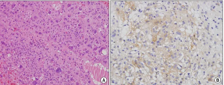
(2) Min Soo Kim, et al:Extragastic Pedunculated Giant Gastrointestinal Stromal Tumor of the Stomach. 269. 소는 5.8 g/dl로 낮아져 있었으나, 그 외 특이소견은 없었다.. 어 있었으며, 폐기능검사(pulmonary function test)에서 중증. 또한 CEA, CA19-9, AFP 등의 종양표지자 역시 정상 범위였. 도의 폐쇄폐병(obstructive lung disease)이 의심되는 상태였. 다. 복부 컴퓨터 단층 촬영(computed tomograpgy, CT) 및 자. 다. 저자들은 종양의 용적 축소를 통한 증상의 완화와 정확. 기공명영상(magnetic resonance imaging, MRI)을 시행한 결. 한 진단을 목적으로 개복술을 시행하였다. 종양은 복막과. 과, 막이 형성되어 있는 거대한 낭성 종양이 복강 전체를. 분리되어 있었고, 피막이 잘 형성되어 있는 낭종이었으며,. 차지하고 있으며, 종양은 부분적으로 고형성분을 가지고. 그물막(omentum)에 의해 부분적으로 둘러싸여 있었고, 복. 있었다. 종양의 크기는 약 40×35 cm였고, 복강 내 주요 장. 강전체를 차지하고 있었다. 종양은 위 몸통의 작은굽이. 기는 모두 종양에 의해 복부 뒤쪽으로 밀려나 있었다. CT. (lesser curvature of gastric body)에 2 cm 길이의 짧은 줄기. 및 MRI 소견으로 보아 중피종(mesothelioma)이 가장 의심되. (pedicle)와 연결되어 있었으며, 간 및 복막 전이 등의 이상. 었다(Fig. 1). 흉부 CT 소견에서는 좌하엽에 폐렴이 동반되. 소견은 발견되지 않았다. 수술은 종양의 완전 절제와 함께 줄기가 포함된 위의 작은굽이 부위를 동반 쐐기 절제하였 다. 육안소견에서 종양의 크기는 47×34×23 cm였고 무게는. Fig. 1. Abdominal computed tomography (CT). A huge septated cystic tumor is occupying whole abdomen.. Fig. 2. Gross findings. The surgical specimen is 47×34×23 cm in size, and weighed 40 kg.. Fig. 3. Pathologic findings. (A) Histologically, the tumor is composed of short spindle-shaped and oval cells (H&E, ×200). (B) The tumor cells demonstrate immunoreaction to c-kit..
(3) 270 J Korean Surg Soc. Vol. 75, No. 4 40 kg이었다(Fig. 2). 조직학적 소견으로는 H&E 염색에서. 후가 가장 불량하며, 다음으로 대장 및 위, 식도 순으로 예. 방추세포가 관찰되었고 50 HPF (high power field)당 5∼10. 후가 불량한 것으로 보고하였다.. 개의 유사분열이 관찰되었으며 근육성 분화(myogenic dif-. 위장에서 발생한 위장관 간질종양은 발생모양에 따라,. ferentiation)를 보이고 있었고, 면역조직화학염색에서 c-kit,. 위내형(intragastric type), 위외형(extragastric type), 벽내형. CD34, Vimentin에 양성소견을 보이는 고위험군의 상피모양. (intraluminal type), 혼합형(mixed type)으로 분류하고 있. 형(epithelioid type) 위장관 간질종양으로 진단할 수 있었다. 다.(7) 위외형 위장관 간질종양의 경우 위나 장관 벽에 붙어. (Fig. 3).. 다른 기관 등을 밀어내며 복강 내로 자라는 특징이 있으며,. 수술 후 5일째 측정한 환자의 체중은 56 kg이었으며, 이. 진행이 될 때까지 특별한 증상을 나타내지 않는 경우가 많. 후 특별한 합병증 없이 2주 뒤 퇴원하였고, 8개월간의 추적. 다. 본 증례처럼 위외형 위장관 간질종양 중 유경성의 특징. 기간 동안 재발의 증거는 확인되지 않았다.. 을 가진 종양은 더욱 드물게 보고되고 있으며, 국내에서는 Kim 등(8)이 보고한 1예의 증례뿐이며, 일본에서도 총 5예. 고. 찰. 의 증례만이 보고되고 있다.(9) 대부분의 경우 종양의 크기 는 평균 8.6 cm였고, 특별한 증상이 없이 우연히 발견되었. 위장관 간질종양은 식도에서 직장에 이르기까지 위장관. 다.. 의 어느 부위에서도 발생 가능한 종양으로 50∼60%가 위에. 위장관 간질종양의 크기와 관련하여, 대부분의 종양은 5. 서 발생하며, 그 외 소장, 대장, 식도 순으로 발생한다. 위장. cm 이상으로 보고하고 있으며, 10 cm 이상인 경우 원격전. 관 간질종양의 발생과정에서 KIT는 카할세포의 성숙발달. 이의 가능성이 더욱 높은 것으로 보고되고 있다. 그러나 15. 에 중요한 역할을 담당한다. C-kit gene에 변이(mutation)가. cm 이상의 거대 종양은 소수의 증례보고뿐이며, 모든 증례. 발생하면 줄기세포인자(stem cell factor) 없이도 KIT가 활성. 에서 고령이었으며, 원격전이는 동반되지 않았다. 그리고,. 화되어 카할세포는 증식하고 세포자멸사(apoptosis)가 억제. 고령이라는 측면은 진단 및 치료의 지연을 가져오는 중요. 되어 위장관 간질종양이 발생한다고 보고하고 있다.(2) 따. 한 요소로 보고하였다.(10) 본 증례에서는 고령은 아니더라. 라서, 위장관 간질종양의 진단을 위해서는 KIT에 대한 면. 도 낮은 사회경제적 상태가 환자의 진단이 지연되는 이유. 역조직화학염색이 필요하다.. 가 되었다. 게다가 47 cm의 직경을 가진 거대한 위장관 간. 위장관 간질종양의 악성도를 예측할 수 있는 병리학적. 질종양임에도 불구하고 몇몇의 증례보고와 같이 원격전이. 기준은 현재까지 확립되어 있지 않다. 다만 종양의 크기와. 를 발견할 수 없었고, 또한 8개월의 추적기간 동안 재발 및. 유사 분열 개수가 가장 믿을 만한 예후 인자로 받아들여지. 전이 역시 확인되지 않았다.. 고 있으며,(2,3) 그 외 점막 침윤, 종양 괴사와 고세포 충실. 저자 등은 복부 팽만을 주소로 내원한 고령의 남자 환자. 도, Ki67, PCNA, 유세포 측정기 등이 예후인자로서 보고되. 에서 직경 47 cm, 무게 40 kg의 위외형으로 자란 유경성 거. 고 있다. 그러나, 종양의 괴사나 궤양 형성은 종양의 크기와. 대 위장관 간질종양을 수술적으로 완전 절제를 시행하여. 관계되어 위장관 출혈을 일으킬 수는 있으나 병의 예후에. 치료했던 1예를 경험하여 이에 문헌 고찰과 함께 보고하는. 는 영향을 미치지 않는다고 보고되고 있다. 따라서, 위장관. 바이다.. 간질종양은 종양의 크기와 유사분열개수에 따라서 분류할 수 있으며, 초저위험군, 저위험군, 중간위험군, 고위험군으. REFERENCES. 로 분류하여 예후를 예측하는 합의안이 만들어졌다.(4) 이 합의안에 따르면 저자들의 증례는 고위험군에 포함된다고 할 수 있다. 또한 Yoon 등(5)은 고위험군의 경우 5년 무병 생존율을 27% 미만으로 보고하고 있어 본 증례에서도 주의 깊은 추적관찰이 필요할 것으로 보인다. 추가적으로, Emory 등(6)은 해부학적 위치가 종양의 크기, 유사 분열 개 수, 연령과는 별개로 위장관 간질종양의 독립적인 예후인 자가 될 수 있다고 보고하였으며, 소장에서 발생한 경우 예. 1) Kimura H, Yonemura Y, Kadoya N, Kosaka T, Miwa K, Miyazaki I, et al. Prognostic factors in primary gastrointestinal leiomyosarcoma: a retrospective study. World J Surg 1991;15: 771-6. 2) Heinrich MC, Rubin BP, Longley BJ, Fletcher JA. Biology and genetic aspects of gastrointestinal stromal tumors: KIT activation and cytogenetic alterations. Hum Pathol 2002;33:48495. 3) Miettinen M, El-Rifai W, H L Sobin L, Lasota J. Evaluation.
(4) Min Soo Kim, et al:Extragastic Pedunculated Giant Gastrointestinal Stromal Tumor of the Stomach. 4). 5). 6). 7). of malignancy and prognosis of gastrointestinal stromal tumors: a review. Hum Pathol 2002;33:478-83. Fletcher CD, Berman JJ, Corless C, Gorstein F, Lasota J, Longley BJ, et al. Diagnosis of gastrointestinal stromal tumors: a consensus approach. Hum Pathol 2002;33:459-65. Yoon SJ, Lee SH, Lee SM, Park HC, Koh SH, Hong SW, et al. Diagnosis and prognosis of gastrointestinal stromal tumors in the stomach. J Korean Surg Soc 2005;68:464-70. Emory TS, Sobin LH, Lukes L, Lee DH, O'Leary TJ. Prognosis of gastrointestinal smooth-muscle (stromal) tumors: dependence on anatomic site. Am J Surg Pathol 1999;23:82-7. Skandalakis JE, Gray SW, Shepard D. Smooth muscle tumors. 271. of the stomach. Int Abstr Surg 1960;110:209-26. 8) Kim YJ, Choi JH, Kim ES, Lee DH, Park NS, Koo JS, et al. A case of a pedunculated exoluminal gastrointestinal stromal tumor in the stomach. Korean J Gastrointest Endosc 2007;35:332-6. 9) Naitoh I, Okayama Y, Hirai M, Kitajima Y, Hayashi K, Okamoto T, et al. Exophytic pedunculated gastrointestinal stromal tumor with remarkable cystic change. J Gastroenterol 2003;38:1181-4. 10) Dal Corso HM, Solej M, Nano M. Giant gastrointestinal stromal tumor of the stomach in an elderly patient. J Gastrointest Surg 2007;11:804-6..
(5)
수치
관련 문서
Synchronous Gastrointestinal Stromal Tumor and Ampullary Neuroendocrine Tumor in Association with Neurofibromatosis Type 1: A Report of Three Cases.. Eun Kyu Park, Hee Joon Kim, Yun
Treatment and risk factors for recurrence after curative resection of gastrointestinal stromal tumors of the stomach. Prognostic factors influencing survival in
Gastrointestinal Stromal Tumor of the Appendix Mimicking a Mucinous Cystadenocarcinoma: A Case Report 1.. 점액성 낭선암종으로 오인한 충수돌기의 위장관간질종양:
Dose escalation of imatinib after failure of standard dose in Korean patients with metastatic or unresectable gastrointestinal stromal tumor. Demetri GD, van Oosterom AT, Garrett
Metastatic Cutaneous Duodenal Gastrointestinal Stromal Tumor: A Possible Clue to Multiple Metastases
We report a case of a 69-year-old man with metastatic cuta- neous duodenal gastrointestinal stromal tumor, which led to find multiple metastases on orbital muscle and scalp.. The
Malignant gastrointestinal stromal tumors (GISTs) are rare non-epithelial, mesenchymal neoplasms of the gastrointestinal tract that metastasize or recur in 30% of patients who
Although GIST occurs mainly in the gastrointestinal tract, it also occurs, rarely, in non-gastrointestinal tract and in this case, it is often named as extra-gastrointestinal
We report a case of extragastrointestinal stromal tumor originating from the prostate diagnosed after retropubic open prostatectomy.. The patient underwent additional
