submit.radiology.or.kr 대한영상의학회지 2011;65(1):81-83
81
서론충수돌기에서 발생하는 원발성 종양은 흔하지 않으며 유암 종, 점액성 혹은 비점액성의 낭선종이나 낭선암종, 림프종 등이 발생한다. 충수돌기에서 발생하는 위장관간질종양은 매우 드 물며 지금까지 영상 소견을 중점으로 자세히 기술한 보고가 없 다. 저자들은 점액성 낭선암종으로 오인한 충수돌기의 위장관 간질종양 1예를 경험하였기에 영상 소견을 문헌 고찰과 함께 보고하고자 한다.
증례 보고
67세 남자 환자가 일주일간 지속된 우하복부 통증을 주소로 내원하였다. 특별한 과거력은 없었으며 이학적 검사에서 우하복 부에 동통은 있었으나 반발 동통은 없었다. 검사 소견이나 단순 복부촬영에서 특이 소견은 없었다.
복부 전산화단층촬영에서 맹장과 인접한 타원형의 낭성 종괴 가 관찰되었다. 종괴벽은 조영증강 전 영상에서 석회화가 관찰 되었고 조영증강 후 영상에서 조영증강이 잘 되었고 불규칙한 두 께를 가지고 있었다. 종괴 내부에 불균질하게 조영증강되는 연조 직을 포함하고 있었다. 종괴의 말단에는 관모양의 연조직 구조
물이 이어져 있어 종괴가 충수돌기에서 기원한 것으로 생각하였 으며 인접한 맹장벽에도 부분적 비후가 동반되어 있었다(Fig. 1).
충수돌기에서 생긴 점액성 낭선암종을 의심하여 말단 회장부 를 포함한 우측 결장반절제술을 시행하였다. 육안 소견에서는 6.0 × 5.5 × 3.0 cm 크기의 경계가 좋은 타원형 종괴가 충수 돌기 중간 내측 장막하부에 위치하고 있었다. 단면은 육질이며 광범위한 출혈과 낭성 변화를 동반하고 있었다(Fig. 2). 조직학 적으로는 주로 호산성의 세포질을 갖는 방추형 세포들이 다발 을 이루거나 소용돌이 모양의 성장을 보이고 있었고 괴사나 유 사분열은 관찰되지 않았다. 면역조직화학염색에서 종양세포들 은 C-Kit, CD34, 액틴에 양성을 보이고 데스민과 S-100 단백에 는 음성을 보여 위장관간질종양으로 확진되었다(Fig. 3). 수술 후 환자는 증상이 호전되어 퇴원하였다.
고찰
충수돌기에서 발생하는 원발성 종양은 흔하지 않으며 충수 돌기 절제술 시행 후 병리 검사에서 0.5~1.0% 정도가 발견된 다. 유암종이 약 80% 정도로 가장 흔하며 점액성 혹은 비점액 성의 낭선종이나 낭선암종, 림프종 등이 발생한다. 드물게 발생 하는 종양으로는 신경내분비종과, 신경원성 종양이 있으며, 평
Case Report
pISSN 1738-2637
J Korean Soc Radiol 2011;65(1):81-83
Received April 6, 2011; Accepted June 20, 2011 Corresponding author: Hae Kyung Lee, MD Department of Radiology, Bucheon Hospital, Soonchunhyang University College of Medicine, 1174 Jung-dong, Wonmi-gu, Bucheon 420-767, Korea.
Tel. 82-32-621-5851 Fax. 82-32-621-5874 E-mail: [email protected]
Copyrights © 2011 The Korean Society of Radiology
A gastrointestinal stromal tumor of the appendix is a rare entity. Only a few cases have been reported in this location to date. We present here a case of a pathologi- cally confirmed gastrointestinal stromal tumor of the appendix mimicking a muci- nous cystadenocarcinoma in a 67-year-old man.
Index terms
Gastrointestinal Stromal Tumor Appendix
CT
Mucinous Cystadenocarcinoma
Gastrointestinal Stromal Tumor of the Appendix Mimicking a Mucinous Cystadenocarcinoma: A Case Report
1점액성 낭선암종으로 오인한 충수돌기의 위장관간질종양: 증례 보고
1Min Hee Lee, MD
1, Hae Kyung Lee
1, MD, Boem Ha Yi, MD
1, Hee Kyung Kim, MD
2Departments of 1Radiology, 2Pathology, Bucheon Hospital, Soonchunhyang University College of Medicine, Bucheon, Korea
점액성 낭선암종으로 오인한 충수돌기의 위장관간질종양
submit.radiology.or.kr
대한영상의학회지 2011;65(1):81-83
82
발현된다. 위장관간질종양을 이루는 세포가 Cajal 세포와 면역 조직화학염색에서 유사한 모습을 보이고 kit 단백에 양성을 보 이는 것과 전자현미경 소견에서 유사점이 확인되어 위장관간질 종양이 Cajal 세포에서 기원했을 것으로 추정된다(7). 그러나 Cajal 세포가 존재하지 않는 장간막이나 그물막에서 발생하기 도 하는 것으로 보아 Cajal 세포로 분화할 수 있는 간엽성 줄기 세포(pluripotent mesenchymal stem cell)에서 기인하는 것으로 추정되기도 한다(8). 바깥근육층에서 주로 병발하기 때문에 외 장성 성장을 하기 쉬우며 대부분이 장관벽에서 발생하여 복강 내로 돌출하는 형태를 보이게 된다. 수 mm부터 30 cm 이상 크 기까지 보고 된 바가 있으며 전형적으로 피막이 없고 주변 조직 을 압박하는 경계가 좋은 종괴로 보인다. 특히 큰 종괴의 경우 내부에 출혈이나 낭성 변성, 괴사 등이 있을 수 있으며 광범위 출혈이나 괴사로 인해 형성된 공동이 장관 내강과 교통하기도 한다(9).
활근종과 위장관간질종양 등이 있다(1).
위장관간질종양은 위에서 가장 많이 발생하며 약 60~70%
가 위에서 발견되고 충수돌기에서 발생하는 경우는 전체 사례 의 0.1% 정도로 매우 드물다(2). 지금까지 문헌 보고된 충수돌 기의 위장관간질종양은 대부분 3 cm 미만으로 급성 충수돌기 염의 임상 증상을 보이거나, 출혈이 있거나, 주요 장기 수술 중 우연히 충수돌기 절제를 한 후 병리 소견에서 진단된 경우였다 (3-5). 충수돌기 주변에 커다란 농양을 형성하였던 악성 위장관 간질종양이 보고된 바 있지만(6) 처음에는 충수돌기염으로 진 단되었던 사례로 지금까지 충수돌기에 발생한 종괴로서 위장관 간질종양의 영상 소견을 자세히 기술한 보고는 없었다.
위장관간질종양은 위장관의 가장 흔한 중간엽 조직 기원 종 양으로 특유의 조직 소견을 가진 kit 단백(CD117) 양성인 종양 으로 정의된다. 정상 위장관에서 kit 단백은 비만세포 외에 장 운동을 조절하는 자율신경계의 속도조절세포인 Cajal 세포에
Fig. 1. A. Pre-contrast axial CT image shows a cystic mass with internal soft-tissue component in the right lower quadrant abdominal cavity, which appeared to abut to the cecum in the other CT images. Note the wall calcification (arrows).
B, C. Post-contrast axial (B) and coronal (C) CT images show the same cystic mass with irregular, enhancing wall thickening. The internal soft- tissue component (arrowhead) enhances heterogeneously. Note the tubular structure (arrow) abutting the mass, which was connected with the mass in other CT images, representing that the mass arises from the appendix.
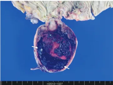
Fig. 2. The cut surface of the surgical specimen shows a tan-white mass with extensive areas of hemorrhage and cystic degeneration (ar- rows).
Fig. 3. Photomicrograph of histopathologic specimen shows that the tumor cells are strongly reactive for C-Kit in immunohistochemical stain (× 200).
A B C
이민희 외
submit.radiology.or.kr 대한영상의학회지 2011;65(1):81-83
83
the appendix: a clinicopathologic and immunohistochem- ical study of four cases. Am J Surg Pathol 2001;25:1433- 1437
4. Yap WM, Tan HW, Goh SG, Chuah KL. Appendiceal gastro- intestinal stromal tumor. Am J Surg Pathol 2005;29:1545- 1547
5. Kim KJ, Moon W, Park MI, Park SJ, Lee SH, Chun BK. Gas- trointestinal stromal tumor of appendix incidentally diag- nosed by appendiceal hemorrhage. World J Gastroenterol 2007;13:3265-3267
6. Elazary R, Schlager A, Khalaileh A, Appelbaum L, Bala M, Abu-Gazala M, et al. Malignant appendiceal GIST: case re- port and review of the literature. J Gastrointest Cancer 2010;41:9-12
7. Badalamenti G, Rodolico V, Fulfaro F, Cascio S, Cipolla C, Cicero G, et al. Gastrointestinal stromal tumors (GISTs):
focus on histopathological diagnosis and biomolecular features. Ann Oncol 2007;18 Suppl 6:vi136-vi140
8. Miettinen M, Lasota J. Gastrointestinal stromal tumors-- definition, clinical, histological, immunohistochemical, and molecular genetic features and differential diagnosis.
Virchows Arch 2001;438:1-12
9. Levy AD, Remotti HE, Thompson WM, Sobin LH, Miettinen M. Gastrointestinal stromal tumors: radiologic features with pathologic correlation. Radiographics 2003;23:283- 304, 456; quiz 532
10. Gore R, Levine M. Textbook of gastrointestinal imaging.
3rd ed. Philadelphia: Saunders, 2008;593-648 전산화단층촬영에서 양성 위장관간질종양은 대부분 근육과
비슷한 정도의 균일한 음영을 보이며, 악성일 경우 크기가 크고 종종 불균일한 음영을 보이며 내부에 낭성 변성 또는 괴사를 시 사하는 저음영의 부위를 포함한다. 흔하진 않지만 내부에 석회 화를 동반하는 경우도 있다(10).
Miettinen 등(3)의 보고에 따르면, 8예의 충수돌기 중간엽 종양 중 4예가 위장관간질종양으로 확진되어 충수돌기에 발생 하는 점막하 종양의 반수를 차지하였다. 본 사례의 경우 종괴는 내부에는 불균일하게 조영증강되는 고형 부분을 포함하는 낭성 종괴로 보였으며 종괴벽은 비후되었고 일부분은 석회화를 포함 하고 있었다. 종괴가 충수돌기에서 기원하고 있는 것으로 생각 하였으나 크기가 매우 크고 충수돌기의 내강이 따로 구분되지 않아 점막하 종양을 의심하기 어려웠다.
결론적으로 충수돌기에 위치한 낭성 종괴가 연조직을 포함하 고 있을 때 점액성 낭선종이나 낭선암종을 먼저 감별하겠으나, 본 예처럼 내부에 출혈과 낭성 변성이 있는 위장관간질종양의 가능성도 고려해야 할 것으로 생각된다.
참고문헌
1. Pickhardt PJ, Levy AD, Rohrmann CA Jr, Kende AI. Primary neoplasms of the appendix: radiologic spectrum of disease with pathologic correlation. Radiographics 2003;23:645- 662
2. Miettinen M, Lasota J. Gastrointestinal stromal tumors:
pathology and prognosis at different sites. Semin Diagn Pathol 2006;23:70-83
3. Miettinen M, Sobin LH. Gastrointestinal stromal tumors in
점액성 낭선암종으로 오인한 충수돌기의 위장관간질종양: 증례 보고
1이민희1
·
이혜경1·
이범하1·
김희경2위장관간질종양은 대부분 위에서 발생하며 충수돌기에서 발생하는 경우는 매우 드물다. 저자들은 점액성 낭선암종으로 오인한 충수돌기의 위장관간질종양 1예를 경험하였기에 영상 소견을 문헌 고찰과 함께 보고하고자 한다.
순천향대학교 의과대학 부천병원 1영상의학과학교실, 2병리과학교실