□ 증례보고 □
383
Gastrointestinal Stromal Tumor of Prostate
Sung-Woo Park, Wan Lee, Gi-Young Huh1, Moon-Kee Chung
From the Departments of Urology and 1Pathology, College of Medicine, Pusan National University, Busan, Korea
Gastrointestinal stromal tumor (GIST) are the most common mesenchymal malignancy of the gastrointestinal tract. They are diagnosed by combi- nation of c-kit and CD34. We report a case of extragastrointestinal stromal tumor originating from the prostate diagnosed after retropubic open prostatectomy. The patient underwent additional retropubic radical pro- statectomy again after 2 weeks. The possibility of secondary involvement by a rectal GIST was excluded by radiological, intraoperative and patho- logic findings. (Korean J Urol 2008;49:383-385)
Key Words: Gastrointestinal stromal tumors, Prostate, Prostatectomy
대한비뇨기과학회지 제 49 권 제 4 호 2008 부산대학교 의과대학원
비뇨기과학교실, 1병리학교실
박성우ㆍ이 완ㆍ허기영1ㆍ정문기
접수일자:2008년 1월 29일 채택일자:2008년 2월 28일 교신저자: Moon-Kee Chung
Department of Urology, College of Medicine, Pusan National University, 10, Ami-dong, 1-ga, Seo-gu, Busan 602-739, Korea TEL: 051-240-7351 FAX: 051-247-5443 E-mail: mkchung@
pusan.ac.kr
Gastrointestinal stromal tumor (GIST) is the designation for a specific group of mesenchymal neoplasms that express the KIT protein (CD117). GISTs almost exclusively occur in the gastrointestinal tract and comprise most gastrointestinal mesen- chymal tumors outside the gastrointestinal tract. But, GISTs rarely if ever occur as primary tumors outside the gastroin- testinal tract.
Two hypothesis are presented by Miettinen et al.1 as a origin of extragastrointestinal stromal tumor. One is that GISTs are tumors of the Cajal cells, the gastrointestinal pacemaker cells, and that they should be renamed as gastrointestinal pacemaker cell tumors. Another hypothesis is that Cajal cells differentiate from the common interstitial precursor cells, which also give rise to smooth muscle cells. Therefore, GISTs also could be considered tumors of intestinal mesenchymal precursor cells.
The latter hypothesis could explain also why GISTs can arise outside of the gastrointestinal tract.
We report on an unusual case of extragastrointestinal stromal tumor originating from prostate. The patient underwent supra- pubic open prostatectomy followed by retropubic radical prostatectomy 2 weeks later.
CASE REPORT
A 58 years old man presented with intermittent bloody stool, constipation and urologic symptoms for a month. On colono- scopic examination, there was friable ulcerative rectal mucosa
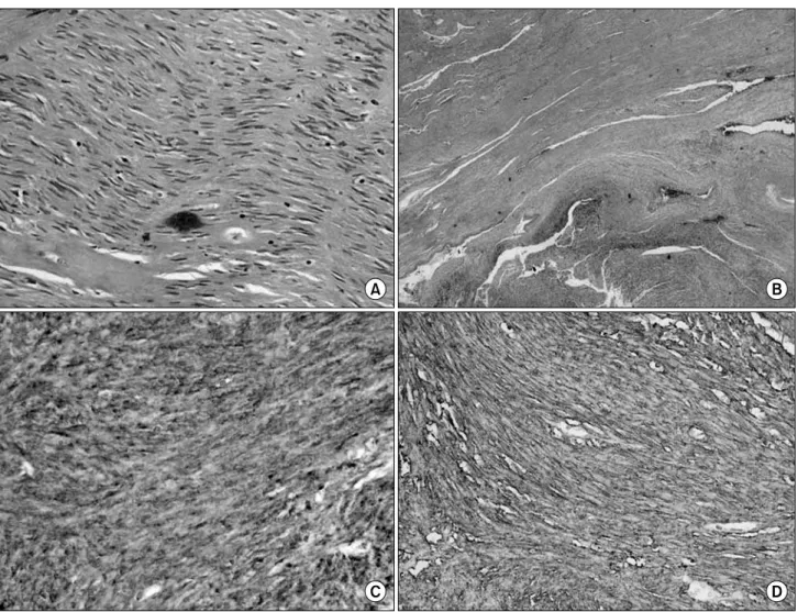
covered huge round elevated lesion above anus. Endoscopic biopsy examination revealed a chronic inflammation. Trans- rectal ultrasonography revealed the 65x75x70 mm sized round, heterogenous and hypoechoic prostate with intact prostatic capsule (Fig. 1). His serum prostate-specific antigen (PSA) level was 2.16 ng/ml. Thus, This patient underwent suprapubic open prostatectomy. Because hardness of prostatic adenoma has been decreased, Prostatic adenoma was fragmented into lots of pie- ces. On microscopic findings, prostatic adenoma was replaced by cellular tumor mass and tumor cells compressed prostatic glands without involvement of prostatic capsule. The tumor cells were spindle shaped with little atypia and showed mainly fasciculuar arrangement. The tumor cells were strongly positive for c-kit and CD34, weakly positive for smooth muscle actin
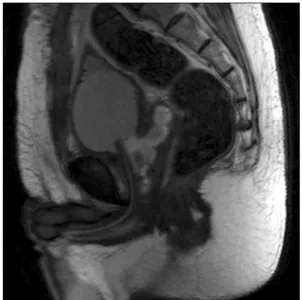
(SMA) and negative for desmin (Fig. 2). Mitotic counts were fewer than 5 per 50 high-power fields. Staging studies by MR prostate and abdominal CT scan showed no other areas of involvement. On MR prostate findings, fat strand between prostatic capsule and rectum was intact, rectal invasion or origi- nating from rectum were excluded (Fig. 3). We concluded that tumor cells originated from the prostate. Additional imaging studies including chest roentgenogram and whole body bone scan showed no metastatic disease. The patient underwent addi- tional retropubic prostatectomy again after 2 weeks. The speci- men was excised with clear resection margin to rectal wall. The patient has been observed for 6 months and is in good condition except for experiencing mild urinary incontinence.
384 대한비뇨기과학회지:제 49 권 제 4 호 2008
Fig. 1. Preoperative transrectal and transabdominal ultrasonography. The large 65x75x70 mm sized prostate was well encapsulated.
Fig. 2. (A) Prostate is replaced by cellular tumor mass. The tumor cells are spindle shaped with little atypia. They shows mainly fascicular arrangement. (B) Compressed prostatic glands are seen on upper left field. (C) Tumor cells are strongly positive for C-kit. (D) Tumor cells are strongly positive for CD34.
Sung-Woo Park, et al:Gastrointestinal Stromal Tumor of Prostate 385
Fig. 3. MR prostate image showing open prostatectomy state and definite fat strand between the rectum and the remaining prostate capsule.
DISCUSSION
The discovery of gain-of-function mutations in the KIT proto-oncogene of GIST by Hirota et al.2 in 1998 was crucially importance in terms of the genesis and classification of these tumors. At present, the expression of KIT protein has emerged as the tumors' most important defining feature, and it is probably the gold standard for diagnosing GISTs.3 For that reason, the demonstration of KIT expression in extragastro- intestinal lesions has helped validate their existence, and particularly when finding these tumors in exceptional sites such as the prostate.4-7
Aside from the consistent positivity for CD117, 60-70%, 30-40%, 1-2% of GISTs show immunoreactivity for CD34, smooth muscle actin (SMA), S-100 protein and desmin, respectively.3 But the immunophenotype of true CD117 positive GISTs varies to some degree with their location. The previously reported extragastrointestinal stromal tumor from prostate showed immunoreactivity for CD117, CD34, SMA and desmin were positive, positive, locally positive and negative, respectively.4-7 To distinguish between benign and malignant GISTs, many parameters have been proposed, but tumor size and the mitotic rate have been widely used as histologic criteria. Fletcher et al.3 have recently proposed criteria to separate benign from malignant tumors by tumor size and the mitotic count.By this definition, the tumor larger than 10 cm or having more than
10 mitosis per 50 high power fields belong to the high risk category. In this respect our case belongs to the intermediate risk category.
Complete excision has been accepted as the only and efficacious treatment regimen for nonmetastatic GISTs. Al- though it is necessary to expand the scope of surgical resection, extensive clearance of lymph nodes is not recommended because lymph node metastasis is very rare in extragastro- intestinal stromal tumor. With the recent discovery that the gain-of-function mutation of the c-kit proto-oncogene is an etiologic factor for GIST, many studies have reported that imatinib mesylate inhibiting kit receptor tyrosine kinase is an efficacious agent for patients with malignant, metastatic and/or unresectable GIST. Currently, imatinib mesylate is widely used as neoadjuvant or adjuvant therapy.8
In conclusion, our present case was an exceedingly rare entity occurring in the prostate, and diagnosed following retro- pubic open prostatectomy, and the patient underwent retropubic radical prostatectomy again. Urologists should be aware of this rare entity and adopt appropriate strategies of treatment that can achieve the best outcome for patients.
REFERENCES
1. Miettinen M, Sarlomo-Rikala M, Lasota J. Gastrointestinal stromal tumors: recent advances in understanding of their biology. Hum Pathol 1999;30:1213-20
2. Hirota S, Isozaki K, Moriyama Y, Hashimoto K, Nishida T, Ishiguro S, et al. Gain-of-function mutations of c-kit in human gastrointestinal stromal tumors. Science 1998;279:577-80 3. Fletcher CD, Berman JJ, Corless C, Gorstein F, Lasota J,
Longley BJ, et al. Diagnosis of gastrointestinal stromal tumors:
a consensus approach. Hum Pathol 2002;33:459-65
4. Arce-Lara C, Shah MH, Jimenez RE, Patel VR, Benson DM Jr, Clinton SK, et al. Gastrointestinal stromal tumors involving the prostate: presentation, course, and therapeutic approach.
Urology 2007;69:1209
5. Lee CH, Lin YH, Lin HY, Lee CM, Chu JS. Gastrointestinal stromal tumor of the prostate: a case report and literature review. Hum Pathol 2006;37:1361-5
6. Van der Aa F, Sciot R, Blyweert W, Ost D, Van Poppel H, Van Oosterom A, et al. Gastrointestinal stromal tumor of the prostate. Urology 2005;65:388
7. Yinghao S, Bo Y, Xiaofeng G. Extragastrointestinal stromal tumor possibly originating from the prostate. Int J Urol 2007;14:869-71
8. Dematteo RP, Heinrich MC, El-Rifai WM, Demetri G. Clinical management of gastrointestinal tumors: before and after STI- 571. Hum Pathol 2002;33:466-77