대한외과학회지:제 68 권 제 3 호
□ Case Report □
Vol. 68, No. 3, March, 2005
247 INTRODUCTION
Primary cysts of the spleen are very rare, account- ing for 10% of all nonparasitic cysts of the spleen, usually occurring in children or young adults and possibly arising from cell rests.(1) These lesions are
often asymptomatic; however, when symptoms occur, they are usually nonspecific.(2) Although rare, it is well documented that patients with splenic cysts may present with complications, such as infection or peritonitis.(3-5) Herein, we present a rare complica- tion of spontaneous rupture of a primary splenic cyst presenting as hemoperitoneum.
CASE REPORT
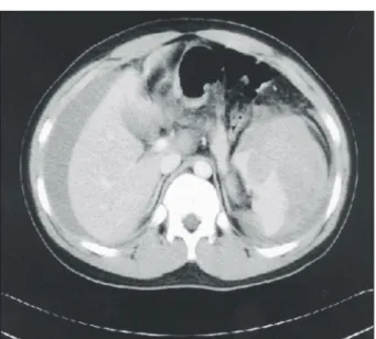
A 14-year-old male patient, who had been in good health, was admitted with an 11-hour history of abdominal pain which had originated in the left hypochondrium. On admission, his blood pressure was 100/60 mmHg; pulse rate, 120 beats/min; and on physical examination, there was tenderness with guarding in the left hypochondrium and in the left flank. The patient had no recent history of trauma and no systemic disease. Laboratory examination re- vealed leukocytosis and anemia, with hemoglobin level of 7.0 g/dl and hematocrit level of 20.4%. Other blood chemistry parameters were unremarkable. He was stabilized with intravenous fluid and blood transfusion. Computed tomographic scan showed multiple round shaped low density masses in spleen with a hematoma and hemoperitoneum (Fig. 1). A presumptive diagnosis of splenic cyst rupture with hemoperitoneum was considered and a laparotomy was performed.
At operation, 1,000 ml of blood and blood clots were evacuated from the peritoneal cavity. The spleen was found to be ruptured at the lower pole from one of the multiple cyst-like lesions, measuring about 2 cm in diameter. A total splenectomy was performed. Surgical specimen showed four variable
Spontaneous Rupture of a Primary Splenic Cyst Causing Hemoperitoneum
Departments of Surgery, 1Diagnostic Radiology and 2Diagnostic Pathology, University of Ulsan College of Medicine, Gangneung Asan Hospital, Republic of Korea
Yong Pil Cho, M.D., Seung Mun Jung, M.D.1, Gil Hyun Kang, M.D.2, Myoung Sik Han, M.D., Hyuk Jai Jang, M.D., Yong Ho Kim, M.D., Jin-Ho Kwak, M.D. and Youn Baik Choi, M.D.
원발성 비장낭종의 자발적인 파열에 의한 복 강내 출혈
조용필․정승문1․강길현2․한명식․장혁재․김용
호․곽진호․최윤백
A 14-yr-old male presented with a hemoperitoneum caused by a spontaneous rupture of a primary splenic cyst. The laparotomy showed a ruptured spleen at the lower pole from one of the multiple cyst-like lesions, measuring approxi- mately 2 cm in diameter. After the splenectomy, a micros- copic examination confirmed the diagnosis of a primary splenic cyst lined with a mature, well-differentiated squamous epithelium. A hemoperitoneum caused by a spontaneous rupture of a primary splenic cyst is a rare but potentially lethal complication, because most patients with a splenic rupture present with some degree of hypovolemia. (J Korean Surg Soc 2005;68:247-248)
Key Words: Splenic cyst, Rupture, Hemoperitoneum 중심 단어: 비장 낭종, 파열, 복강내 출혈
ꠏꠏꠏꠏꠏꠏꠏꠏꠏꠏꠏꠏꠏꠏꠏꠏꠏꠏꠏꠏꠏꠏꠏꠏꠏꠏꠏꠏꠏꠏꠏꠏꠏꠏꠏꠏꠏꠏꠏꠏꠏꠏꠏꠏꠏꠏꠏꠏꠏꠏꠏꠏꠏ 울산대학교 강릉아산병원 외과, 1진단방사선과, 2진단병 리과
Corresponding to: Yong Pil Cho, Department of Surgery, Gangneung Asan Hospital, 415 Bangdong-ri, Sacheon-myeon, Gangneung 210- 711, Republic of Korea. (Tel) 033-610-3229, (Fax) 033-641-8120, (E-mail) [email protected]
Received September 13, 2003, Accepted November 25, 2004
248 대한외과학회지 : 제 68 권 제 3 호 2005
ꠏꠏꠏꠏꠏꠏꠏꠏꠏꠏꠏꠏꠏꠏꠏꠏꠏꠏꠏꠏꠏꠏꠏꠏꠏꠏꠏꠏꠏꠏꠏꠏꠏꠏꠏꠏꠏꠏꠏꠏꠏꠏꠏꠏꠏꠏꠏꠏꠏꠏꠏꠏꠏꠏꠏꠏꠏꠏꠏꠏꠏꠏꠏꠏꠏꠏꠏꠏꠏꠏꠏꠏꠏꠏꠏꠏꠏꠏꠏꠏꠏꠏꠏꠏꠏꠏꠏꠏꠏꠏꠏꠏꠏꠏꠏꠏꠏꠏꠏꠏꠏꠏꠏꠏꠏꠏꠏꠏꠏꠏꠏꠏꠏꠏꠏ
sized cysts in spleen (Fig. 2). The luminal surfaces were smooth with slight trabeculations. Pathologi- cally, they were typical epithelial cysts of the spleen lined with mature, well-differentiated squamous epithelium. The postoperative course was unevent- ful.
DISCUSSION
Spontaneous rupture of the spleen is rare, and when it occurs, there is usually some underlying splenic pathology.(2) The most common cause of splenic cysts is parasitic infestation with Echino- coccus granulosus.(1-6) Nonparasitic splenic cysts are rare.(1-6) Nonparasitic splenic cysts are classified as primary, or epithelial cysts, and secondary, or pseu- docysts. The etiology of primary splenic cysts re- mains unknown and they are encountered more commonly in children or young adults. Symptoms of splenic cysts are vague and are caused primarily by mass effect, compression of adjacent viscera, and diaphragmatic irritation.(1-6) Occasionally, a patient may present with complications of a splenic cyst, such as infection, abscess formation, intracystic he- morrhage or peritonitis from rupture of splenic
cyst.(3-5) Hemoperitoneum caused by spontaneous rupture of a primary splenic cyst is rare but po- tentially lethal, because most patients with splenic rupture present with some degree of hypovolemia.
We present a rare case of spontaneous rupture of a primary splenic cyst presenting as an abdominal emergency.
REFERENCES
1) Spence RAJ, Dane TEB. Spontaneous rupture of an epithelial cyst of the spleen. Postgrad Med J 1983;59:65.
2) Lam CM, Yuen ST, Yuen WK. Hemoperitoneum caused by spontaneous rupture of a true splenic cyst. Hepatoga- stroenterology 1998;45:1884-6.
3) Robbins FG, Yellin AE, Lingua RW, Craig JR, Turrill FL, Mikkelsen WP. Splenic epidermoid cysts. Ann Surg 1978;
187:231-5.
4) Rathaus V, Zissin R, Goldberg E. Spontaneous rupture of an epidermoid cyst of spleen: preoperative ultrasonographic diagnosis. J Clin Ultrasound 1991;19:235-7.
5) Panossian DH, Wang N, Reeves CD, Weeks DA. Epidermoid cyst of the spleen presenting as a generalized peritonitis.
Am Surg 1990;56:295-8.
6) Sirinek KR, Evans WE. Nonparasitic splenic cysts-case report of epidermoid cyst with review of the literature. Am J Surg 1973;126:8-13.
Fig. 1. Computed tomographic scan shows multiple round shaped low density masses in spleen with a hematoma and hemo- peritoneum.
Fig. 2. The spleen was ruptured at the lower pole from one of the multiple cyst-like lesions.