Spontaneous rupture of the liver is rare, but it can be the most serious and potentially life-threatening compli- cation associated with preeclampsia or eclampsia. The patient’s survival generally depends on the early recog- nition of the characteristic signs and symptoms and prompt surgical intervention. Even with surgical inter- vention, the maternal mortality had been reported to be as high as 59-86% (1, 2). We describe here a case of suc- cessful treatment of spontaneous hepatic rupture due to preeclampsia of pregnancy by performing transcatheter hepatic arterial embolization, and we also review the relevant literature.
Case Report
A 41-year-old woman (gravida 3, para 2) at 34 weeks
gestation presented with epigastric pain. Her blood pres- sure was noted to be 178/100 mmHg with 2+ protein- uria, and we observed signs of fetal distress. So, she un- derwent an emergency cesarean section and she deliv- ered a live healthy female infant.
Five hours after delivery, she developed severe epigas- tric pain without any vaginal bleeding. Her hematocrit fell from 48% to 20%. Her blood pressure fell from 178/100 mmHg to 80/40 mmHg. Massive transfusions were begun and an emergency CT scan was performed.
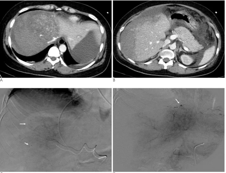
An emergency CT scan showed a large, poorly circum- scribed, low density area in the both lobes of the liver, which contained irregular frond-like branches of normal hepatic density. A moderate amount of adjacent subcap- sular fluid was present, as well as a moderate amount of free intraperitoneal fluid (Figs. 1A, B). Massive transfu- sions were begun and an emergency hepatic angiogram was obtained.
The emergency hepatic angiogram demonstrated dis- placement of hepatic vessels due to a large subcapsular hematoma and multiple small punctate areas of extrava- sation in both lobes of the liver (Figs. 1C, D). We then performed angiographic embolization of both hepatic ar- teries with using a 2.7-French microcatheter (Progreat,
J Korean Soc Radiol 2010;63:29-32
─ 29 ─
Spontaneous Hepatic Rupture Associated with Preeclampsia: Treatment with Hepatic Artery Embolization1
Seung Boo Yang, M.D., Dong Erk Goo, M.D., Yun Woo Chang, M.D., Yong Jae Kim, M.D., In Cheol Hwang, M.D.2, Hyo Sang Han, M.D.2,Jong Hyun Yoon, M.D.3, Tae Il Lee, M.D.4
1Department of Radiology, Soonchunhyang University Hospital
2Department of Obstetrics and Gynecology, Soonchunhyang University Hospital
3Department of Urology, Soonchunhyang University Hospital
4Department of Internal Medicins, Soonchunhyang University Hospital Received October 18, 2009 ; Accepted March 17, 2010
Address reprint requests to : Seung Boo Yang, M.D., Department of Radiology, Soonchunhyang University Gumi Hospital, 250, Gongdan- dong, Gumi-si, Kyungbuk 730-030, Korea.
Tel. 82-54-468-9391 Fax. 82-54-464-9300 E-mail: [email protected]
Spontaneous rupture of the liver due to preeclampsia is a rare condition of pregnant women, and it can be very dangerous if not treated. We report here on a case of suc- cessfully treating spontaneous liver rupture associated with preeclampsia by perform- ing transcatheter hepatic arterial embolization. A 41-year-old woman with sponta- neous rupture of the liver associated with preeclampsia was treated by hepatic arterial embolization.
Index words :Pregnancy Complication Rupture, Spontaneous Liver Disease
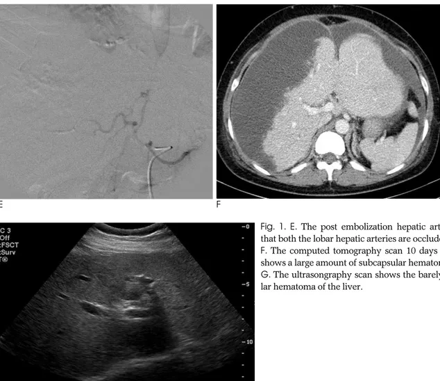
Terumo, Tokyo, Japan). Gelfoam pledge were injected into both the hepatic arteries until the arterial blood flow to the liver was completely blocked (Fig. 1E).
After embolization of the hepatic arteries, the patient’s vital signs stabilized and her hematocrit remained sta- ble.
A computed tomographic (CT) scan 10 days after em- bolization showed a large amount of subcapsular hematoma of the liver and some intraperitoneal free flu- id (Fig. 1F). Eleven days after embolization, she had a low-grade fever and abdominal fullness, so insertion of pigtail drainage catheter was performed and the exten- sive subcapular hematoma from the liver was drained.
The patient recovered gradually from her abdominal fullness and her persistent low grade fever. Her recov- ery from this procedure was uneventful, and she was
discharged 4 weeks later in good condition. The follow- up ultrasonogram that was done 8 weeks after emboliza- tion in the outpatient department showed the barely vis- ible subcapsular hematoma of the liver and the intraab- dominal hematoma (Fig. 1G).
Discussion
Spontaneous rupture of liver is extremely rare, and it can occur in association with hepatocelluar carcinoma, hepatic hemangioma, hepatic abscess or rarely pregnan- cy (1). When it is associated with pregnancy, it has espe- cially occurred combined with eclampsia or preeclamp- sia.
Rupture of liver associated with eclampsia or preeclampsia generally occurs in a multiparous patient
Seung Boo Yang, et al: Spontaneous Hepatic Rupture Associated with Preeclampsia
─ 30 ─
A B
C D
Fig. 1. A 41-year-old woman with hepatic rupture associated with preeclampsia.
A, B. The computed tomography scan through the liver with intravenous contrast enhancement on the first postpartum day. A poorly circumscribed low density area in the both lobes of the liver with a large amount of subcapsular hematoma.
C, D. The hepatic arteriogram shows displacement of hepatic vessels due to a large subcapsular hematoma with multiple small pseudoaneurysms (arrows) in both lobes.
with an average age of the late 20s or early 30s. The vast majority of cases have occurred in the third or late sec- ond trimester of pregnancy, although approximately 15% of the reported cases have occurred in the puer- perium, and usually within 24 hours of delivery, as in the present case (2). Generally, there are signs of preeclampsia present when the patient experiences a sudden onset of epigastric pain or moderate to severe right upper quadrant abdominal pain and eventually hy- povolemic shock (3).
The pathogenesis of spontaneous rupture of liver asso- ciated with eclampsia or preeclampsia is unclear.
Eclamptic patients are generally prone toward sponta- neous hemorrhage and they frequently show fibrin de- position in the hepatic sinusoids and arterioles.
Microangiopathic changes that are the consequence of fibrin deposition result in the occlusion of the hepatic si- nusoids and necrosis of the small arterioles within the liver. As a result, infarction with vascular disruption may occur, leading to intrahepatic hemorrhage, rupture of the parenchyma, production of a subcapsular
hematoma and ultimately rupture of Glisson’s capsule with intraperitoneal hemorrhage (4). Some of the hepat- ic lesions in eclampsia have been attributed to dissemi- nated intravascular coagulation (DIC) (5). McKay de- scribes that eclampsia is actually a form of disseminated intravascular coagulation (6), yet others feel that DIC is not the primary event. In our case and in other reported cases, multiple small pseudoaneurysms were present in the area of hemorrhage, which is consistent with the suggestion that a toxic vasculopathy is the cause of this rare complication of pregnancy (7).
Survival appears to be closely related to making a rapid diagnosis and prompt surgical intervention. Of the currently available imaging techniques such as high res- olution ultrasound examination and computed tomogra- phy examination for confirming the diagnosis of hepatic rupture, computed tomography is by far the most useful if the patent is relatively stable.
Conservative treatment of the contained intra- parenchymal or subcapsular hemorrhage without free intraperitoneal rupture has been performed without
J Korean Soc Radiol 2010;63:29-32
─ 31 ─
E F
G
Fig. 1. E. The post embolization hepatic arteriogram showing that both the lobar hepatic arteries are occluded.
F. The computed tomography scan 10 days after embolization shows a large amount of subcapsular hematoma of the liver.
G. The ultrasongraphy scan shows the barely visible subcapsu- lar hematoma of the liver.
subsequent death, but surgical intervention is thought to improve survival. A number of methods, including packing, hepatic resection, hepatic artery occlusion by ligation, hepatic lobectomy and liver transplantation have been employed to achieve hemostasis. But there is a difficulty to achieve liver hemostasis surgically be- cause of the presence of multiple areas of infarction and hematoma, and especially in the presence of coagulopa- thy.
Transcatheter embolization of the hepatic artery for controlling hemorrahge and for treating hepatic neo- plasms has been well described (8, 9). Several recently reported cases of successful management with hepatic artery embolization of hepatic rupture associated with pregnancy have been described (3, 10, 11). We per- formed transcatheter embolization of the hepatic artery as a mean of obtaining hemostasis. We used 1 mm cubes of Gelfoam sponge for our patient. Gelfoam seems to be the ideal embolic material as it is a gelatin sponge that is resorbed over several weeks, and so it usually allows partial recanalization of the occluded ves- sels. We performed embolization with the catheter tip in the proximal right hepatic artery and left hepatic artery.
Because there was diffuse bleeding in both lobes, selec- tive catheterization was not attempted. Embolization of the hepatic artery is a relatively safe procedure because the liver has a dual blood supply from the portal vein and hepatic artery.
In conclusion, transcatheter hepatic artery emboliza- tion is a safe, effective alternative to surgical treatment
and it can play a major role in controlling hepatic rup- ture associated with preeclampsia.
References
1. Hibbard LT. Spontaneous rupture of the liver in pregnancy: report of eight cases. Am J Obstet Gynecol 1976;126:334-338
2. Bis KA, Waxman B. Rupture of the liver associated with pregnan- cy: a review of the literature and report of 2 cases. Obstet Gynecol Surv 1976;31:763-773
3. Gyang AN, Srivastava G, Asaad K. Liver capsule rupture in eclampsia: treatment with hepatic artery embolisation. Arch Gynecol Obstet 2006;274:377-379
4. Rolfes DB, Ishak KG. Liver disease in toxemia of pregnancy. Am J Gastroenterol 1986;81:1138-1144
5. Killam AP, Dillard SH, Patton RC, Pederson PR. Pregnancy-in- duced hypertension complicated by acute liver disease and dis- seminated intravascular coagulation. Am J Obstet Gynecol 1975;123:823-828
6. McKay DG. Hematologic evidence of disseminated intravascular coagulation in eclampsis. Obstet Gynecol Surv 1972;27:399-417 7. Wagner WH, Lundell CJ, Donovan AJ. Percutaneous angiographic
embolization for hepatic hemorrhage. Arch Surg 1985;120:1241- 1249
8. Hirai K, Kawazoe Y, Yamashita K, Kumagai M, Nagata K, Kawaguchi S, et al. Transcatheter arterial embolization for sponta- neous rupture of hepatocelluar carcinoma. Am J Gastroenterol 1986;81:275-279
9. Chuang VP, Wallace S. Hepatic artery embolization in the treat- ment of hepatic neoplasm. Radiology 1981;140:51-58
10. Terasaki KK, Guinn MF, Lundell CJ, Finck EJ, Pentecost MJ.
Spontaneous hepatic hemorrhage in preeclampsis: treatment with hepatic arterial embolization. Radiology 1990;174:1039-1041 11. Loevinger EH, Vujic I, Lee WM, Anderson M. Hepatic rupture as-
sociated with pregnancy: treatment with transcatheter emobolotherapy. Obstet Gynecol 1985;65:281-284
Seung Boo Yang, et al: Spontaneous Hepatic Rupture Associated with Preeclampsia
─ 32 ─
대한영상의학회지 2010;63:29-32
임신성 고혈압과 관련된 자발성 간 파열: 간동맥색전술에 의한 치료1
`1순천향대학교 영상의학과학교실
2순천향대학교 산부인과학교실
3순천향대학교 비뇨기과학교실
4순천향대학교 내과학교실
양승부∙구동억∙장윤우∙김용재∙황인철2∙한효상2∙윤종현3∙이태일4
전자간증과 관련되어 발생한 자발적 간 파열은 임신과 관련되어 매우 드물며 치료되지 않으면 매우 위험하다. 저 자들은 전자간증과 관련되어 발생한 자발적 간 파열을 간동맥색전술을 이용하여 성공적으로 치료한 예를 보고하고 자 한다.