• 검색 결과가 없습니다.

Massive Spontaneous Diaphragmatic Rupture Induced by a Squatting Position

N/A
N/A
Protected

Academic year: 2021

Share "Massive Spontaneous Diaphragmatic Rupture Induced by a Squatting Position"

Copied!
4
0
0

로드 중.... (전체 텍스트 보기)

전체 글

(1)

Korean J Thorac Cardiovasc Surg 2013;46:230-233 □ Case Report □ http://dx.doi.org/10.5090/kjtcs.2013.46.3.230 ISSN: 2233-601X (Print) ISSN: 2093-6516 (Online)

− 230 −

Department of Thoracic and Cardiovascular Surgery, Jeju National University Hospital, Jeju National University School of Medicine

†This article was presented at the 44th Autumn Scientific Meeting of The Korean Society for Thoracic and Cardiovascular Surgery.

Received: November 6, 2012, Revised: December 17, 2012, Accepted: December 18, 2012

Corresponding author: Seogjae Lee, Department of Thoracic and Cardiovascular Surgery, Jeju National University Hospital, Jeju National University School of Medicine, 15 Aran 13-gil, Jeju 690-767, Korea

(Tel) 82-64-717-2081 (Fax) 82-64-757-8276 (E-mail) sj1106@jejunu.ac.kr

C

The Korean Society for Thoracic and Cardiovascular Surgery. 2013. All right reserved.

CC
This is an open access article distributed under the terms of the Creative Commons Attribution Non-Commercial License (http://creative- commons.org/licenses/by-nc/3.0) which permits unrestricted non-commercial use, distribution, and reproduction in any medium, provided the original work is properly cited.

Massive Spontaneous Diaphragmatic Rupture Induced by a Squatting Position

Su Wan Kim, M.D., Seogjae Lee, Ph.D.

While a diaphragmatic rupture commonly results from trauma to the abdomen and chest, a spontaneous dia- phragmatic rupture is very rare. A 68-year-old male presented with chest pain that had originated while doing farm work in a squatting position. Images revealed a 5 cm defect of the left diaphragmatic dome, and the entire stom- ach was displaced into the thorax. The diaphragmatic defect was round and half had a well-demarcated margin.

The remaining fragile tissue was completely excised and was closed primarily. The patient was uneventfully dis- charged and resumed with a normal diet 10 days after the operation.

Key words: 1. Diaphragm 2. Chest pain

3. Computed tomography

CASE REPORT

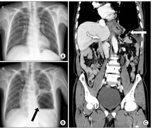
A 68-year-old male presented with chest pain similar to acute coronary syndrome. The patient had felt a substernal pain from a period of four days prior to examination, which had begun while carrying out farm work in a squatting position. The severity of the chest pain had fluctuated, and then the patient felt intolerable chest pain that lasted for two hours. An electrocardiogram showed normal sinus rhythm, and cardiac enzymes were normal. However, compared with another chest X-ray four months earlier (Fig. 1A), a second chest X-ray revealed left diaphragmatic hernia with a mark- edly distended stomach and passive atelectasis of the left lower lung (Fig. 1B). An abdomen computed tomography showed that the herniated stomach was significantly distended by ingested air due to severe pyloric compression. The dia-

phragmatic defect was approximately 5 cm in diameter (Fig.

1C).

We performed an emergency operation because the patient

suffered from continuing chest pain, which suggested a stran-

gulation of the stomach. A left lateral thoracotomy was per-

formed through the eighth intercostal space after intubation

with a double-lumen endotracheal tube and insertion of a na-

sogastric tube for decompressing the stomach. We carefully

inspected the stomach, which was entirely displaced into the

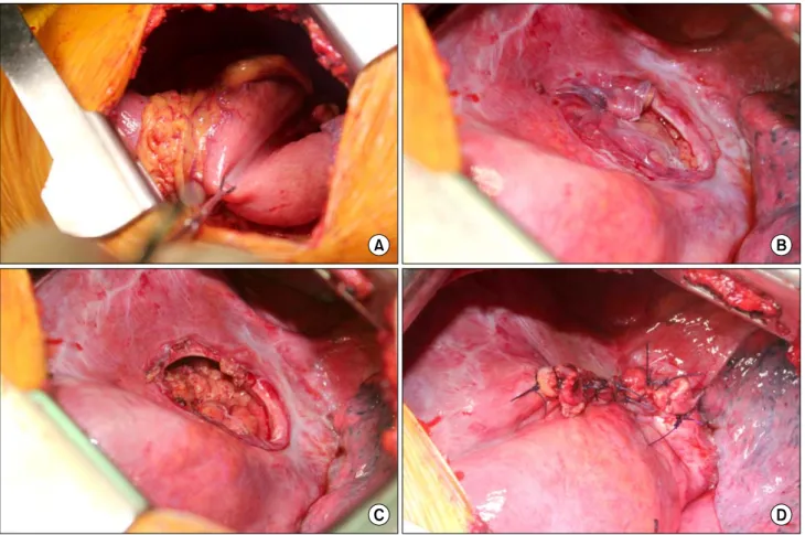
thoracic cavity, including the greater omentum (Fig. 2A), and

we then reduced the stomach into the peritoneal cavity. A di-

aphragmatic defect was identified on the highest central ten-

don and had a round shape, half of which had a well-demar-

cated margin (Fig. 2B). The diaphragm was thinned and un-

evenly rough owing to senile change. The remaining tissue of

the fragile margin was completely excised (Fig. 2C) and was

(2)

Spontaneous Diaphragmatic Rupture

− 231 −

Fig. 1. The patient had a normal chest X-ray four months earlier (A).

A new chest X-ray (B) and com- puted tomography (C) revealed that the stomach had migrated into the thoracic cavity. A black arrow (B) points to the stomach, which is ex- tremely inflated by ingested air. A white arrow (C) points to a 5 cm defect of the diaphragm. The dia- phragmatic dome is suspected to be a perforated site.

closed primarily using continuous running sutures and multi- ple interrupted sutures (Fig. 2D). The patient was extubated before leaving the operating room. The chest X-rays before discharge showed complete reduction of the stomach and full re-expansion of the left lung (Fig. 3). The patient was un- eventfully discharged and resumed a normal diet 10 days af- ter the operation.

DISCUSSION

Diaphragmatic rupture (DR) is commonly observed after a blunt or penetrating trauma to the abdomen and chest.

Several reports of traumatic DR appear in the Korean literature. Recently, Hwang et al. [1] investigated 40 patients and Cho et al. [2] investigated 37 patients with traumatic DR.

The mortality rates of these patients were 17.5% and 16.2%, respectively, and the patients had similar prognostic factors affecting mortality, such as a high injury severity score, high revised trauma score, and hypotension. However, spontaneous DR has not been reported in Korea.

Losanoff et al. [3] reported one case of spontaneous DR

that had been induced by a recent bout of coughing and re- viewed the world literature about spontaneous DR of 28 pa- tients from 1956 to 2009. Coughing was the precipitating event in 9 patients (32%), physical exercise in 6 patients (21%), vaginal delivery in 4 patients (14%), vomiting in 2 patients (7%), and massage in 1 patient (4%). On the other hand, there have been sporadic case reports of secondary spontaneous DR since 2009. Hamaoui et al. [4] reported a case of spontaneous DR in a 35-year-old male patient with Ehlers-Danlos syndrome, a genetic disorder that causes abnor- malities in the synthesis and structure of collagen and can lead to multiple anatomical defects. Pehar et al. [5] reported a case of spontaneous DR related to local invasion by retro- peritoneal liposarcoma. Diaphragmatic eventration is thought to be a secondary cause of spontaneous DR during preg- nancy. Servais et al. [6] suggested that symptomatic dia- phragmatic eventration during pregnancy should be repaired during the third trimester once fetal organogenesis is com- plete in order to prevent further herniation from the enlarging uterus and the risk of hernia strangulation.

One case of spontaneous DR in a Korean patient was re-

(3)

Su Wan Kim and Seogjae Lee

− 232 −

Fig. 2. The entire stomach including the greater omentum is displaced into the thorax (A). The diaphragmatic defect is observed on the highest central tendon, and has a round shape, half of which has a well-demarcated margin (B). The remaining tissue of the fragile mar- gin was completely excised (C) and was closed primarily (D).

Fig. 3. (A) The diaphragm became flat after the repair. (B) Postoperatively, a small amount of pneumoperitoneum was observed, (C) but

the pneumoperitoneum spontaneously disappeared after discharge.

(4)

Spontaneous Diaphragmatic Rupture

− 233 − ported by Yang et al. [7] in a United States journal. They re- ported a spontaneous DR in a 29-year-old woman and sug- gested that spontaneous DR might be caused by a static sport activity, such as pilates. We, however, report the first case of spontaneous DR in the Korean Journal of Thoracic and Cardiovascular Surgery. Furthermore, we present several pho- tographs of high quality that clearly point out the dia- phragmatic defect. No pictures grossly showing a dia- phragmatic defect definitively like that of our patient have been published before.

Diaphragmatic injuries that are diagnosed promptly should be explored from the abdomen with either a laparotomy or laparoscopy because of the frequent association of other ab- dominal organ injury. In those cases with a significant delay in diagnosis, injuries generally should be explored from the thorax, with either thoracotomy or thoracoscopy, because the herniated abdominal structure is frequently adhered to the ip- silateral lung [8]. Additionally, it is recommended that the di- aphragm be restored by simple interrupted suture or double layer suture methods with non-absorbable suture materials [1,2,8]. In our case, we performed a left thoracotomy because the patient had no abdominal trauma history and the suspi- cious rupture site was the central tendon, which could be ob- served by direct vision through the thoracotomy.

In summary, we report the first description of massive spontaneous DR arising while in the squatting position, prob- ably resulting from increased intra-abdominal pressure. This and similar reports emphasize that in cases with high clinical suspicion of DR, diagnosis should be pursued even in the ab- sence of a preceding traumatic event.

CONFLICT OF INTEREST

No potential conflict of interest relevant to this article was reported.

REFERENCES

1. Hwang SW, Kim HY, Byun JH. Management of patients with traumatic rupture of the diaphragm. Korean J Thorac Cardiovasc Surg 2011;44:348-54.

2. Cho S, Lee EB, Seok YK. The prognostic factors of trau- matic diaphragmatic rupture. Korean J Thorac Cardiovasc Surg 2010;43:47-52.

3. Losanoff JE, Edelman DA, Salwen WA, Basson MD.

Spontaneous rupture of the diaphragm: case report and comprehensive review of the world literature. J Thorac Cardiovasc Surg 2010;139:e127-8.

4. Hamaoui K, Riaz A, Hay A, Botha A. Massive spontaneous diaphragmatic rupture in Ehlers-Danlos syndrome. Ann R Coll Surg Engl 2012;94:e5-7.

5. Pehar M, Vukoja I, Rozic D, Miskovic J. Spontaneous dia- phragmatic rupture related to local invasion by retro- peritoneal liposarcoma. Ann R Coll Surg Engl 2012;94:

e18-9.

6. Servais EL, Stiles BM, Finnerty BM, Paul S. Ruptured dia- phragmatic eventration: a rare cause of acute postpartum dyspnea. Ann Thorac Surg 2012;93:e143-4.

7. Yang YM, Yang HB, Park JS, Kim H, Lee SW, Kim JH.

Spontaneous diaphragmatic rupture complicated with perfo- ration of the stomach during Pilates. Am J Emerg Med 2010;28:259.e1-3.

8. Daniel CE, Michael TJ. Surgery of the diaphragm: a de- ductive approach. In: Sellke FW, Del Nido PJ, Swanson SJ, Sabiston DC, Spencer FC. Sabiston & Spencer surgery of the chest. 7th ed. Philadelphia: Elsevier Saunders; 2004.

p.501-15.

수치

Fig. 1. The patient had a normal  chest X-ray four months earlier (A).
Fig. 2. The entire stomach including the greater omentum is displaced into the thorax (A)

참조

관련 문서

A 27-year-old man with spondyloarthropathy, oblique coronal fat-saturated T2-weighted (A) and oblique coronal postcontrast fat-saturated T1-weighted (B) images show

 store value (read from the Register File during decode) written to the Data Memory.  load value, read from the Data Memory, written to

 The disk arm starts at the first I/O request on the disk, and moves toward the last I/O request on the other end, servicing requests until it gets to the other extreme

 allow instructions to contain immediate operands..  Good design

 Assuming a cache hit, the new data is written from the store buffer into the cache on the next unused cache access cycle.  Many write-back caches also include write buffers

• This determination is facilitated by the introduction of the equilibrium constant for the reaction, and the relation between this constant and the standard Gibbs free

Typical trace of pacemaker potentials in current clamping mode (A) and spontaneous inward pacemaker currents in voltage clamping mode at a holding potential of -70 mV

The feed is commonly a solution in a solvent like ethanol or t-butanol, and the nonsolvent is water..