• 검색 결과가 없습니다.

Bradykinin-induced $Ca^{2+}$ signaling in human oral squamous cell carcinoma HSC-3 cells

N/A
N/A
Protected

Academic year: 2021

Share "Bradykinin-induced $Ca^{2+}$ signaling in human oral squamous cell carcinoma HSC-3 cells"

Copied!
7
0
0

로드 중.... (전체 텍스트 보기)

전체 글

(1)

Copyright ⓒ 2009, The Korean Academy of Oral Biology

73

Journal of Oral Biology

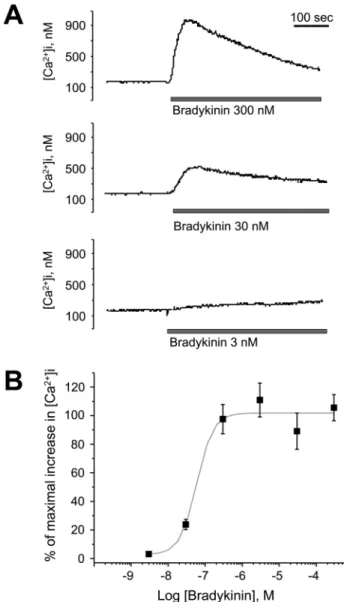
Bradykinin-induced Ca 2+ signaling in human oral squamous cell carcinoma HSC-3 cells

Byung Jin Sohn

1

, Ji-Ah Kang

1

, Su-Hyun Jo

2

, and Se-Young Choi

1

*

1

Department of Physiology and Dental Research Institute, Seoul National University School of Dentistry, Seoul 110-749, Republic of Korea,

2

Department of Physiology, Kangwon National University School of Medicine, Chuncheon 200-701, Korea.

(received April 14, 2009 ; revised April 22, 2009 ; accepted April 24, 2009)

Cytosolic Ca

2+

is an important regulator of tumor cell proliferation and metastasis. Recently, the strategy of blocking receptors and channels specific to certain cancer cell types has emerged as a potentially viable future treatment. Oral squamous cell carcinoma is an aggressive form of cancer with a high metastasis rate but the receptor- mechanisms involved in Ca

2+

signaling in these tumors have not yet been elucidated. In our present study, we report that bradykinin induces Ca

2+

signaling and its modulation in the human oral squamous carcinoma cell line, HSC-3.

Bradykinin was found to increase the cytosolic Ca

2+

levels in a concentration-dependent manner. This increase was inhibited by pretreatment with the phospholipase C- β inhibitor, U73122, and also by 2-aminoethoxydiphenyl borate, an inhibitor of the inositol 1,4,5-trisphosphate receptor. Pretreatment with extracellular ATP also inhibited the peak bradykinin-induced Ca

2+

rise. In contrast, the ATP-induced rise in cytosolic Ca

2+

was not affected by pretreatment with bradykinin. Pretreatment of the cells with either forskolin or phorbol 12-myristate 13-acetate (activators of adenylyl cyclase and protein kinase C, respectively) prior to bradykinin application accelerated the recovery of cytosolic Ca

2+

to baseline levels. These data suggest that bradykinin receptors are functional in Ca

2+

signaling in HSC-3 cells and may therefore represent a future target in treatment strategies for human oral squamous cell carcinoma.

Key words : Bradykinin; oral squamous cell carcinoma;

HSC-3 cells; phospholipase C

Introduction

Head and neck squamous cell carcinoma or oral squamous cell carcinoma show high incidence world wide, with poor prognosis and low survival rate; less than 40% of those diagnosed survive >5 years (Parkin et al., 1999). Oral squamous cell carcinoma, especially is known to show a progressive increase in metastatic potential due to the loss of basement membranes, subsequent lymph node metastasis and local invasion (Kurahara et al., 1999). Thus, it is of great therapeutic value to be able to modulate tumor progress and metastasis of oral squamous cell carcinoma. Multiple cellular steps are involved in the migration, invasion and metastasis of oral squamous cell carcinoma (Son et al., 2006; Min et al., 2007). At each step, tumor cells decide their fate according to signals from the external environment.

Communication with the environment is mediated by membrane and/or intracellular receptors, which may then initiate a host of intracellular signaling cascades. The level of cytosolic free Ca

2+

([Ca

2+

]

i

) is one such important signal transduction mechanism in tumor cells.

[Ca

2+

]

i

modulates the proliferation of tumor cells by affecting the cell cycle and other proliferative and apoptotic mechanisms (Roderick and Cook, 2008). In oral squamous cell carcinoma, increased [Ca

2+

]

i

is also known to affect the tumor growth and chemotherapy resistance (Thomas et al., 2003; Kokoska et al., 2000). In addition to dynamic increases in [Ca

2+

]

i

via Ca

2+

influx, elevated basal [Ca

2+

]

i

levels are also considered to modulate the tumorigenesis (Lee et al., 2002; Usachev et al., 2001).

*Corresponding author: Se-Young Choi, Ph.D. Department of Physiology, Seoul National University School of Dentistry.

Tel.: +82-2-740-8650, Fax.: +82-2-762-5107, E-mail: [email protected]

(2)

The machinery required for an increase in [Ca

2+

]

i

in tumor cells could be characteristic for each cancer, depending on the originating tissue. For better chemotherapy with higher specificity and target selectivity, it has been suggested to use antagonists to target G-protein coupled receptors (GPCRs) expressed selectively on tumors subtypes (Stewart et al., 2002; Stewart et al., 2005). Thus, in order to develop a better treatment for oral squamous cell carcinoma, it is urgently required to identify and characterize the signal mechanisms of these cells. Oral squamous cell carcinomas are derived from epithelial cells, which are non-excitable and lack voltage-sensitive channels to allow Ca

2+

influx. Instead, signaling induced by GPCR activation is likely to serve a crucial role in the regulation of [Ca

2+

]

i

. However, it is not yet known what families of GPCRs are functional in oral squamous cell carcinoma nor how their [Ca

2+

]

i

signals are modulated.

To address this question, we used the HSC-3 cell line as a model of human oral squamous carcinoma cells (Momose et al., 1989). These cells are known to exhibit tumor migration behavior (Myoung et al., 2003; Shen and Kramer, 2004). We report that HSC-3 cells have functional bradykinin receptors, which are linked to the phospholipase C- β (PLC-β) pathway.

Materials and Methods

Materials

Bradykinin, adenosine 5’-triphosphate (ATP), U73122, 2- aminoethoxydiphenyl borate (2-APB), forskolin, and phorbol 12-myristate 13-acetate (PMA), ethylene glycol-bis ( β- aminoethyl ether)-N,N,N’,N’-tetraacetic acid (EGTA), dimethyl sulfoxide (DMSO) and sulfinpyrazone were purchased from Sigma (St. Louis, MO, USA). Fura-2AM was obtained from Molecular Probes (Eugene, OR, USA).

Dulbecco’s Modified Eagle’s Medium (DMEM), bovine calf serum and penicillin/streptomycin were purchased from GIBCO (Grand Island, NY, USA). Fetal bovine serum was obtained from HyClone Laboratories (Logan, UT, USA).

Cell Culture

HSC-3 cells were maintained at 37

o

C in DMEM supplemented with 10% (v/v) heat-inactivated bovine calf serum and 1% (v/v) penicillin-streptomycin in a humidified atmosphere of 95% O

2

and 5% CO

2

. The culture medium was changed every day and the cells were subcultured weekly. All cells were cultured in a humidified atmosphere of 95% O

2

and 5% CO

2

.

[Ca

2+

]

i

measurement

The level of intracellular Ca

2+

was determined using fura- 2/AM as we have previously described (Choi et al., 2001).

Briefly, the cell suspension was incubated in fresh serum- free DMEM with 3 µM fura-2/AM at 37

o

C for 60 minutes with continuous stirring. Sulfinpyrazone (250 µM) was

added to prevent dye leakage. Changes in the fluorescence ratio were measured at dual excitation wavelengths of 340 nm and 380 nm and emission wavelength of 500 nm.

Calibration of the fluorescent signal in terms of [Ca

2+

]

i

was performed as described by Grynkiewicz et al. (1985) using the following equation:

[Ca

2+

]i = K

D

[(R - R

min

)/(R

max

- R)](S

f2

/S

b2

)

where R is the ratio of fluorescence emitted after excitation at 340 and 380 nm. S

f2

and S

b2

are the proportionality coefficients at 380 nm excitation of Ca

2+

-free fura-2 and

Fig. 1. Bradykinin-induced changes in intracellular Ca

2+

concen-

tration in human oral squamous cell carcinoma HSC-3 cells. (A)

Fura-2-loaded HSC-3 cells were challenged with bradykinin at

various concentrations (3 nM, 30 nM and 300 nM) and the changes

in cytosolic Ca

2+

level were monitored. Typical Ca

2+

transients

from more than three separate experiments are presented. (B)

Concentration-response curve for HSC-3 cells challenged with

rising concentrations of bradykinin. Values represent ratio of peak

changes in fluorescence relative to maximum response. Data

points indicate mean ± SEM. All results were reproducible.

(3)

Ca

2+

-saturated fura-2, respectively.

Data Analysis

All quantitative data are expressed as the mean ± SEM.

The results were analyzed for differences using an unpaired Student’s t-test. The half maximal effective concentration (EC

50

) of bradkykinin was calculated using Microcal Origin software (Northampton, MA, USA).

Results

We tested bradykinin-induced cellular responses in human oral squamous cell carcinoma HSC-3 cells. Bradykinin triggered a rise in [Ca

2+

]

i

in fura-2-loaded HSC-3 cells (Fig 1A). The [Ca

2+

]

i

response reached a peak concentration of about 900 nM within 50 s and then fell back to the baseline level within 600 s. Bradykinin elevated [Ca

2+

]

i

in a concentration-dependent manner with an EC

50

of 59.2 ± 3.6 nM (n = 4); the maximal response was observed at 300 nM (Fig 1C). Repeated application of bradykinin did not show any increase in [Ca

2+

]i level (data not shown).

Bradykinin signaling is known to be mediated via the PLC- β pathway. We analyzed the characteristics of the bradykinin-induced increase in [Ca

2+

]

i

using the PLC- β antagonist, U73122, and inositol 1,4,5-trisphosphate (IP

3

)

receptors antagonist, 2-APB. Pretreatment of the cells with U73122 inhibited the bradykinin-induced [Ca

2+

]

i

increase in HSC-3 cells (Fig 2A). 2-APB reduced the bradykinin- induced [Ca

2+

]

i

increase (Fig 2B).

We compared the effect of bradykinin on Ca

2+

signaling with that of purinergic receptors using application of ATP

Fig. 2. U73122 and 2-aminoethoxydiphenyl borate (2-APB) inhibit the bradykinin-induced [Ca

2+

]i increase in HSC-3 cells. (A) Fura- 2-loaded HSC-3 cells were treated with bradykinin (300 nM), with (black trace) or without (gray trace) the preincubation of U73122 (10 µM; phospholipase C-β inhibitor). Typical Ca

2+

transients from more than three separate experiments are presented. (B) Cells were treated with bradykinin (300 nM), with (black trace) or without (gray trace) the preincubation of 2-APB (50 µM; IP

3

receptors inhibitor). Typical Ca

2+

transients from more than three separate experiments are presented. All results were reproducible.

Fig. 3. Bradykinin-induced Ca

2+

signaling is occluded by extracel- lular ATP in HSC-3 cells. (A) Fura-2-loaded HSC-3 cells are treated with ATP (300 µM), with (black trace) or without (gray trace) the preincubation of bradykinin (300 nM). Typical Ca

2+

tran- sients from more than three separate experiments are presented. (B) Cell are treated with bradykinin (300 nM) with (black trace) or without (gray trace) the preincubation of ATP (300 µM). Typical Ca

2+

transients from more than three separate experiments are pre- sented. The results were reproducible.

Fig. 4. Forskolin and phorbol 12-myristrate 13-acetate (PMA) inhibit the bradykinin-induced [Ca

2+

]

i

increase in HSC-3 cells. (A) Fura-2-loaded HSC-3 cells were treated with bradykinin (300 nM;

solid bar), in the presence (black trace) or absence (gray trace) of

forskolin (1 µM). Typical Ca

2+

transients from more than three sep-

arate experiments are presented. (B) Cells were treated with brady-

kinin (300 nM) in the presence (black trace) or absence (gray trace)

of PMA (50 µM). Typical Ca

2+

transients from more than three

separate experiments are presented. All results were reproducible.

(4)

(300 µM). Extracellular ATP alone evoked a [Ca

2+

]

i

increase that did not differ significantly from that evoked after pretreatment with bradykinin (Fig. 3A). However, pretreat- ment with ATP (300 µM) inhibited a following bradykinin- evoked [Ca

2+

]

i

increase (Fig. 3B). The results suggest that bradykinin receptor-mediated Ca

2+

signaling is occluded by a purinergic Ca

2+

signal pathway.

We investigated the possibility of functional crosstalk between GPCR signaling by monitoring changes in bradykinin-induced Ca

2+

signaling by pre-challenge with either forskolin (adenylyl cyclase activator) or PMA (protein kinase C activator). Preincubation with forskolin increased the rate of recovery of the bradykinin-induced Ca

2+

increase, but did not affect the peak amplitude of the Ca

2+

increase (Fig. 4A). PMA decreased the peak [Ca

2+

]

i

level and increased the rate of the recovery phase of the bradykinin- induced Ca

2+

increase (Fig. 4B).

Discussion

Bradykinin is an important peptide hormone involved in various physiological responses such as inflammation, pain, vasodilation, vascular permeability, smooth muscle contraction, synthesis of prostaglandins, and hormone secretion (Bhoola et al., 1992). In oral pathology, bradykinin serves a significant physiological function in the nervous system by acting in pain signaling (Wang et al., 2005; Lai et al., 2006) and is involved in the regulation of pathological conditions such as ischemia and epilepsy (Ongali et al., 2003; Lumenta et al., 2006). In tumor cells, bradykinin is known to modulate tumor growth and migration (Mahabeer and Bhoola, 2000; Stewart, 2003). It is also reported that bradykinin receptors are overexpressed in certain cancers, including astrocytic tumor and prostate cancer (Raidoo 1999; Barki-Harrington, 2003; Taub 2003). Bradykinin shows not only a direct effect on cancer cells, but also has indirect effects on tumor metastasis by vascular endothelial growth factor (VEGF) secretion and promoting angiogenesis (Hosoi et al., 1993; Wu et al., 1998).

In this study, we have found for the first time that bradykinin serves to induce Ca

2+

signaling in the HSC-3 human oral squamous cell carcinoma line. The time course of the [Ca

2+

]

i

response (reaching peak level within 50 s and then falling back to the baseline level within 600 s) was general characteristic of GPCR activation. The concentration- dependent manner of the bradykinin-induced Ca

2+

increase and nanomolar EC

50

value suggest that the Ca

2+

increase is likely mediated by the response of bradykinin receptors and not due to the activation of other neuropeptide hormones (e.g. neurotensin) which have cross-reactivity in the micromolar concentration level. Bradykinin receptors are coupled through various G proteins to at least two separate phospholipid-mediated signaling pathways: PLC (Chuang et al., 2001) and phospholipase A

2

(Hsieh et al., 2006). To

confirm this, we used the selective PLC- β inhibitor, U73122, which prevents G-protein interactions with PLC- β, thereby blocking subsequent intracellular signaling. The pretreatment with U73122 showed a dramatic inhibition of the bradykinin-induced [Ca

2+

]

i

increase, which suggests that bradykinin signaling occurs in a PLC- β dependent manner.

The activation of PLC- β triggers the breakdown of membrane phosphatidyl 4,5-bisphosphate into diacylglycerol and IP

3

, which then induces Ca

2+

release from intracellular Ca

2+

stores and store-operated Ca

2+

entry by the activation of TrpC channels. We tested the effect of 2-APB, which is known to block the interaction between IP

3

-receptors and TrpC channels and thereby inhibit the downstream signaling of IP

3

. We found that treatment with 2-APB also decreased the bradykinin-induced [Ca

2+

]

i

response. These results suggest that bradykinin induces an increase of [Ca

2+

]

i

in HSC-3 cells through a PLC- β and IP

3

receptor pathway.

Cytosolic Ca

2+

serves as a significant signal for tumor cells. Intracellular Ca

2+

modulates tumor proliferation via regulation of the mammalian cell cycle by its effect on G1/S progression, separation, and centrosome duplication (Roderick et al., 2008). Tumor cells are thought to change their PCL2 and PKB pathway in order to utilize cytosolic Ca

2+

for enhancing proliferation and blocking apoptosis (Roderick et al., 2008). Especially, cytosolic Ca

2+

modulates the Ca

2+

level and membrane permeability of mitochondria (in other term, mitochondrial permeability transition), which is also regulated by Bcl-2 pathway and trigger the cancer cell death (Tsujimoto et al., 2006; Roy et al., 2009). There are two features of cytosolic Ca

2+

which are significant for the tumor development. One factor is the increased basal level of cytosolic [Ca

2+

]. If the cytosolic Ca

2+

is not efficiently cleared after Ca

2+

increase, the proliferation and migration of tumor cells may be promoted. Sarcoplasmic reticulum Ca

2+

-ATPase 2 (SERCA2) and plasma membrane Ca

2+

- ATPase 1 (PMCA1), proteins responsible for returning cytosolic Ca

2+

to baseline, have been shown to be down- regulated in oral squamous cell carcinoma (Endo et al., 2004; Kato et al., 2006a; Saito et al., 2006. In addition, mice heterozygote for Atp2a2 (the gene coding the SERCA2 pump) show serious squamous cell tumors in the tongue, oral mucosa, esophagus and fore-stomach (Liu et al., 2001;

Prasad et al., 2005). The second factor affecting cytosolic Ca

2+

is Ca

2+

influx through plasma membrane ion channels or GPCR. Voltage sensitive Ca

2+

channels (VGCCs), such as T-type Ca

2+

channels, are thought to be expressed in certain cancer cells, and blockers of VGCCs have shown potential as cancer treatments (Taylor et al., 2008). However, in the case of tumors with an epithelial cell origin, which lack VGCCs and other Ca

2+

-permeable ion channels, PLC- linked Ca

2+

signaling may be of greater clinical importance.

Thus it is important to identify the receptors involved in

intracellular Ca

2+

signaling in tumors. In HSC-3 cells,

hyaluronic acid is reported to serve in Ca

2+

signaling and has

been suggested as a treatment for oral squamous cell

(5)

carcinoma (Wang and Bourguignon, 2006). Potential targets in tumor cell Ca

2+

signaling are not restricted on receptors or channels; effector enzymes in down-stream mechanisms also show important roles in tumor proliferation. For example, IP

3

-kinase A protein was found in the search for tumor-specific associated proteins using 2D-PAGE and MALDI-TOF mass spectrometry (Kato et al., 2006b).

Finally, PLC is important for the resistant mechanism of cisplatin, which is one of the most common treatments of oral squamous cell carcinoma (Ferguson et al., 1999). Thus it bears significance to modulate the Ca

2+

signaling triggered by the activation of GPCRs such as the bradykinin receptor.

It is common for certain receptor-mediated signals to be affected by preactivation of other receptors (Werry et al., 2003); therefore, we investigated whether the bradykinin Ca

2+

response could be modulated by other receptor signals.

Interestingly, we found bradykinin signaling was inhibited by pretreatment with extracellular ATP, however, in the converse experiment, purinergic signaling was not significantly altered by pretreatment with bradykinin. These results imply not only that both bradykinin and extracellular ATP provide Ca

2+

signaling in HSC-3 cells, but purinergic receptor activation can modulate the bradykinin-response in HSC-3 cells. We found that forskolin (adenylyl cyclase activator) and PMA (protein kinase C activator) decreased the bradykinin-induced [Ca

2+

]

i

increase. These results suggest that the preceding activation of adenylyl cyclase and PLC may negatively regulate bradykinin signaling and, furthermore, that bradykinin may show negative crosstalk between other receptor systems that utilize adenylyl cyclase or PLC-linked pathways. The results of this study imply that the modulation of bradykinin-induced Ca

2+

signaling can also be achieved by activation of other receptors. Further studies based on our results (for example, an investigation into the difference in inhibitory targets between protein kinase A and C signaling) could suggest a combinatorial strategy for the bradykinin signal modulation.

Ca

2+

pump inhibitors have been considered for cancer treatment (Monteith et al., 2007) but the ubiquitous distribution of PMCA and SERCA in cells other than carcinoma presents a challenge for the specificity of such drugs. Bradykinin receptor antagonists are currently being developed for treatment of small cell lung cancer and prostate cancer; these tumor cells originate from neuroendocrine cells and express bradykinin receptors (Chan et al., 2002; Barki-Harington et al., 2003). We expect our results could answer how the ‘oral’

cancer communicates with its environment and would be helpful to develop more reliable treatment for this type of cancer. Thus further investigation for the role of bradykinin such as proliferation, metastasis, or apoptic modulation, would be helpful to design the bradykinin-related treatment of oral squamous cell carcinoma. Our results show that HSC-3 cells display functional bradykinin Ca

2+

signaling and raise the possibility of targeting bradykinin receptor- signaling in future treatments of oral squamous cell

carcinoma.

Acknowledgements

We thank Dr. Alexander Davies for reading the manuscript. This work was supported by the Korea Science

& Engineering Foundation grant (R13-2008-008-01001-0) through the Oromaxillofacial Dysfunction Research Center for the Elderly (ODRCE) at Seoul National University.

Reference

Barki-Harrington L, Bookout AL, Wang G, Lamb ME, Leeb- Lundberg LM, Daaka Y. Requirement for direct cross-talk between B1 and B2 kinin receptors for the proliferation of androgen-insensitive prostate cancer PC3 cells. Biochem J.

2003;371:581-7.

Bhoola KD, Figueroa CD, Worthy K. Bioregulation of kinins:

kallikreins, kininogens, and kininases. Pharmacol Rev.

1992;44:1-80.

Chan DC, Gera L, Stewart JM, Helfrich B, Zhao TL, Feng WY, Chan KK, Covey JM, Bunn PA Jr. Bradykinin antagonist dimer, CU201, inhibits the growth of human lung cancer cell lines in vitro and in vivo and produces synergistic growth inhibition in combination with other antitumor agents. Clin Cancer Res. 2002;8:1280-7.

Choi SY, Choi BH, Suh BC, Chae HD, Kim JS, Shin MJ, Kang SS, Negishi M, Kim KT. Potentiation of PGE

2

-mediated cAMP production during neuronal differentiation of human neuroblastoma SK-N-BE(2)C cells. J Neurochem. 2001;79:

303-10.

Chuang HH, Prescott ED, Kong H, Shields S, Jordt SE, Basbaum AI, Chao MV, Julius D. Bradykinin and nerve growth factor release the capsaicin receptor from PtdIns(4,5) P

2

-mediated inhibition. Nature 2001;411:957-62.

Endo Y, Uzawa K, Mochida Y, Shiiba M, Bukawa H, Yokoe H, Tanzawa H. Sarcoendoplasmic reticulum Ca

2+

ATPase type 2 downregulated in human oral squamous cell carcinoma.

Int J Cancer. 2004;110:225-31.

Ferguson PJ, Currie C, Vincent MD. Enhancement of platinum- drug cytotoxicity in a human head and neck squamous cell carcinoma line and its platinum-resistant variant by liposomal amphotericin B and phospholipase A2-II. Drug Metab Dispos. 1999;27:1399-405.

Grynkiewicz G, Poenie M, Tsien RY. A new generation of Ca

2+

indicators with greatly improved fluorescence properties. J Biol Chem. 1985;260:3440-50.

Hosoi K, Kurihara K, Ueha T. Bradykinin-stimulated transient modulation of epidermal growth factor receptors in A-431 human epidermoid carcinoma cells. J Cell Physiol. 1993;157:

1-12.

Hsieh HL, Wu CY, Hwang TL, Yen MH, Parker P, Yang CM.

BK-induced cytosolic phospholipase A

2

expression via

sequential PKC-δ, p42/p44 MAPK, and NF-κB activation in

rat brain astrocytes. J Cell Physiol. 2006;206:246-54.

(6)

Kato H, Uzawa K, Onda T, Kato Y, Saito K, Nakashima D, Ogawara K, Bukawa H, Yokoe H, Tanzawa H. Down- regulation of 1D-myo-inositol 1,4,5-trisphosphate 3-kinase A protein expression in oral squamous cell carcinoma. Int J Oncol. 2006b;28:873-81.

Kato Y, Uzawa K, Saito K, Nakashima D, Kato M, Nimura Y, Seki N, Tanzawa H. Gene expression pattern in oral cancer cervical lymph node metastasis. Oncol Rep. 2006a;16:1009- 14.

Kokoska ER, Wolff AB, Smith GS, Miller TA. Epidermal growth factor-induced cytoprotection in human intestinal cells involves intracellular calcium signaling. J Surg Res.

2000;88:97-103.

Kurahara S, Shinohara M, Ikebe T, Nakamura S, Beppu M, Hiraki A, Takeuchi H, Shirasuna K. Expression of MMPS, MT-MMP, and TIMPs in squamous cell carcinoma of the oral cavity: correlations with tumor invasion and metastasis.

Head Neck. 1999;21:627-38.

Lai J, Luo MC, Chen Q, Ma S, Gardell LR, Ossipov MH, Porreca F. Dynorphin A activates bradykinin receptors to maintain neuropathic pain. Nat Neurosci. 2006;9:1534-40.

Lee WJ, Roberts-Thomson SJ, Holman NA, May FJ, Lehrbach GM, Monteith GR. Expression of plasma membrane calcium pump isoform mRNAs in breast cancer cell lines. Cell Signal. 2002;14:1015-22.

Liu LH, Boivin GP, Prasad V, Periasamy M, Shull GE. Squamous cell tumors in mice heterozygous for a null allele of Atp2a2, encoding the sarco(endo)plasmic reticulum Ca

2+

-ATPase isoform 2 Ca

2+

pump. J Biol Chem. 2001;276:26737-40.

Lumenta DB, Plesnila N, Kläsner B, Baethmann A, Pruneau D, Schmid-Elsaesser R, Zausinger S. Neuroprotective effects of a postischemic treatment with a bradykinin B

2

receptor antagonist in a rat model of temporary focal cerebral ischemia. Brain Res. 2006;1069:227-34.

Mahabeer R, Bhoola KD. Kallikrein and kinin receptor genes.

Pharmacol Ther. 2000;88:77-89.

Min JH, Kim DK, Lee MH, Bae MK, Um KI, Kwak HH, Park BS, Kim GC. Mutantional analysis of tumor suppressor gene p53 in human oral squmous carcinoma cell line YD-9.

Int J Oral Biol. 2007;32:79-84.

Momose F, Araida T, Negishi A, Ichijo H, Shioda S, Sasaki S.

Variant sublines with different metastatic potentials selected in nude mice from human oral squamous cell carcinomas. J Oral Pathol Med. 1989;18:391-5

Monteith GR, McAndrew D, Faddy HM, Roberts-Thomson SJ. Calcium and cancer: targeting Ca

2+

transport. Nat Rev Cancer. 2007;7:519-30.

Myoung H, Hong SP, Yun PY, Lee JH, Kim MJ. Anti-cancer effect of genistein in oral squamous cell carcinoma with respect to angiogenesis and in vitro invasion. Cancer Sci.

2003;94:215-20.

Ongali B, Campos MM, Bregola G, Rodi D, Regoli D, Thibault G, Simonato M, Couture R. Autoradiographic analysis of rat brain kinin B

1

and B

2

receptors: normal distribution and alterations induced by epilepsy. J Comp Neurol. 2003;461:506-19.

Parkin DM, Pisani P, Ferlay J. Global cancer statistics. CA Cancer J Clin. 1999;49:33-64.

Prasad V, Boivin GP, Miller ML, Liu LH, Erwin CR, Warner BW, Shull GE. Haploinsufficiency of Atp2a2, encoding the sarco(endo)plasmic reticulum Ca

2+

-ATPase isoform 2 Ca

2+

pump, predisposes mice to squamous cell tumors via a novel mode of cancer susceptibility. Cancer Res. 2005;65:8655-61.

Raidoo DM, Sawant S, Mahabeer R, Bhoola KD. Kinin receptors are expressed in human astrocytic tumour cells.

Immunopharmacology. 1999;43:255-63.

Roderick HL, Cook SJ. Ca

2+

signalling checkpoints in cancer:

remodelling Ca

2+

for cancer cell proliferation and survival.

Nat Rev Cancer. 2008;8:361-75.

Roy SS, Madesh M, Davies E, Antonsson B, Danial N, Hajnóczky G. Bad targets the permeability transition pore independent of Bax or Bak to switch between Ca

2+

-dependent cell survival and death. Mol Cell. 2009;33:377-88.

Saito K, Uzawa K, Endo Y, Kato Y, Nakashima D, Ogawara K, Shiba M, Bukawa H, Yokoe H, Tanzawa H. Plasma membrane Ca

2+

ATPase isoform 1 down-regulated in human oral cancer.

Oncol Rep. 2006;15:49-55.

Shen X, Kramer RH.Adhesion-mediated squamous cell carcinoma survival through ligand-independent activation of epidermal growth factor receptor. Am J Pathol. 2004;165:

1315-29.

Son HJ, Chu JY, Cho ES, Lee DG, Min MG, Lee SK, Cho NP.

Methylation status and expression of E-cadherin in oral squamous cell carcinomas compared to benighn oral epithelial lesions. Int J Oral Biol. 2006;31:27-32.

Stewart JM, Gera L, Chan DC, Bunn PA Jr, York EJ, Simkeviciene V, Helfrich B. Bradykinin-related compounds as new drugs for cancer and inflammation. Can J Physiol Pharmacol. 2002;80:275-80.

Stewart JM, Gera L, Chan DC, York EJ, Simkeviciene V, Bunn PA Jr, Taraseviciene-Stewart L. Combination cancer chemotherapy with one compound: pluripotent bradykinin antagonists. Peptides. 2005;26:1288-91.

Taub JS, Guo R, Leeb-Lundberg LM, Madden JF, Daaka Y.

Bradykinin receptor subtype 1 expression and function in prostate cancer. Cancer Res. 2003;63:2037-41.

Taylor JT, Huang L, Pottle JE, Liu K, Yang Y, Zeng X, Keyser BM, Agrawal KC, Hansen JB, Li M. Selective blockade of T-type Ca

2+

channels suppresses human breast cancer cell proliferation. Cancer Lett. 2008;267:116-24.

Thomas SM, Coppelli FM, Wells A, Gooding WE, Song J, Kassis J, Drenning SD, Grandis JR. Epidermal growth factor receptor-stimulated activation of phospholipase Cgamma-1 promotes invasion of head and neck squamous cell carcinoma.

Cancer Res. 2003;63:5629-35.

Tsujimoto Y, Nakagawa T, Shimizu S. Mitochondrial membrane permeability transition and cell death. Biochim Biophys Acta. 2006;1757:1297-300.

Usachev YM, Toutenhoofd SL, Goellner GM, Strehler EE, Thayer SA. Differentiation induces up-regulation of plasma membrane Ca

2+

-ATPase and concomitant increase in Ca

2+

efflux in human neuroblastoma cell line IMR-32. J Neurochem.

2001;76:1756-65.

Wang SJ, Bourguignon LY. Hyaluronan and the interaction

between CD44 and epidermal growth factor receptor in

oncogenic signaling and chemotherapy resistance in head

(7)

and neck cancer. Arch Otolaryngol Head Neck Surg.

2006;132:771-8.

Wang H, Kohno T, Amaya F, Brenner GJ, Ito N, Allchorne A, Ji RR, Woolf CJ. Bradykinin produces pain hypersensitivity by potentiating spinal cord glutamatergic synaptic transmission.

J Neurosci. 2005;25:7986-92.

Werry TD, Wilkinson GF, Willars GB. Mechanisms of cross-

talk between G-protein-coupled receptors resulting in enhanced release of intracellular Ca

2+

. Biochem J. 2003;374:281-96.

Wu J, Akaike T, Maeda H. Modulation of enhanced vascular

permeability in tumors by a bradykinin antagonist, a

cyclooxygenase inhibitor, and a nitric oxide scavenger. Cancer

Res. 1998;58:159-65.

수치

Fig. 1. Bradykinin-induced changes in intracellular Ca 2+  concen- concen-tration in human oral squamous cell carcinoma HSC-3 cells
Fig. 4. Forskolin and phorbol 12-myristrate 13-acetate (PMA) inhibit the bradykinin-induced [Ca 2+ ] i  increase in HSC-3 cells

참조

관련 문서

Impact of elective neck dissection on regional recurrence and survival in cN0 staged oral maxillary squamous cell carcinoma. Cervical metastasis of maxillary squamous

In conclusion, VEGF gene expression was more highly increased in progressed oral squamous cell carcinoma than in normal tissue cells or intraepithelial carcinoma, and the

These results suggest that bilobalide inhibits cell proliferation and induces apoptosis in FaDu human pharyngeal squamous cell carcinoma via both the death receptor-mediated

The MTT assay was used to measure the effect of icariin, icaritin, and icariside II on the viability of OSCC cell lines (HSC-2, HSC-3, and HSC-4).. First, the effects of

CK17: Cytokeratin 17; IHC: Immunohistochemistry; OSCC: Oral squamous cell carcinoma; p53: Protein 53; PD-L1: Programmed cell death ligand 1; TCs: Tumor cells; TILs:

Oral Squamous Cell Carcinoma (OSCC) is a common malignant tumor of the head and neck, and recurrence is an important prognostic factor in patients with OSCC, and takes

Key words: Oral squamous cell carcinoma, Lymph node metastasis, Metastasis-related factors, Mouth

Michi Y : Human oral squamous cell carcinoma cell lines promote angiogenesis via expression of vascular endothe- lial growth factor and upregulation of KDR/flk-1 expres-