35
Journal of Oral Biology
Recombinant Azurin from Pseudomonas aeruginosa Induces Apoptotic Cell Death in Oral Squamous Carcinoma Cells
Uk-Kyu Kim, Hyun-Jun Jeon, Moo-Hyung Lee
1, Gyoo-Cheon Kim
*1Department of Oral and Maxillofacial Surgery, School of Dentistry, Pusan National University, Yangsan 626-870, Korea
1Department of Oral Anatomy, School of Dentistry, Pusan National University, Yangsan 626-870, Korea (received March 31, 2010 ; revised June 7, 2010 ; accepted June 11, 2010)
The use of bacteria in the treatment of cancer has a long and interesting history. The use of live bacteria in this way however has a number of potential problems including toxicity. Purified low molecular weight bacterial proteins have therefore been tested as anticancer agents to avoid such complications. Oral cancer is a widely occurring disease around the world and these lesions are typically very resistant to anticancer agents. In our present study we investigated the effects of purified recombinant azurin from Pseudomonas (P.) aeruginosa against YD-9 (p53-positive) human oral squamous carcinoma cells. Azurin showed cytotoxic effects against these cells in a dose dependent manner. The cell death accompanied by this treatment was found to be characterized by chromatin condensation and apoptotic bodies. Azurin treatment was further found to increase the expression of p53 The stabilization of p53 and induction of apoptosis in YD-9 cells by azurin suggests that it has potentially very strong anticancer properties in oral squamous carcinoma.
Key words: Pseudomonas aeruginosa , azurin, oral squa- mous carcinoma, apoptosis
Introduction
Scientists fighting cancer have focused in powerful bacteria to overcome various cancers. The idea is far from new, but researchers are now resurrecting it. Reports on reduction of
human cancer and animals infected with microbial pathogens data back more 100 years, originating with the initial report by William B Coley (Coley, 1893; Bickels et al ., 2002;
Agrawal et al ., 2004). He took advantage of spontaneous tumor regression followed bacterial infection, developing a killed bacterial vaccine for cancer in the late 1800s. It offers a rare opportunity for the development of a broadly applicable, relatively inexpensive, yet effective treatment for cancer (Hoption et al ., 2002).
A few years ago, azurin secreted by Pseudomonas aeruginosa was isolated which can kill cancer cells but appears to have no harmful side effects. Tested in tumor-bearing mice, azurin shrank the malignancies. Azurin was isolated from the growth medium of P. aeruginosa .
Among bacteria inducing apoptosis in host, P. aeruginosa infects uncompromised hosts, and occurs naturally almost everywhere in lakes, streams, soil and even our drinking water. P. aeruginosa , a prevalent opportunistic human patho- gen, is a gram-negative bacterium found in nosocomial infec- tions (Filon et al ., 2002; Sadikot et al ., 2005; Macé et al ., 2008). Cystic fibrosis (CF) patients are characteristically susceptible to chronic infection by P. aeruginosa , which is responsible for high rates of illness and death in this population (Greenberg, 2000; Patricia and Jay, 2007). Many extracellular virulence factors were secreted by P. aeruginosa (Ryo et al ., 2003; Martin et al ., 2008). P. aeruginosa is often resistant to antibiotics and causes serious respiratory infections in people who are particularly susceptible, such as patients with cystic fibrosis or severe burns. P. aeruginosa protects itself from destruction by killing macrophages, the immune system's first line of attack against a foreign body.
It was reported that P. aeruginosa secretion protein azurin shrink human melanoma tumors in immunodeficient mice by 60%, suggesting that azurin is an entirely new source of
*Corresponding author: Gyoo-Cheon Kim, Department of Oral
Anatomy, School of Dentistry, Pusan National University, Yangsan
626-870, Korea. Tel: +82-51-510-8243, Fax: +82-51-510-8241,
E-mail: [email protected]
anticancer agents. Furthermore, it was found that redox protein, azurin from P. aeruginosa triggered apoptosis in cancer cells and associated with tumor-suppression protein (Yang et al ., 2005; Chaudhari et al ., 2007). The induction of apoptosis in bacterial infection results from a complex interaction of bacterial proteins with mammalian proteins finally mediating apoptosis. Bacterial proteins are able to activate pro-apoptotic proteins, e.g. caspase.
Azurin is type I blue copper containing protein with a molecular mass of a small 14 kDa and it acts as electron transfer during denitrification in common condition (Yamada et al ., 2009). An important inducer of mammalian cell apoptosis is the tumor suppressor protein p53 (Jin et al ., 2005; Gu et al ., 2005; Jang et al ., 2009). Azurin can form complex with tumor-suppression protein p53 and generate high reactive oxygen species (ROS) level. Besides, azurin enhanced intra- cellular p53 level and it was stabilized (Goto et al ., 2003).
Stabilization and consequent higher levels of intracellular p53 can be achieved by DNA damaging agents, lead to regulate the level of apoptotic proteins such as Bax.
The oral cancer is one of the 10 most frequently occurring cancers world-wide, and its incidence in Europe and the United States ranges from 2% to 6% among all cancer patients.
Treatment of oral cancer has primarily relied on classical modalities encompassing surgery, radiation, and chemotherapy or combination of these methods (Hsu et al ., 2004). Prevention and early detection/treatment of oral cancer could signifi- cantly improve the quality of life for individuals at risk.
Recently, the targeted elimination of oral squamous carcinoma cells by inducing apoptosis has emerged as a valued strategy to combat oral cancer. Studies utilizing a variety of chemical or biological interactions demonstrated promising results for induction of apoptosis in oral malignant cells.
This study was performed to purify azurin from P. aeruginosa and to investigate its apoptotic effect on YD-9 human squamous carcinoma cells. Purified azurin from P. aeruginosa have a cytotoxic effect in YD-9 cells.
Materials and Methods
Amplification of azurin gene
Azurin gene was amplified by PCR method. A template was purified P. aeruginosa genomic DNA. The PCR premix kit (Bioneer, Korea) was used. PCR reaction mixture was template bacterial genomic DNA 50 ng, specific forward primer (5'-GCC CAA GCT TAC CTA GGA GGC TGC TCC ATG CTA-3') 20 pmol, and specific reverse primer (5'-TGA GCC CCT GCA GGC GCC CAT GAA AAA GCC CGG C- 3') 20 pmol. Cycling conditions were (i) 94
oC for 5 min, (ii) 94
oC for 30 sec denaturation, 60
oC for 30 sec annealing, 72
oC for 1 min extension, 35 cycles of this step, and (iii) 72
oC for 10 min. The mixture was placed in a thermocycler (Corbett Research, Palm Cycler, Australia). After reaction, 20
µl of PCR product was analyzed with 1.0% agarose gel electro-
phoresis in 0.5
×TBE buffer. PCR products were eluted from the 1.0% agarose gel and the gene was inserted into pGEM- T vector. As a host, JM109 [ rec A1 sup E44 end A1 hsd R17 gyr A96 rel A1 thi
∆( lac - pro AB)] was selected and transformed white colony was inoculated LB liquid medium containing ampicillin (100
µg/ml) and insert DNA was confirmed by enzyme digestion reaction. Plasmid DNA was purified with Wizard plus SV minipreps DNA purification system (Promega, USA).
Plasmid construction for recombinant azurin production The azurin gene was constructed into pGEX-4T-2 (GST fusion) vector (Amersham bioscience, United kingdom) and pET-28a(+) (His·tag) vector (Novagen, Germany. For sub- cloning into each expression vector, the vector DNA was prepared upstream Bam HI and downstream Xho I enzyme digestion. In order to vector-insert ligation reaction, recom- binant azurin gene was amplified with forward primer (5'- GGA TCC ATG CTA CGT AAA CTC-3') and reverse primer (5'-CTC GAG TCA CTT CAG GGT CAG-3') containing enzyme site. The plasmid DNA inserted into BL21 (DE3) [ hsd S gal (
λc I ts 857 ind I Sam7 nin 5 lac UV5-T7 gene I )].
The cells were cultured overnight in 10 ml of LB containing antibiotics at 37
oC in shaking incubator. The cell was induced by adding IPTG (isopropyl-
β-D-thiogalactopyranoside) to a final concentration of 1 mM at mid-log growth (A
550of 0.5-1.0).
Purification of recombinant azurin protein by FPLC Recombinant protein was purified by ÄKTA FPLC system (Amersham Biosciences, UK) equipped with GSTrap HP column (Amersham biosciences) and HisTrap HP column (Amersham bioscience). The GSTrap column was equili- brated with 5 volume of binding buffer (140 mM NaCl, 2.7 mM KCl, 10 mM Na
2HPO
4, 1.8 mM KH
2PO
4, pH 7.3).
The sample was applied in this column with a flow rate of 0.5 ml/min. Then, the column was washed 5 volume of binding buffer to remove unbinding protein. In order to native azurin protein purification, thrombin solution (80 units, Amersham biosciences) was loaded the same column with flow rate 1 ml/min. This column was incubated at room temperature for 16 hours, and then cleaved GST and azurin was flow through HiTrap Benzamidine FF column (Amersham biosciences) with flow rate 1 ml/min.
In another hand, to harvest (His)
6tagging azurin purification, a binding buffer (20 mM sodium phosphate, 0.5 M NaCl, 20 mM imidazole, pH 7.4) was used for column equilibration.
And then, sample was loaded in column with flow rate 1 ml/
min. For column washing, the column was filtrated with
20 ml washing buffer (20 mM sodium phosphate, 0.5 M
NaCl, 60 mM imidazole, pH 7.4). The sample elution step
was carried out imidazole gradient of elution buffer (20 mM
sodium phosphate, 0.5 M NaCl, 500 mM imidazole, pH
7.4). This purified recombinant histidine tagged azurin was
concentrated and dialyzed with PBS buffer.
Analysis of purified azurin on SDS-Polyacrylamide gel electrophoresis (SDS-PAGE)
Brad-Ford protein assay method was used in measuring the concentration of purified protein determination. The 20
µg of purified protein was resuspended in 10
µl of SDS- loading buffer (50 mM Tris-Cl, pH 6.8, 10% glycerol, 2.0%
SDS, 100 mM dithiothreitol, 0.1% bromophenol blue), and heated the samples to 100
oC for 3 min. The sample was cen- trifuged 14,000 rpm for 1 minute. And the sample was stored on ice until used. The sample was loaded on a 15% SDS- polyacrylamide gel (15% separating gel, pH 8.8 overlaid 4%
stacking gel, pH 6.8). SDS-PAGE was carried out at 200 mA about 4 hours. The gel was stained with buffer containing coomassie brilliant blue and destained with buffer (7% acetic acid, 15% methanol).
Cell culture
The human osteosarcoma cell line MG-63 (ATCC, CRL- 1427, HTB 96, Manassas, VA, USA) and YD-9 cell line that established from oral squamous carcinoma patient was used. The cells were cultured in Dulbecco's modified Eagles medium (Gibco, NY) and nutrient mixture F12 (DMEM/F12, 3:1) supplement with 10% heat-inactivated fetal bovine serum (FBS), 1% glutamine, 100
µg/ml penicillin/streptomycin at 37
oC in a humidified atmosphere containing 5% CO
2condition.
Azurin treatment of cells
The cultured human cells were exposed to azurin at dif- ferent concentrations (0-500
µg/ml) and for different time (0-72 h).
Cell growth assay and IC
50value by MTT method The viability of cultured cells were estimated by MTT [3- (4,5-dimethythiazol-2-yl-2, 5-diphenyl tetrazolium bromide)]
assay. Cells were treated with 1 mg/ml of MTT in growth medium. Cells were incubated at 37
oC, 5% CO
2for 4 h and were dissolved in 100 ml DMSO. Cell viability was evaluated in comparison to the control culture (taken as 100%) by measuring the intensity of the blue color (OD at 570 nm) by a multi-well reader (Quant, Bio-Tek, Highland Park, USA).
Fluorescence microscopy observation
The Hoechst 33258 staining was used to observe the apoptotic morphology of cells. Cells were fixed with 4%
paraformaldehyde in phosphate buffered saline (PBS) for 10 min, stained by Hoechst 33258 (10
µg/
µl) for 1 h, and then observed with fluorescence microscopy.
Hemacolor staining
Cells were centrifuged on to slides using cytospin. The harvested cells were washed with cold PBS. The prepared cells were dried in air, and then, the slides were up and down into fixative solution and allowed to reagent red solution, blue solution by turns. Cells were washed twice with PBS and mounted in 40% glycerol in PBS. The morphological char-
acteristics of cells were identified with the aid of a light microscope.
DNA fragmentation assay
2
×10
6cells were resuspended in 1.5 ml of lysis buffer [10 mM Tris (pH 7.5), 10 mM EDTA (pH 8.0), 10 mM NaCl and 0.5% SDS] containing proteinase K (200
µg/ml).
After incubation overnight at 48
oC, the suspended cells were added ice cold 5M NaCl 200
µl and the supernatant con- taining fragmented DNA was collected by centrifugation.
The DNA was precipitated overnight at -20
oC in 50%
isopeopanol and treated with RNase A for 1 h at 37
oC. The DNA was precipitated by centrifugation. The sample was washed 70% ethanol and eluted TE buffer. For electrophoresis of fragmented DNA, The sample was mixed a loading buffer (containing 100 mM EDTA, 0.5% SDS, 40% sucrose, and 0.05% bromophenol blue). Fragmented DNA separation was achieved in 2% agarose gels containing 0.5
µg/ml EtBr (Ethidium Bromide) in Tris-acertic acid/EDTA buffer at 50 mA for 1.5 h.
Western blot analysis
Cells were lysed with cold buffer (RIPA solution; 10 mM Tris, 150 mM NaCl, 1% Triton X-100, 1% deoxycholate, 0.1% SDS, 5 mM EDTA, pH 7.4). Cell lysats (50
µg) were loaded onto SDS-polyacrylamide gel, transferred onto nitro- cellulose membrane at 100 mA for 16 hours in transfer buffer (25 mM Tris, 120 mM glycine, 20% methanol). The nitrocellulose membrane was washed with TTBS buffer (20 mM Tris-Cl, pH 7.4, 0.5 M NaCl, 2.5 mM KCl, 0.1%
Twin-20) and blocked with TBS buffer containing 5% skin milk for 1.5 hour. The membrane was washed for 5 minutes in TTBS buffer and treated for 1.5 hour with primary antibodies which in TTBS. Primary antibodies using were as follows; Mouse monoclonal anti-human p53, Cdc2, Cyclin B1, and rabbit polyclonal anti-human Pin1 were from Santa Cruz Biotechnology (Santa Cruz, CA). After washing three times for 5 minutes, the membrane was treated for 1 hour with anti-mouse or anti-rabbit HRP-conjugated secondary IgG (Amersham Biosciences, UK). The membrane was washed three times for 5 min in TTBS. Membrane was enhanced reagent (ECL) kit (Amersham Biosciences, UK).
Statistical Analysis
Statistical analysis was performed by using Student t test.
Differences with P values < 0.05 were considered to be statistically significant.
Results
Sequences of azurin from
P. aeruginosaAzurin gene from P. aeruginosa was inserted into pGEM-
T vector. The clone was confirmed by sequencing analysis
with NCBI (National Center for Biotechnology Information)
blast search (http://www.ncbi.nlm.nih.gov). The ORF (Open Reading Frame) of azurin from P. aeruginosa was 447 bp.
Azurin was translated 148 amino acid residues (Fig. 1).
Purification of recombinant azurin
GST-azurin fusion protein (about 40 kDa) was purified by affinity chromatography method. The GST protein was about 26 kDa and native azurin was small molecule, about 14 kDa.
The GST-azurin fusion protein was digested by thrombin (Fig. 2). Histidin tagging azurin was purified by Histrap HP column. These steps and final purified azurin were described in (Fig. 3).
Cytotoxic effect of azurin on MG-63 and YD-9 cells MG-63 (human osteosarcoma) cells and YD-9 (human squamous carcinoma) cells were treated with various con- centration of azurin. After 48 h, viable cells were measured
by MTT assay. Inhibitory effect of azurin on YD-9 cells proliferation was in a dose dependent manner. High cytotoxic activity of azurin was observed in YD-9 cells compared with MG-63 cells. The IC
50at 48 h exposure of azurin was 200
µg/ml in YD-9 cells (Fig. 4, 5). A morphological feature in YD-9 cells and floating dead cells were shown under light microscope (Fig. 6).
Fig. 1.
Sequencing analysis of acquiring azurin clone. The sequences were match up to azurin coding sequences. The arrows were forward and reverse primer sequences. ORF (open reading frame) of azurin was 447 base pairs.
Fig. 2.
Purification of native azurin on GSTrap column. M: low molecular weight markers, lane 1:
P. aeruginosagrowth medium supernatant, lane 2: total protein extract of host BL21(DE3), lane 3:
cytoplasmic extract of BL21 containing IPTG induced pGEX-4T- 2-azurin total protein, lane 4: flow-through from GSTrap column, lane 5: eluate containing GST-azurin fusion protein, lane 6: eluated fraction cleavage GST-azurin by thrombin, lane 7: Purified native azurin by benzamidine column.
Fig. 3.
Purification of azurin on Histrap column. (A) Chromatogram of each step. Azurin was purified by binding, washing and elution steps, (B) The purified histidine tagging azurin on SDA-PAGE.
Fig. 4.
Effect of different azurin concentrations on viability of YD- 9 cells. The viability of YD-9 cells treated azurin decreased dose dependently.
Fig. 5.
Effect of azurin on the MG-63 and YD-9 cells viability at
different time. The viability of YD-9 cells treated azurin decreased
time dependently whereas the viability of MG-63 cells was not
changed.
Azurin induced apoptosis in YD-9 cells
YD-9 cells after treated with 200
µg/ml azurin were ana- lyzed by hoechst 33258 staining. YD-9 cells showed typical apoptosis morphology characterized by volume reduction, chromatin condensation, nuclear fragmentation and appear- ance of apoptotic bodies (Fig. 7).
DNA isolated from YD-9 cells cultured with azurin 200
µg/
ml for 48 h showed the characteristic "ladder" pattern of apoptosis (Fig. 8).
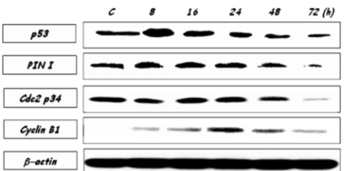
Effect of azurin on cell cycle regulatory proteins.
YD-9 cells were treated with azurin for various times (0, 8, 16, 24, 48, and 72 h). Expression of cell cycle regulatory factors in YD-9 cells treated with 200
µg/ml of azurin was
analyzed. The change in expression of p53, Cdc2, Pin1, and Cyclin B1 were observed. Expression of p53 was markedly increased at 8 h treated with 200
µg/ml azurin. The expression of Pin1 showed to be reduced in 72 h exposure to azurin. After treatment with azurin, Cyclin B1 was progressively increased till 24 h and then decreased. The expression of Cdc2 was rarely changed (Fig. 9).
Discussion
Bacterial protein azurin from P. aeruginosa has been reported to show anticancer effect in a few cancer cells (Yang
Fig. 6.
(I) Light microphotograph of YD-9 cells (
×200 magnification) and (II) Light microphotograph of YD-9 cells using hemacolor stain- ing (
×400 magnification) after treatment with azurin for 48 h. (A) Normal cells morphology was observed in azurin-untreated cells. (B) Con- densed nuclei and apoptotic bodies were observed in azurin-treated cells.
Fig. 7.
Hoechst 33258 staining of YD-9 cells. (A) azurin-untreated YD-9 cells depicted homogeneous staining of their nuclei. In con- trast, (B) apoptotic cells showed asymmetric staining of their nuclei as a result of chromatin condensation and nuclear fragmen- tation (
×200).
Fig. 8.
DNA fragmentation assay. YD-9 cells were exposed to
azurin at various times (8-72 h). Cells treated with 200
µg/ml
azurin for 24, 48 and 72 h clearly were showed DNA degradation
characteristics of apoptosis with a ladder pattern of DNA frag-
ments.
et al ., 2005). To test the potentiality of azurin to damage oral cancer cells, YD-9 (human oral squamous carcinoma) cells and MG63 (human osteosarcoma) cells were used. The cytotoxic activity of azurin was not observed in MG-63 cells.
But in YD-9 cells, small molecule azurin enters, triggering apoptotic cell death. It was demonstrated that this azurin, which it may use as an anticancer drug, possibly depends itself against YD-9 cells preferentially.
The use of bacteria in the treatment of cancer has been reported that bacterial infections can sometimes elicit regression in certain forms of cancer. Much effort has been spent over the years in developing wide type or modified bacterial and viral strains to treat the disease (Crum- Cianflone 2008; Kim et al ., 2009). Although various bacteria are believed to mediate tumor regression through selective proliferation in the anaerobic zone of the tumors or through inhibition of angiogenesis, very little is known about the detailed mechanism of tumor regression by such pathogens.
In particular, little is known about the production of soluble or secreted factors by bacteria that may specifically act on cancer cells, resulting in their death and regression.
Because the use of live bacteria has produced the problems such as toxicity, purified low molecular weight bacterial proteins as anticancer agents were used to bypass the pro- blems. Chakrabarty et al . discovered that azurin set off death sequence by forming a complex with the well known tumor suppressor protein p53, stabilizing it, and activating caspases that induces apoptosis in cancer cells (Yamada et al ., 2002).
It was suggested that this small molecule could potentially be used as a vehicle for cancer targeted chemotherapy (Yamada et al ., 2005).
Azurin DNA was cloned by using molecular technique from the P. aeruginosa genomic DNA and then expressed in E. coli . Azurin was purified by FPLC equipped with affinity column. The purified azurin was applied in YD-9 cells and MG-63 cells. The purified azurin have a cytotoxic effect in YD-9 cells and leads to apoptosis, but not in MG-63 cells.
The p53 is a tumor suppressor protein. It was damaged or missing in many cancer cells. Mutations in p53 are found in most tumor cells, and so contribute to the complex network of molecular events leading to tumor formation. p53 plays a
role in triggering apoptosis (Schuler et al ., 2001; Lahiry et al ., 2009; Galluzzi et al ., 2010). Apoptosis is an important process in a wide variety of different biological systems, including embryonic development, metamorphosis and cell death (Penaloza et al ., 2006). It can be induced by stimulation of plasma membrane death receptors and by perturbation of intracellular homeostasis via activation of specific organelle- mediated death cascades (Jayanthi et al ., 2004).
It was reported that azurin killed human p53 positive melanoma cells and induced apoptosis, but much less have efficiency in p53-null melanoma cells. Azurin was intern- alized and complexes with p53 in the cytosol, leading to stabilize p53 and raise its activity level in p53 positive melanoma cells (Yamada et al ., 2002; Yamada et al ., 2003;
Yamada et al ., 2005). Therefore, the applicability of azurin in cancer therapy will depend on p53 (Apiyo and Stafshede, 2005).
In this study, expression of p53 was markedly increased at early time in YD-9 cells treated with azurin. A lack of p53 in the MG-63 cells lead to a comparative loss of cytotoxicity of azurin. These results indicate that complex formation with p53 may be the primary reason for azurin mediated apoptosis.
Pin1 is also a critical regulator of the tumor suppressor p53 during DNA damage response. It is an indispensable positive regulator of p53 in response to DNA damage induced. Pin1 could represent a new anti cancer target (Ryo et al ., 2003).
Pin1 accelerates apoptosis by enhancing pro-apoptotic genes downstream of p53. In DNA damage, overexpression of activating p53 promoted its interaction with Pin1 (Takahashi et al ., 2006). The expression of Pin1 was also increased at early time exposure to azurin in YD-9 cells. The expression of Pin1 was corresponding to the accumulation pattern of p53 in YD-9 cells treated with azurin. These results suggested that Pin1 was accompanied with p53 in azurin-induced apoptosis of YD-9 cells.
In most cases, activated Cdc2-Cyclin B1 complex is required for progression from G2 into mitosis. The Cdc2 kinase activity is dependent on Cyclin B1. In human mela- noma cells,
γ-irradiation causes G2-M arrest and inhibits Cyclin B1 synthesis (Okada and Mak, 2004). In contrast, Cyclin B1 accumulates in HeLa cells arrested by UV light (Liu et al ., 2008) or etoposide (Lee et al ., 2007). Drug induced DNA damage results in cell cycle arrest before the onset of apoptosis (Le et al ., 2005). Furthermore, transient activation of Cdc2 has been reported as an early event in DNA damage- induced apoptosis (Gil-Gómez, 2004). In this experiment, the expression of Cdc2 was rarely changed in oral squamous carcinoma cells treated with azurin. But Cyclin B1 was progressively increased after treatment with azurin. It is likely that azurin might affect the expression of Cdc-2 and thus, the small amounts of Cdc-2 molecule rarely bind to Cyclin B1, resulting in cell cycle arrest. In summary, azurin purified from P. aeruginosa induces DNA damage as evidence of apoptosis and modulate cell cycle regulatory molecules in oral carcinoma YD-9 cells.
Fig. 9.