• 검색 결과가 없습니다.

Anticancer effects of D-pinitol in human oral squamous carcinoma cells

N/A
N/A
Protected

Academic year: 2021

Share "Anticancer effects of D-pinitol in human oral squamous carcinoma cells"

Copied!
10
0
0

로드 중.... (전체 텍스트 보기)

전체 글

(1)

Introduction Introduction

The term “head and neck cancer” is used to describe various types of cancers, including cancer of the oral cavity, parana- sal sinuses, pharynx, and larynx. Additionally, the term “oral cancer” broadly encompasses tumors arising in the lips, hard palate, alveolar ridges, tongue, sublingual region, and buccal mucosa [1,2]. More than 90% of all oral cancers are squamous cell carcinomas that arise from the squamous epithelial cells that make up the mucous membranes of the mouth [3,4].

In addition, sarcoma of the facial soft tissue and malignant melanoma, and more rarely lymphoma, can appear in cases of

salivary gland carcinoma [4]. The overall five-year survival rate, including all the stages of oral cancer, is approximately 50%, and both local and distant metastases are found in more than 70% of patients at the time of diagnosis [5]. The incidence of oral cancer has been increasing worldwide for many years, and most cases are treated by means of oral surgery because the facial area is an exposed and thus easily accessible part of the body. The use of cosmetic surgery for reconstruction purposes following oral surgery is increasing, although it remains dif- ficult to fully restore patients’ quality of life due to the resultant functional and aesthetic impairments [6]. In recent years, nat- ural substances have increasingly been used in the treatment Int J Oral Biol 45:152-161, 2020

pISSN: 1226-7155 • eISSN: 2287-6618 https://doi.org/10.11620/IJOB.2020.45.4.152

Anticancer effects of D-pinitol in human oral squamous carcinoma cells

Hyun-Chul Shin

1

, Tea-Hyun Bang

1

, Hae-Mi Kang

1

, Bong-Soo Park

1,2

, and In-Ryoung Kim

1,2

*

1

Department of Oral Anatomy, School of Dentistry, Pusan National University, Yangsan 50612, Republic of Korea

2

BK21 FOUR Project, School of Dentistry, Pusan National University, Yangsan 50612, Republic of Korea

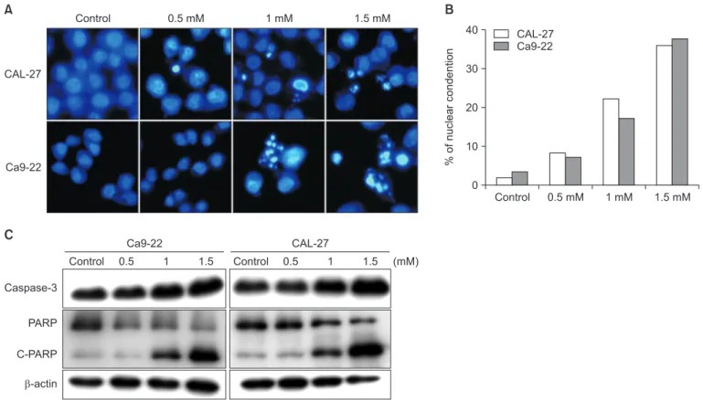
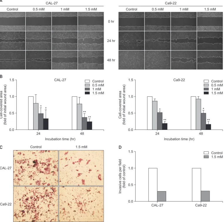
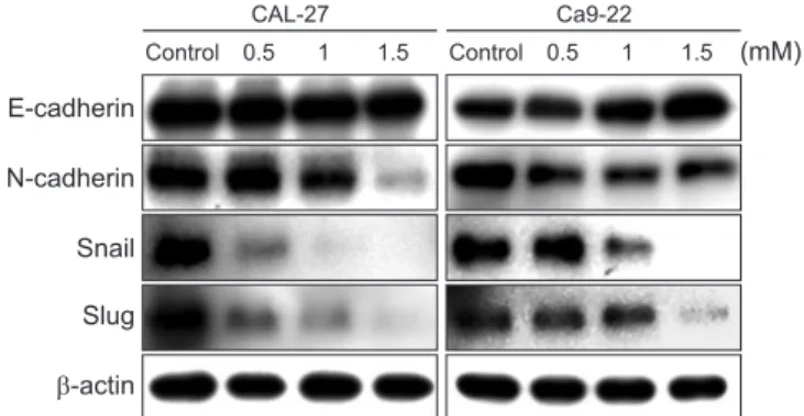
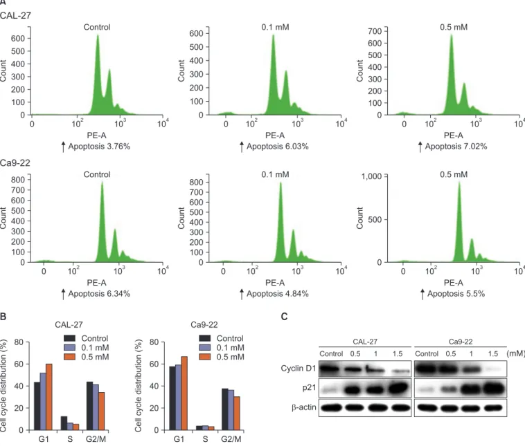
D-pinitol is an analog of 3-methoxy-D-chiro-inositol found in beans and plants. D-pinitol has anti-inflammatory, antidiabetic, and anticancer effects. Additionally, D-pinitol induces apoptosis and inhibits metastasis in breast and prostate cancers. However, to date, no study has investigated the anticancer effects of D-pinitol in oral cancer. Therefore, in this study, whether the anticancer effects of D-pinitol induce apoptosis, inhibit the epithelial- to-mesenchymal transition (EMT), and arrest cell cycle was investigated in squamous epithelial cells. D-pinitol decreased the survival and cell proliferation rates of CAL-27 and Ca9-22 oral squamous carcinoma cells in a concentration- and time-dependent manner. Evidence of apoptosis, including nuclear condensation, poly (ADP-ribose) polymerase, and caspase-3 fragmentation, was also observed. D-pinitol inhibited the migration and invasion of both cell lines. In terms of EMT-related proteins, E-cadherin was increased, whereas N-cadherin, Snail, and Slug were decreased. D-pinitol also decreased the expression of cyclin D1, a protein involved in the cell cycle, but increased the expression of p21, a cyclin-dependent kinase inhibitor. Hence, D-pinitol induces apoptosis and cell cycle arrest in CAL-27 and Ca9-22 cells, demonstrating an anticancer effect by decreasing the EMT.

Keywords: D-pinitol, Epithelial-mesenchymal transition, Cell cycle checkpoints, Oral squamous carcinoma cells

Received October 14, 2020; Revised December 10, 2020; Accepted December 10, 2020

*Correspondence to: In-Ryoung Kim, E-mail: biowool@pusan.ac.kr https://orcid.org/0000-0003-0232-0385 Copyright © The Korean Academy of Oral Biology

CC

This is an open-access article distributed under the terms of the Creative Commons Attribution Non-Commercial License (http://creativecommons.org/licenses/by- nc/4.0/), which permits unrestricted non-commercial use, distribution, and reproduction in any medium, provided the original work is properly cited.

Original Article IJOB

(2)

of various diseases, including cancer [7-9]. Such substances have both anti-inflammatory and anti-oxidant effects, and their use results in only limited toxicity and physiological activ- ity within the human body [7,10]. Therefore, a clear need ex- ists to develop anticancer drugs using natural substances.

D-pinitol (3-0-methyl-D-chiro-inositol) is an active low- molecular substance composed of a soybean oil and methyl ether compound. It has received considerable research atten- tion due to its diverse biological activities, including its antiviral, anti-inflammatory, anti-hyperlipidemic, and anti-oxidant ef- fects [11]. In addition, D-pinitol is structurally related to phos- phatidylinositol phosphate, which participates in the insulin signaling pathway that stimulates glucose transport, and it is reported to be helpful in the treatment of diabetes [12].

Recent studies have demonstrated the potential chemo- therapeutic efficacy of lung, bladder, and breast cancer [13,14].

Further, D-pinitol has been reported to reduce the metastasis of human lung cancer [15]. However, the effects of D-pinitol on oral squamous carcinoma cell metastasis remain largely un- known.

Metastasis is caused by uncontrolled cell proliferation, the stimulation of angiogenesis, segregation, motility, invasion into the bloodstream, and the stimulation of a new microenviron- ment [16]. More specifically, cancer cells separate from the primary site, migrate through the extracellular matrix, invade the bloodstream or lymphatic system, propagate distantly, and then proliferate [17]. This process is known as epithelial-mes- enchymal transition (EMT), and it is caused by the reduction in adhesion between cancer cells and the increased expression of N-cadherin and vimentin during tumor progression due to the disappearance of E-cadherin, which promotes renal cell and mesenchymal transition [18]. This process is associated with various tumorigenic pathways in various types of cancers, including oral cancer [19]. The present study was designed to elucidate the effect of D-pinitol on human oral squamous car- cinoma cells through apoptotic cell death, EMT, and the mo- lecular mechanism.

Materials and Methods Materials and Methods

1. Drug and reagents

The D-pinitol used in this study was purchased from Sigma- Aldrich (St. Louis, MO, USA). It was diluted in dimethyl sulfox- ide (DMSO) and then stored at –80℃. The specific antibodies for E-cadherin, N-cadherin, Snail, Slug, Caspase-3, and PARP

were purchased from Cell Signaling Technology (Danvers, MA, USA), while the β-actin was purchased from Santa Cruz Bio- technology (Santa Cruz, CA, USA). The secondary antibodies of mouse anti-rabbit IgG and the rabbit anti-mouse IgG antibod- ies were obtained from Enzo Biochem (Farmingdale, NY, USA).

2. Cell culture

The human oral squamous cancer cell (OSCC) lines used in this study, namely CAL-27 and Ca9-22, were purchased from the American Tissue Culture Collection (Rockville, MD, USA).

The CAL-27 and Ca9-22 were cultured in Dulbecco’s Modi- fied Eagle Medium (DMEM) with 10% fetal bovine serum (FBS;

Hyclone, Logan, UT, USA) and 1% penicillin-streptomycin (GIBCO-BRL, Rockville, MD, USA). Both cell lines were incu- bated in a humidified atmosphere of 5% CO

2

at 37℃.

3. Cell viability assay

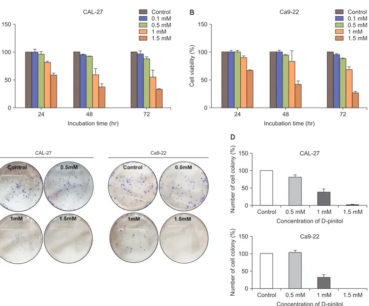
The cell viabilities of the CAL-27 and Ca9-22 cell lines were determined by means of a 3-[4,5-dimethythiazol-2-yl]-2,5- diphenyltetrazoliumbromide (MTT) assay. Both types of cells were seeded in a 96-well plate at 1 × 10⁴ cells/well, and they were treated with different concentrations of D-pinitol (0–1.5 mM). After treatment for 24, 48, and 72 hours, the medium was removed and 0.5 mg/mL of MTT solution was added to each well and then incubated at 37℃ for 4 hours. The forma- zan crystals that formed were dissolved in DMSO, and the resultant solution was measured using an ELISA reader (Tecan, M änedorf, Switzerland).

4. Colony formation assay

The cells were seeded in a six-well plate at 3 × 10² cells/

well, and they were treated with different concentrations of D- pinitol (0–1.5 mM) for 7 days. The colonies were fixed using 100% methanol, dyed with crystal violet for 10 minutes, and then washed using distilled water. The number of colonies was counted using an optical microscope.

5. Hoechst 33342 staining assay

Interms of determining the nuclear morphological change,

Hoechst 33342 was used for the nuclear staining. The cells

were seeded in an eight-well Lab-TekII chambered coverglass

(Invitrogen, Thermo Fisher Scientific, Waltham, MA, USA), and

(3)

they were treated with D-pinitol for 24 hours. Afterwards, the cells were fixed with 4% paraformaldehyde for 10 minutes.

Next, the cells were stained with 1 µg/mL of Hoechst 33342 solution for 10 minutes in a 37℃ incubator and then washed three times with phosphate-buffered saline (PBS). The fluo- rescent images were observed under a Zeiss LSM700 laser- scanning confocal microscope (CalZeiss, G öettingen, Germa- ny).

6. Wound healingassay

The cells were cultured in a six-well plate at 1 × 10

6

cells and then incubated with 5% CO

2

at 37℃. When the cells were 90–95% confluent, each well was scratched with pipette tip.

All the wells were washed twice with PBS and treated with D- pinitol. Images were acquired after 24 hours and 48 hours, and the widths of the cell-covered areas were examined using the Image J program (version 1.41o, Java 1.6.0_10, Wayen Ras- band, US National Institutes of Health, Bethesda, MD, USA).

24 150

100

Cell viability (%) 50

Incubation time (hr) 0

48 72

Control 0.1 mM 0.5 mM 1 mM 1.5 mM CAL-27

24 150

100

Cell viability (%) 50

Incubation time (hr) 0

48 72

Control 0.1 mM 0.5 mM 1 mM 1.5 mM Ca9-22

CAL-27 Ca9-22

Control 150

100

50

Number of cell colony (%)

Concentration of D-pinitol 0

CAL-27

0.5 mM 1 mM 1.5 mM

Control 150

100

50

Number of cell colony (%)

Concentration of D-pinitol 0

Ca9-22

0.5 mM 1 mM 1.5 mM

A B

D C

Fig. 1. D-pinitol reduced both the cell viability and the cell proliferation in the CAL-27 and Ca9-22 cells. (A, B) The CAL-27 and Ca9-22 cells were treated

with different concentrations of D-pinitol for 24 hours. The cell viability was examined using a 3-[4,5-dimethythiazol-2-yl]-2,5-diphenyltetrazoliumbromide as-

say. (C, D) The cell proliferation was determined using a colony formation assay. The cells were treated with D-pinitol for 7 days and then stained with a crys-

tal violet solution. The numbers of colonies were converted into percentages and then shown in a histogram. The result is expressed as the mean ± standard

deviation.

(4)

7. Transwell invasion assay

A trans well with an 8.0 µm pore polycarbonate membrane (Corning Costar, Cambridge, MA, USA) was coated with 20 µL of Matrigel at 200 µg/mL and then incubated overnight. The cells were seeded (1 × 10

5

cells in 200 µL) and treated with 1.5 mM D-pinitol in the upper chamber of the transwell with a serum-free medium. The lower chamber was filled with 800 µL of a medium containing 10% FBS. The cells were incubated at 37℃ with 5% CO

2

. After 48 hours, the cells were fixed in methanol and then stained with hematoxylin and eosin for 30 minutes. The number of cells that invaded the lower chamber through the pores was counted under an inverted microscope (Olympus, Tokyo, Japan).

8. Flow cytometry

The cell cycle analysis of the D-pinitol-treated cells were performed by means of flow cytometry. The CAL-27 and Ca9- 22 cells were seeded in a 60 mm dish and then treated with D-pinitol for 24 hours. The cells were harvested by trypsin-

ization and fixed with ice-cold 70% ethanol, including 0.5%

Tween20, for 24 hours. The fixed cells were pelleted and washed with 1% bovine serum albumin (BSA)-PBS solution.

The cells were suspended in 1 mL of PBS containing 20 µg/

mL RNaseA, incubated on ice for 30 minutes, and resus- pended in a propidiumiodide (PI) solution. Next, the cell cycle was analyzed using a BD FACSverse

TM

flow cytometer (Becton, Dickinson and Company, Franklin Lakes, NJ, USA).

9. Western blot analysis

The cells were lysed with a 150 µL ice-cold radio immu- noprecipitation assay buffer (cell signaling), which included pH7.6 50 mM Tris-Cl, 300 mM NaCl, 0.5% Triton X-100, 2 µL/

mL of a protinin, 2 mM PMSF, and 2 µL/mL of leupeptin, for 2 hours. The lysate samples were centrifuged at 13,200 rpm for 30 minutes. The protein amounts were measured using a Bradford protein assay (Bio-Rad, Richmond, CA, USA). The cell proteins were separated by means of 10% sodium dodecylsul- fate-polyacrylamide gel electrophoresis (SDS-PAGE) and then transferred on to a polyvinylidenedifluoride (PVDF) membrane.

Control 40

30

20

% o f nuclear condention

0

0.5 mM 1 mM 1.5 mM CAL-27

Ca9-22

10 CAL-27

Ca9-22

Control 0.5 mM 1 mM 1.5 mM

Caspase-3

PARP

C-PARP

-actin

(mM)

Control 0.5 1 1.5 Control 0.5 1 1.5

Ca9-22 CAL-27

A B

C

Fig. 2. D-pinitol induced apoptosis in the CAL-27 and Ca9-22 cells. The cells were treated with D-pinitol for 24 hours. (A) To indicate a change in the nuclei,

Hoechst staining was used in the CAL-27 and Ca9-22 cells. The cells were treated with various concentrations of D-pinitol for 24 hours. ×200. (B) The num-

bers of apoptosis cells were converted into percentages and then shown in a histogram. (C) To reveal the expression of those proteins closely related to cell

apoptosis, such caspase-3 and PARP, a Western blot analysis was performed. β-actin was used as the loading control.

(5)

After one day, the PVDF membrane was blocked with 5%

nonfat dry milk blocking buffer for 1 hour and incubated with the appropriate primary antibodies for 24 hours at 4℃. Fol- lowing this, the membrane was washed with Tris-NaCl-EDTA

(TNE) buffer and incubated with secondary antibodies for 2 hours at room temperature. Next, the membrane was washed again for 1 hour before SuperSignal

TM

WestFemto (Pierce, Rockford, IL, USA) was used for the detection of the proteins.

0 hr

24 hr

48 hr

Control 0.5 mM 1 mM 1.5 mM Control 0.5 mM 1 mM 1.5 mM

CAL-27 Ca9-22

A

B

24 1.5

1.0

0.5 Cell-covered area (fold of initial wound area)

Incubation time (hr) 0.0

48

Control 0.5 mM 1 mM 1.5 mM CAL-27

Incubation time (hr) 24

1.5

1.0

0.5 Cell-covered area (fold of initial wound area)

0.0

48

Control 0.5 mM 1 mM 1.5 mM Ca9-22

CAL-27

Ca9-22

Control 1.5 mM

CAL-27 1.5

1.0

0.5 Invasive cells per field (fold of control)

0.0

Ca9-22

Control 1.5 mM

C D

* *

**

**

*

**

*

**

Fig. 3. D-pinitol reduced the cell migration and invasion in the CAL-27 and Ca9-22 cells. (A) The cells were treated with D-pinitol for 24 hours and 48 hours. (B) The fold of the initial wound area is shown in the histogram. (C) A transwell invasion assay was used to examine the invasion ability of the D-pinitol-treated cells. Hematoxlin, ×100. (D) The numbers of invasive cells were converted into percentages and then shown in a histogram.

*p < 0.05 and **p < 0.01 for the difference between the control and the treatment groups for each group.

(6)

10. Statistical analysis

The results of the present study were expressed as the mean ± standard deviation. Either a Student’s t-test was used for multiple comparisons during the statistic alanalysis. The statistical analysis was performed using GraphPad PrismVer- sion 5.0 (GraphPad Software, San Diego, CA, USA).

Results Results

1. D-pinitol reduced the cell viability and proliferation in OSCC cells

We performed an MTT assay to determine the cytotoxic ef- fect of D-pinitol in CAL-27 and Ca9-22 cells. The cells were treated with various concentrations (0–1.5 mM) of D-pinitol for 24–72 hours. The cell viability was suppressed by 33% and 27% in the CAL-27 and Ca9-22 cells, respectively, when they were treated with 1.5 mM D-pinitol for 72 hours. Hence, D- pinitol reduced the cell viability in a dose- and time-dependent manner in both cell lines (Fig. 1A and 1B). To confirm the in- hibitory effect of D-pinitol on cell proliferation, a colony forma- tion assay was performed for 7 days. In both the CAL-27 and Ca9-22 cells, the number and the size of the colonies were decreased in a dose-dependent manner (Fig. 1C and 1D).

These results revealed that D-pinitol attenuated the cell viabil- ity proliferative activity in OSCC cells.

2. D-pinitol induced apoptosis in OSCC cells

To determine whether D-pinitol induces apoptosis, we used Hoechst staining to investigate the nuclear morphological changes in CAL-27 and Ca9-22 cells that were treated with different concentrations of D-pinitol. As shown in Fig. 2A and 2B, the D-pinitol treatment caused a significant change in the treated cells’ nuclei when compared to the non-treated cells. The cells that had been treated with D-pinitol showed condensed and fragmented nuclei in a dose-dependent man- ner. We conducted a Western blot analysis to determine the molecules that were closely involved in the cell apoptosis. We treated the CAL-27 and Ca9-22 cells with various concentra- tions of D-pinitol for 24 hours. The D-pinitol activated cas- pase-3 and PARP in a dose-dependent manner in both cells (Fig. 2C). Therefore, it could be concluded that the D-pinitol treatment induced apoptosis in OSCC cells.

3. D-pinitol inhibited the cell migration and invasion in OSCC cells

We confirmed the effect whereby D-pinitol suppressed cell migration in CAL-27 and Ca9-22 cells. The cells were treated with several concentrations of D-pinitol for 24 hours and 48 hours so that the migratory effects could be examined using a wound healing assay. As shown in Fig. 3A and 3B, the cell- covered area was reduced in a dose-dependent manner. To investigate the inhibitory effect of D-pinitol on cell invasion, a transwell invasion assay was conducted. The CAL-27 and Ca9-22 cells were treated with 1.5 mM D-pinitol for 48 hours.

The D-pinitol caused a decrease in the invasive attribute of approximately 0.38-fold in the CAL-27 cells and of approxi- mately 0.36-fold in the Ca9-22 cells when compared to the non-treated cells (Fig. 3C and 3D). These results revealed that D-pinitol inhibited the migratory and invasive properties in OSCC cells.

4. D-pinitol regulated the EMT-related protein expression in OSCC cells

Next, we explored the molecular mechanism by which D- pinitol suppressed the EMT in CAL-27 and Ca9-22 cells. The EMT transforms epithelial cells through the down-regulation of E-cadherin expression and the resultant mesenchymal mark- ers, such as N-cadherin, Snail, and Slug, which promote tumor progression and metastasis in epithelial cells. As shown in Fig.

4, the D-pinitol-treated cells showed the up-regulation of E- cadherin in a dose-dependent fashion, while the N-cadherin,

E-cadherin

N-cadherin

Snail

Slug

-actin

(mM) Control 0.5 1 1.5

CAL-27

Control 0.5 1 1.5 Ca9-22

Fig. 4. D-pinitol changed the expression of the epithelial-mesenchymal

transition (EMT)-related markers in the CAL-27 and Ca9-22 cells. The cells

were treated with different concentrations of D-pinitol for 24 hours. The

expression of the EMT-related markers, such as E-cadherin, N-cadherin,

Snail, and Slug, was verified by means of a Western blot analysis. β-actin

was used as the loading control.

(7)

Snail, and Slug protein levels were all down-regulated. These results revealed that D-pinitol exerted an anti-metastatic ef- fect on OSCC cells through the EMT process.

5. D-pinitol induced G1 cell arrest in OSCC cells

The arrest of the cell cycle during the G1 phase means that the cells will not be able to begin DNA synthesis and hence will remain arrested. We investigated the effect of G1 arrest in D-pinitol-treated cells using flow cytometry. In the CAL-27 and Ca9-22 cells, the flow cytometry analysis indicated that a

statically significant arrest was induced during the G1 phase in a dose-dependent manner when compared to the non-treated cells, and it was accompanied by a reduction during the S and G2/M phases, too (Fig. 5A and 5B). Next, we estimated the effects of D-pinitol on the expression of the cell cycle molecu- lar cyclin D1 and the cyclin-dependent kinase inhibitor (CKI) p21 in the CAL-27 and Ca9-22 cells using a Western blot analysis. The D-pinitol-treated cells showed the reduced ex- pression of cyclin D1 in a dose-dependent fashion, while the p21 protein expression was induced (Fig. 5C). These results indicated that D-pinitol induced G1 arrest in OSCC cells.

0 600 500 400 300 200 100

Count

PE-A 0

10

4

10

3

10

2

Apoptosis 6.03%

0.1 mM

0 700 500 400 300 200 100

Count

PE-A 0

10

4

10

3

10

2

Apoptosis 4.84%

0.1 mM 0

600 500 400 300 200 100

Count

PE-A 0

10

4

10

3

10

2

Apoptosis 3.76%

CAL-27

Control

0 700 500 400 300 200 100

Count

PE-A 0

10

4

10

3

10

2

Apoptosis 6.34%

Ca9-22

Control

600 800

600 800

0 600 500 400 300 200 100

Count

PE-A 0

10

4

10

3

10

2

Apoptosis 7.02%

700 0.5 mM

0

Count

PE-A 0

10

4

10

3

10

2

Apoptosis 5.5%

1,000 0.5 mM

500

A

G1 80

60

40

20

Cell cycle distribution (%)

0

CAL-27

S G2/M G1

80

60

40

20

Cell cycle distribution (%)

0

Ca9-22

S G2/M Control

0.1 mM 0.5 mM

Control 0.1 mM 0.5 mM

B C

Cyclin D1 p21

-actin

(mM) Control 0.5 1 1.5

CAL-27

Control 0.5 1 1.5 Ca9-22

Fig. 5. D-pinitol induced G1 arrest in the CAL-27 and Ca9-22 cells. Cells were treated with D-pinitol (0.1 and 0.5 mM) for 24 hours. (A) To analyze the cell

cycle, the CAL-27 and Ca9-22 cells were treated different concentrations of D-pinitol and flow cytometry was performed. (B) The percentages during the G1, S,

and G2/M phases were calculated and then presented in a histogram. (C) The expression of the cyclin D1 and p21 protein levels, which was closely linked to

the G1 arrest, was observed by Western blot analysis. β-actin was used as the loading control.

(8)

Discussion Discussion

The leaves of Bougainvillea spectabilis have long been used as traditional diabetes remedies in Asia and the West Indies, and D-pinitol is a component extracted from the leaves [20].

D-pinitol is structurally related to phosphatidylinositol phos- phate, which is known to participate in the insulin signaling pathway that stimulates glucose transport [21,22]. The term

“diabetes mellitus” refers to a group of metabolic diseases characterized by persistent hyperglycemia, and both the preva- lence and the mortality rates of these diseases are increasing.

Prior investigations have reported a strong association between diabetes (especially type 2 diabetes) and carcinogenesis, which results in hyperinsulinemia, hyperglycemia, and fat-in- duced chronic inflammation [23]. Several studies have identi- fied the anticancer effects of anti-diabetic drugs. For instance, metformin, a well-known drug for the treatment of type 2 diabetes, has been reported to inhibit breast, pancreas, liver, colon, ovarian, and prostate cancer [24-26]. Recently, studies have been conducted to determine whether D-pinitol has an- ticancer effects aside from merely lowering the blood glucose level in prostate and breast cancer cells [11,27]. However, the effect of D-pinitol on human oral squamous cell carcinoma has not yet been elucidated. Hence, in this study, we investigated the antitumor effect of D-pinitol on human oral squamous cell carcinoma CAL-27 and Ca9-22 cells.

The induction of apoptosis is a well-known regulatory meth- od in cancer therapy, and it is characterized by cell shrinkage, nuclear condensation, and cell cycle arrest [28]. These cell reactions are important in terms of determining the toxicity and the response to current cancer therapies because most of them target DNA [29]. It has been reported that D-pinitol induces apoptosis through the mitochondrial pathway [11] and promotes cell death via interleukin and hormones through the inhibition of nuclear factor kappa B (NF- κB) so as to mitigate tumor growth in MCF-7 cells [30]. However, no studies have previously been conducted on the cell cycle arrest caused by D-pinitol in any other cancer cells, including OSCCs. In our study, we found that D-pinitol induced nuclear condensation, which led to the fragmentation of caspase-3 and PARP, repre- sentative proteins of apoptosis, and increases in the rate of G1

arrest.

The EMT is a crucial process for acquiring the malignant phenotype, aggression, and metastatic capacity in neoplasms.

It is characterized by the loss of epithelial markers (E-cadherin) and the gain of mesenchymal markers (N-cadherin). Studies concerning the potential link with the EMT are rare in OS- CCs [31]. The EMT is further characterized by increased cell migration and invasion as well as by increased resistance to apoptosis [18]. In addition, the EMT process is regulated by several transcription factors, including Snail and Slug [18,32].

Thus, the inhibition of the EMT represents a way to overcome the increased resistance on the part of apoptosis and hence to actively prevent cancer metastasis. Recently, D-pinitol has been reported to inhibit prostate cancer metastasis through in- hibiting the αVβ3 integrins by modulating the FAK, c-Src, and NF-κB pathways [27], although unfortunately, save for this paper, no other studies have yet been published in this regard.

In this study, we found that D-pinitol reduced the CAL-27 and Ca9-22 cell migration and invasion through the regula- tion of the EMT-associated marker protein expression. The E- cadherin expression increased and the N-cadherin expression decreased in a dose-dependent fashion. In addition, the Snail and Slug transcription factors decreased following treatment with D-pinitol. These results suggest that D-pinitol induces cancer resistance and inhibits metastasis, thereby showing promising potential as an anticancer agent. The results further suggest the possibility of identifying therapeutic targets for fundamental oral cancer treatment.

Acknowledgements Acknowledgements

This work was supported by the National Research Founda- tion of Korea (NRF) grant funded by the Korea government (No.

NRF-2018R1D1A1B07047739 and NRF-2019R1A2C10840 5712).

Conflicts of Interest Conflicts of Interest

No potential conflict of interest relevant to this article was reported.

References References

1. Fritz AG. International classification of diseases for oncology:

ICD-O. 3rd ed. Geneva: World Health Organization; 2013.

2. Vokes EE, Weichselbaum RR, Lippman SM, Hong WK. Head

and neck cancer. N Engl J Med 1993;328:184-94. doi: 10.

(9)

1056/NEJM199301213280306.

3. Laramore GE, Scott CB, al-Sarraf M, Haselow RE, Ervin TJ, Wheeler R, Jacobs JR, Schuller DE, Gahbauer RA, Schwade JG, Campbell BH. Adjuvant chemotherapy for resectable squamous cell carcinomas of the head and neck: report on Intergroup Study 0034. Int J Radiat Oncol Biol Phys 1992;23:

705-13. doi: 10.1016/0360-3016(92)90642-u.

4. Casiglia J, Woo SB. A comprehensive review of oral cancer.

Gen Dent 2001;49:72-82.

5. Liao S, Xia J, Chen Z, Zhang S, Ahmad A, Miele L, Sarkar FH, Wang Z. Inhibitory effect of curcumin on oral carcinoma CAL- 27 cells via suppression of Notch-1 and NF- κB signaling pathways. J Cell Biochem 2011;112:1055-65. doi: 10.1002/

jcb.23019.

6. Lei Y, Zhang D, Yu J, Dong H, Zhang J, Yang S. Targeting au- tophagy in cancer stem cells as an anticancer therapy. Cancer Lett 2017;393:33-9. doi: 10.1016/j.canlet.2017.02.012.

7. Nobili S, Lippi D, Witort E, Donnini M, Bausi L, Mini E, Ca- paccioli S. Natural compounds for cancer treatment and prevention. Pharmacol Res 2009;59:365-78. doi: 10.1016/

j.phrs.2009.01.017.

8. Scotece M, Conde J, Abella V, Lopez V, Pino J, Lago F, Smith AB 3rd, G ómez-Reino JJ, Gualillo O. New drugs from ancient natural foods. Oleocanthal, the natural occurring spicy com- pound of olive oil: a brief history. Drug Discov Today 2015;20:

406-10. doi: 10.1016/j.drudis.2014.10.017.

9. Matias I, Buosi AS, Gomes FC. Functions of flavonoids in the central nervous system: astrocytes as targets for natural compounds. Neurochem Int 2016;95:85-91. doi: 10.1016/

j.neuint.2016.01.009.

10. Jagtap S, Meganathan K, Wagh V, Winkler J, Hescheler J, Sa- chinidis A. Chemoprotective mechanism of the natural com- pounds, epigallocatechin-3-O-gallate, quercetin and curcum- in against cancer and cardiovascular diseases. Curr Med Chem 2009;16:1451-62. doi: 10.2174/092986709787909578.

11. Rengarajan T, Nandakumar N, Rajendran P, Haribabu L, Nish- igaki I, Balasubramanian MP. D-pinitol promotes apoptosis in MCF-7 cells via induction of p53 and Bax and inhibition of Bcl-2 and NF- κB. Asian Pac J Cancer Prev 2014;15:1757-62.

doi: 10.7314/apjcp.2014.15.4.1757.

12. Bates SH, Jones RB, Bailey CJ. Insulin-like effect of pinitol. Br J Pharmacol 2000;130:1944-8. doi: 10.1038/sj.bjp.0703523.

13. Zhan T, Lou H. Synthesis of azole nucleoside analogues of D- pinitol as potential antitumor agents. Carbohydr Res 2007;

342:865-9. doi: 10.1016/j.carres.2007.01.004.

14. Rengarajan T, Nandakumar N, Balasubramanian MP. D-Pinitol

attenuates 7, 12 dimethylbenz [a] anthracene induced hazards through modulating protein bound carbohydrates, adenosine triphosphatases and lysosomal enzymes during experimental mammary carcinogenesis. J Exp Ther Oncol 2012;10:39-49.

15. Sethi G, Ahn KS, Sung B, Aggarwal BB. Pinitol targets nuclear factor-kappaB activation pathway leading to inhibition of gene products associated with proliferation, apoptosis, invasion, and angiogenesis. Mol Cancer Ther 2008;7:1604-14. doi: 10.

1158/1535-7163.MCT-07-2424.

16. Noguti J, De Moura CF, De Jesus GP, Da Silva VH, Hossaka TA, Oshima CT, Ribeiro DA. Metastasis from oral cancer: an overview. Cancer Genomics Proteomics 2012;9:329-35.

17. Irani S. Distant metastasis from oral cancer: a review and mo- lecular biologic aspects. J Int Soc Prev Community Dent 2016;

6:265-71. doi: 10.4103/2231-0762.186805.

18. Kalluri R, Weinberg RA. The basics of epithelial-mesenchymal transition. J Clin Invest 2009;119:1420-8. doi: 10.1172/

JCI39104.

19. Mk H, Prince S, Mohan AM, Krishnan KV, Devi A. Association of Notch4 with metastasis in human oral squamous cell car- cinoma. Life Sci 2016;156:38-46. doi: 10.1016/j.lfs.2016.05.

019.

20. Narayanan CR, Joshi DD, Mujumdar AM, Dhekne VV. Pinitol- a new anti-diabetic compound from the leaves of Bougainvil- lea spectabilis. Curr Sci 1987;56:139-41.

21. White MF. The insulin signalling system and the IRS pro- teins. Diabetologia 1997;40 Suppl 2:S2-17. doi: 10.1007/

s001250051387.

22. Holman GD, Kasuga M. From receptor to transporter: insulin signalling to glucose transport. Diabetologia 1997;40:991- 1003. doi: 10.1007/s001250050780.

23. Wojciechowska J, Krajewski W, Bolanowski M, Kr ęcicki T, Zato ński T. Diabetes and cancer: a review of current knowl- edge. Exp Clin Endocrinol Diabetes 2016;124:263-75. doi:

10.1055/s-0042-100910.

24. Franciosi M, Lucisano G, Lapice E, Strippoli GF, Pellegrini F, Nicolucci A. Metformin therapy and risk of cancer in patients with type 2 diabetes: systematic review. PLoS One 2013;8:

e71583. doi: 10.1371/journal.pone.0071583.

25. Garrett CR, Hassabo HM, Bhadkamkar NA, Wen S, Baladan- dayuthapani V, Kee BK, Eng C, Hassan MM. Survival advan- tage observed with the use of metformin in patients with type II diabetes and colorectal cancer. Br J Cancer 2012;106:1374- 8. doi: 10.1038/bjc.2012.71.

26. Lee MS, Hsu CC, Wahlqvist ML, Tsai HN, Chang YH, Huang

YC. Type 2 diabetes increases and metformin reduces total,

(10)

colorectal, liver and pancreatic cancer incidences in Taiwan- ese: a representative population prospective cohort study of 800,000 individuals. BMC Cancer 2011;11:20. doi: 10.1186/

1471-2407-11-20.

27. Lin TH, Tan TW, Tsai TH, Chen CC, Hsieh TF, Lee SS, Liu HH, Chen WC, Tang CH. D-pinitol inhibits prostate cancer metas- tasis through inhibition of αVβ3 integrin by modulating FAK, c-Src and NF- κB pathways. Int J Mol Sci 2013;14:9790-802.

doi: 10.3390/ijms14059790.

28. Evan GI, Vousden KH. Proliferation, cell cycle and apoptosis in cancer. Nature 2001;411:342-8. doi: 10.1038/35077213.

29. Kastan MB, Bartek J. Cell-cycle checkpoints and cancer. Na- ture 2004;432:316-23. doi: 10.1038/nature03097.

30. Rengarajan T, Nandakumar N, Rajendran P, Ganesh MK,

Balasubramanian MP, Nishigaki I. D-pinitol mitigates tumor growth by modulating interleukins and hormones and induces apoptosis in rat breast carcinogenesis through inhibition of NF- κB. J Physiol Biochem 2015;71:191-204. doi: 10.1007/

s13105-015-0397-9.

31. Angadi PV, Patil PV, Angadi V, Mane D, Shekar S, Hallikeri- math S, Kale AD, Kardesai SG. Immunoexpression of epithelial mesenchymal transition proteins E-cadherin, β-catenin, and N-cadherin in oral squamous cell carcinoma. Int J Surg Pathol 2016;24:696-703. doi: 10.1177/1066896916654763.

32. Micalizzi DS, Farabaugh SM, Ford HL. Epithelial-mesenchymal

transition in cancer: parallels between normal development

and tumor progression. J Mammary Gland Biol Neoplasia

2010;15:117-34. doi: 10.1007/s10911-010-9178-9.

수치

Fig. 1. D-pinitol reduced both the cell viability and the cell proliferation in the CAL-27 and Ca9-22 cells
Fig. 2.  D-pinitol induced apoptosis in the CAL-27 and Ca9-22 cells. The cells were treated with D-pinitol for 24 hours
Fig. 3.  D-pinitol reduced the cell migration and invasion in the CAL-27 and Ca9-22 cells
Fig. 4. D-pinitol changed the expression of the epithelial-mesenchymal  transition (EMT)-related markers in the CAL-27 and Ca9-22 cells
+2

참조

관련 문서

Effects of Anthriscus sylvestris Hoffmann aqueous layer (ASAL) on cell apoptosis and PARP activity in NIH/3T3 fibroblast and KB, FaDu oral cancer cells by

In this study, the expression profiles of miRNAs were compared and analyzed for establishment of miRNAs related cancer cell growth inhibition in normal human oral

Anticancer activities of EF-24, a novel curcumin analog, on KB human. oral

Concentration-Dependent inhibition of cell viability by adenosine in human oral fibroblast and FaDu human head and neck squamous cell carcinoma · · ·

It also suggest that cell proliferation inhibits by a novel signal transduction for adenosine effect in human FaDu hypopharynx squamous cell carcinoma cells....

Differential Expression of Desmoglein1, Desmoglein3, Epithelial Membrane Antigen, Ber-EP4 and CD10 in Basal Cell Carcinoma and Squamous Cell Carcinoma

Objective: The purpose of this study was to analyze recent trend in incidence of basal cell carcinoma and squamous cell carcinoma in patients from the Gwangju City

In contrast, cells expressing wild-type form or constitutively active form of RapC (GFP-RapC and GFP-RapC G13V ) showed decreased cell area and cell adhesion, whereas rapC