김용관*∙이재훈∙김철환
*관동대학교 의과대학 치과학교실 구강악안면외과, 단국대학교 치과대학 구강악안면외과학교실
cDNA Microarray를 이용한 구강편평세포암종 세포주에서 Taxol
�과 Cyclosporin A로 유도된 유전자 발현양상
GENE EXPRESSION PATTERNS INDUCED BY TAXOL
�AND CYCLOSPORIN A IN ORAL SQUAMOUS CELL CARCINOMA CELL LINE USING CDNA MICROARRAY
Yong-Kwan Kim * , Jae-Hoon Lee, Chul-Hwan Kim
*Dept. of Oral & Maxillofacial Surgery, College of Medicine, Kwandong University Dept. of Oral & Maxillofacial Surgery, School of Dentistry, Dankook University
It is well-known that paclitaxel(Taxol
�), which is extracted from the pacific and English yew, has been used as a chemotherapeutic agent for ovarian carcinoma and advanced breast carcinoma and Cyclosporin A, which is highly lipophilic cyclic peptide and isolated from a fungus, has been also used as an useful immunosuppressive drug after transplantation and is associated with cellular apoptosis.
Since 1953, in which James Watson, Rosalind Franklin and Francis Crick discovered the double helical structure of DNA, a few kinds of techniques for identifying gene expression have been developed. In post- genomic period, many of researchers have used the DNA microarray which is high throughput screening technique to screen large numbers of gene expression simultaneously. In this study, we searched and screened the gene expression in the oral squamous cell carcinoma cell lines treated with Taxol
�, cyclosporin or cyclosporin combined with Taxol
�using cDNA microarray.
The results were as following;
1. It was useful that the appropriate concentration of Cyclosporin A and Taxol
�used in oral squamous cell carcinoma cell line was under 1 μg/ml and 3 μg/ml.
2. In the experimental group in which Taxol
�and Taxol
�+ Cyclosporin A were used, the cell growth was extremely decreased.
3. In the group in which Cyclosporin A was used, the MTT assay was rarely decreased which means the activity of succinyl dehydrogenase is remained in mitochondria but in the group in which the mixture of Cyclosporin A and Taxol
�were used, the MTT assay was extremely decreased.
4. In the each group in which Cyclosporin A(3 μg/ml) and Taxol
�(1 μg/ml) were used, the cell arrest was appeared in G
2/M phase and in the group in which Taxol
�(3 μg/ml) was used, the cell arrest was appeared in both S phase and G
2/M phase.
5. In the oral squamous cell carcinoma cell line treated with Taxol
�, several genes including ANGPTL4, RALBP1 and TXNRD1, associated with apoptosis, SUI1, MAC30, RRAGA and CTGF, related with cell growth, HUS1 and DUSP5, related with cell cycle and proliferation, ATF4 and CEBPG, associated with transcription factor, BTG1 and VEGF, associated with angiogenesis, FDPS, FCER1G, GPA33 and EPHA4 associated with signal transduction and receptor activity and AKR1C2 and UGTA10 related with carcinogenesis were detected in increased levels. The genes that showed increaced expression in the oral squamous cell carcinoma cell line treated with Cyclosporin A were CYR61, SERPINB2, SSR3 and UPA3A which are known as genes associated with cell growth, carcinogenesis, receptor activity and transcription factor. The genes expressed in the HN22 cell line treated with cyclosporin combined with taxol
�were ALCAM and GTSE1 associated with cancer invasiveness and cell cycle regulation.
Key words : Oral squamous cell carcinoma cell line, Taxol
�, Cyclosporin A, cDNA microarray
Abstract
Ⅰ. 서 론
주목나무과인 Taxus brevifolia에서 추출된 천연물질인 Taxol은 유방암과 난소암에 탁월한 항암효과를 보이는 것 으로 알려져 있다
1). Taxol은 세포의 분화와 성장을 억제하 여 세포를 죽음으로 유도하는 것으로 알려져 있으며
2-5), 이 는 세포분열시 지주역할을 하는 미소관(microtubule)을 안 정화시켜 염색체의 움직임을 정지시키고 세포주기중 G
2/M phase에 세포분열을 억제하여 세포사멸(apoptosis)을 유
도하며
2,4,6,7), 또한 혈관형성(angiogenesis)을 억제하는 작
용이 있어
8)항암효과를 나타내며 방사선치료와 병용 시 항 암효과가 증가되는 것으로 알려져 있다
9-11).
이러한 세포사멸은 세포내에서 프로그램된 신호에 따라 특정유전자가 발현되어 일어나는 능동적인 죽음을 의미하 며, 생리학적 특징은 세포의 형태적인 변화 즉, 세포질 (cytoplasm)의 수축, 핵막주위 염색사의 농축 및 apoptot- ic body 등의 형성과, DNA 분절현상 및 세포주기의 변화 등을 나타낸다. 특히 DNA의 분절현상은 세포내 Ca
2+와 cAMP의 농도증가에 의해 핵내에 존재하는 endogenous endonuclease의 활성화로 나타난다고 보고되고 있다
12,13). 이러한 세포사멸에 관여하는 인자들은 TNFR(tumor necrosis factor receptor), CD95/Fas/Apol, DR(death receptor)3, DR4, DR5, NGFR(nerve growth factor receptor)와 같은 세포막 수용체와 리간드(ligand)들이 있 고, p53, pRb 및 CDK inhibitor와 같은 세포주기 조절인 자와 calpain 또는 caspase와 같은 단백질 분해효소들이 세포사멸과 관련된 세포신호전달과정에 관여한다. 또한 Bcl-2 family에 속하는 단백질중 Bcl-2나 Bcl-XL 등은 세 포사멸을 억제하며 Bax, Bid, Bad, Bcl-XS 등은 세포사멸 을 촉진하는 역할을 하는 것으로 알려져 있다
14). 이외에 세포사멸의 대표적인 신호전달 효소인 caspase의 활성 을 억제하는 IAP(inhibitor of apoptosis protein) 즉 DIAP, HIAP, XIAP 및 NIAP 등도 세포사멸 억제인자들 이다
1,14-15).
Cyclosporin A는 1972년 곰팡이균인 Tolypocladium inflatum과 Cylindrocarpum lucidum에서 처음 추출된 lipophilic cyclic peptide이며
16), 1978년 신장이식 수술 후 면역억제제로서 임상에 사용된 이래로
17)장기이식환자에 이 식거부반응 억제를 위해 스테로이드와 병용 또는 단독으로 사용되어지고 있다. 또한 자가면역질환 및 인슐린의존형 당 뇨에도 이용하고 있으나 신독성 및 간독성의 부작용을 나타 내기도 한다. Cyclosporin A의 면역억제작용은 T임파구세 포 내에서 IL-2와 같은 cytokine의 생성억제를 통해 T임파 구 성장 및 기능을 억제함으로 나타난다
18). 또한 Cyclo- sporin A는 신경종양세포와 골수세포 등의 다양한 세포에 서 세포사멸을 유도한다고 보고되고 있다
33,34).
구강암의 대부분을 차지하고 있는 구강편평세포암종은 타 부위 암종에 비해 발생빈도는 낮으나 인접조직으로의 침투 및 타장기 전이 비율이 높은 것으로 알려져 있으며 수술요 법, 방사선요법 및 항암요법 등의 다양한 치료 방법을 통해 많은 노력을 경주해 왔으나 아직 암종의 발생 및 원인기전, 암종의 성장 그리고 전이에 관여하는 분자생물학적 유전자 조절 기전 등이 규명되지 않아 예후가 극히 불량하며 생존 율이 낮은 편이다
19). 암종의 예후 차이는 종양세포의 분자생 물학적 유전자 특성에 기인된다고 제시되어 항암제치료에 따른 반응도 및 종양발생에 결정적 역할을 하는 유전자의 변화와 세포증식 및 사멸에 관여하는 유전자발현 등과 같은 연구가 활발히 시행되고 있다.
지금까지 사용된 유전자 분석방법은 northern blot 또는 southern blot 및 DNA sequencing 등이 있으나 소수의 유전자 발현만을 분석할 수 있기 때문에 많은 유전자 발현 의 검색은 기술적인 한계로 인해 많은 시간과 비용이 요구 된다. 또한 암세포의 성장 대사를 지배하는 성분들은 서로 복잡한 경로들이 연관되어 있어 어느 한 성분만의 분석으로 전체 암세포의 특성을 확인할 수 없고 한꺼번에 수많은 유 전자의 발현을 분석해야 한다. 이와 같은 문제점을 해소하 기 위해 최근에 개발된 C-DNA microarray 기법이 새롭게 부각되었다
20).
1995년 Brown과 Botstein 등
21)에 의해 개발된 DNA chip(cDNA microarray) 분석기법은 분자생물학적 지식을 바탕으로 반도체 부착기술인 첨단 전자공학기법과 생물정 보학이 접목된 방법으로 poly L-lysine 및 amine과 alde- hyde 등으로 피복시켜 유전자가 결합 될 수 있도록 만든 슬 라이드에 수천 수만의 target DNA를 부착하여 glass microarray를 제작하거나 이미 제작된 DNA microarray 를 사용한다
22). 이때 spot의 직경은 100 mm 내외가 되며 약 1,000 spots/cm
2가 형성된다. 또한 탐침은 DNA chip 의 염기서열과 서로 상보적인 서열을 갖고 있어 이들의 결 합상태가 유전자발현에 영향을 주고 증폭하고자 하는 염기 서열은 다른 유전자와 유사성이 적고 해당유전자에 특이적 인 서열을 선택해야 한다. DNA chip은 붙이는 유전물질의 크기에 따라서 cDNA chip과 oligonucleotide로 나눠지며 cDNA chip에는 최소 500 bp이상의 증폭된 target DNA 가 점적되어있고 oligonucleotide chip에는 약 20여개의 염기들로 이루어진 oligonucleotide가 부착된다.
검사하고자 하는 조직 또는 세포들의 대조군과 실험군에 서 RNA를 추출 후 형광물질(cy3, cy5)로 labelling된 cDNA probe를 target DNA와 hybridization 후 DNA microarray slide를 형광 scanner를 사용하여 scanning 한 후 유전자 발현양상의 컴퓨터 분석을 시행한다
22).
본 연구는 유방암과 난소암에 항암효과를 보이는 Taxol
과, 면역억제제로 많이 사용되며 세포사멸 효과가 있다고
알려진 cyclosporin A를 구강편평세포 암종 세포주에 투여 후 나타나는 세포독성 및 항암효과를 알아보고, cDNA microarray를 통해 발현된 유전자의 작용기전을 관찰함으 로써 향후 구강편평세포암종의 치료에 도움이 되고자 시행 하였다.
Ⅱ. 연구재료 및 방법
1. 세포배양
구강편평세포암종 세포주(HN22, Main state universi- ty, USA) 2×10
5개를 10% FBS와 항생제(페니실린, 스트 렙토마이신)를 포함하는 DMEM(Dulbecco`s minimum essential medium, Hyclone
�USA) 배양액에 배양하였다.
그 후 종양세포 2×10
5개씩 100 mm 배양접시 3개에 분주 하여 계대배양 하였다.
2. 약제내성검사(Drug Resistance Assay)
2-3일간 배양된 HN22 세포주에 Taxol
�(한국BMS제약) 과 Cyclosporin
�(종근당)을 농도(1 ㎍/ml, 3 ㎍/ml, 5 ㎍ /ml)로 투여후 1주일을 배양하여 세포 생존률을 계산하 였다.
3. 성장곡선 (Growth Curve)
종양세포 2×10
5개를 100 mm 배양접시 3개에 분주하고 cyclosporin A(농도 1 ㎍/ml, 3 ㎍/ml, 5 ㎍/ml)와 Taxol
�
(농도 1 ㎍/ml, 3 ㎍/ml, 5 ㎍/ml) 및 cyclosporin A(농 도 1 ㎍/ml)와 Taxol
�(농도 1 ㎍/ml, 3 ㎍/ml, 5 ㎍/ml) 을 각각 병합 투여 후 1,2,3,4,5,6,7일째 DPBS로 세포를 세척하고 각 well에 Trypsin-EDTA 용액(0.5% Trypsin- 0.53mM EDTA 4Na)을 넣고 5분간 기다린 후 세포를 수 거하여 원심분리 하였다. DMEM 배양액 4 ml씩 원심분리 관에 혼합한 후 90 ㎕를 튜브에 옮겨담고 0.4% Trypsin- blue 용액 10 ㎕를 혼합하여 헤모사이로메터로 세포를 측 정하였다. 배양접시 당 총세포수를 투여한 Cyclosporin A 와 Taxol
�의 농도에 따라 환산하여 성장곡선을 표시하였다.
4. MTT(Tetrazolium-based colorimetric) assay
Well 당 세포수를 2×10
4이 되도록 분주하고 24시간 배 양하였다. Taxol
�(1 ㎍/ml, 3 ㎍/ml, 5 ㎍/ml)과 Cyclo- sporin A(1 ㎍/ml, 3㎍/ml, 5 ㎍/ml) 및 cyclosporin A(1
㎍/ml)+Taxol
�(1 ㎍/ml, 3 ㎍/ml, 5 ㎍/ml)를 포함하는 배양액에 48시간 배양하고 MTT(5-diphenyl dimethyl-
tetrazolium bromide) 50 ㎍/ml이 포함된 배양액에 추가 로 3시간 배양하였다. 배양액을 버린후 DMSO 2 ml를 넣 고 실온에 5분간 처리하여 550 nm파장 분광 광도계로 측 정하였다.
5. 유세포분석(Flow cytometry)
다양한 농도의 Cyclosporin A와 Taxol
�로 처리한 세포주 (3~5×10
6cells/ml)를 PBS로 세포를 잘 부유시키고 70% 에탄올(in PBS) 5 ml를 넣고 조심스럽게 혼합하고 4�C에서 12시간 고정시켰다. 고정된 세포를 15,000 rpm 에서 10분간 다시 원심분리하여 상층액을 제거하고 PBS(1% BSA 함유) 5 ml로 세척하였다. 세척후 15,000 rpm에서 10분간 다시 원심분리 하여 상층액을 제거하고 PBS(1%BSA 함유) 0.5 ml를 넣고 현탁시켰다. 여기에 0.1 mg/ml RNase A를 처리하고 37�C에서 30분간 반응 시킨후 0.2 mg/ml Propidium iodide(P.I) 용액을 넣고 4�C에서 빛을 차단하고 30분간 염색한뒤 FACS Calibur flowcytometry(Becton Dickinson, San Diego, U.S.A) 로 측정하여 세포주기를 확인하고 세포사멸유도에 관한 분 석을 시행하였다.
6. cDNA Microarray
추출된 RNA를 이용하여 Target DNA probes의 합성과 hybridization 과정은 다음과 같이 시행하였다
29).
1) 역전사반응을 위해 반응 혼합물은 다음과 같다. 400 unit의 역전사효소(superscript II TM, Invitrogen) 2mg의 oligo-DT(Amersham biosciences. UK) 1`nMdATP, dCTP, dGTP, 0.6nM dTTP 그리고 0.1mM cy3-또는 cy5-dUTP(NEN Life Science Products, Boston, USA)의 혼합물을 42℃에서 2시간 동안 반응시켰다.
2) 역전사반응 동안 total RNA의 각각 50 ㎍을 cy3 또는 cy5-dut로 labelling 시켰다.
3) cy3와 cy5로 labelling된 cDNA를 PCR purification kit(Qiagen, Hilden, Germany)로 정제하였다.
4) 정제된 cDNA를 서로 혼합하여 Microcon YM- 30(Millipore corp, Bedford, MA, USA)을 사용하여 농축 하였다.
5) Hybridization 하기위한 혼합물은 다음과 같다.
39 ul의 labelling된 cDNA target, 25 ul의 20×SSC,
1 ul의 10%SDS, 30 ul의 formamide(sigma), 1 ul의
hmuman cot-1 DNA(20 ㎍/ul, in vitrogen), 2 ul의
yeast tRNA(10 ㎍/ul, invitrogen)과 2 ul의 polyA
RNA(10 ㎍/ul, sigma)
6) Hybridization 혼합물은 100℃에서 2분간 열을 가한 후 즉시 hybridization시켜 17,446개의 인간 cDNA 복 제가(invitrogen)된 cDNA microarray를 만들었다. 이 hybridization은 습기가 있는 Hybchamber X (Genomic Tree, Inc., Korea)로 42℃에서 12-16시간 동안 이루어졌다.
7) Hybridization이 끝난후 laser scanner(Axon 4000B, Axon instruments Inc., Foster CA, USA)로 image scanning을 시행하였다(Fig. 1).
8) 형광강도는 Genepix pro4.0 software(Axon instru- ments Inc, Foster, CA, USA)를 사용하여 각 pixel의 형광강도의 평균을 계산하여 분석하였다.
9) Genespring 6.2(Silicon Genetics, Redwood CA, USA)를 사용하여 data를 분석하였다.
10) 0.01 %의 유의수준에서 single T-test를 시행하였다 (Table 1).
Fig. 1. Gene expression related to Cyclosporin A and Cyclosporin A+Taxol(1 ㎍/ml).
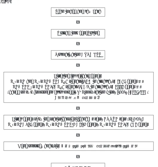
Table 1. Data Analysis
Scan image and raw data
↓
Upload into geneSpring
↓
Normalization : LOWESS
↓
Filtering for reliable genes:
C vs CA and C vs CA+TA: Cy3 signal>215.9 in all arrays: 4937 genes 선별 CA+TA5 vs CA+TA10: Cy3 signal> 199.1 in all arrays: 5224 genes 선별 (based on two component error model, J. Computation Biol., 2001, 8:557-569)
: 재현성이 낮은 유전자 제거
↓
Filter genes showing statistical significance: t test, ANOVA test (p<0.01):
C vs CA: 127 genes, C vs CA+TA5.0 : 420 genes, C vs CA+TA10: 394 genes
↓
Gene ontology analysis: 통계적으로 의미있는 유전자들을 대상으로 분석
↓ 공통유전자 분석
Treat CA Treat CA+TA 5
Ⅲ. 연구결과
1. 약제내성검사 (Drug Resistance Assay)
각 농도에 따른 Taxol
�과 Cyclosporin A 투여 시 농도 증가에 따라 세포수가 감소하였다.
Taxol
�을 투여한 경우에서 농도증가에 따라 세포수의 급 격한 감소를 보였으며, 5 ㎍/ml 이상의 농도에서는 거의 모 든세포가 죽어 Taxol 농도는 1 ㎍/ml, Cyclosporin A 농 도는 3 ㎍/ml 이하에서 투여 시행하는 것이 적절하다고 판 단하였다(Fig. 2).
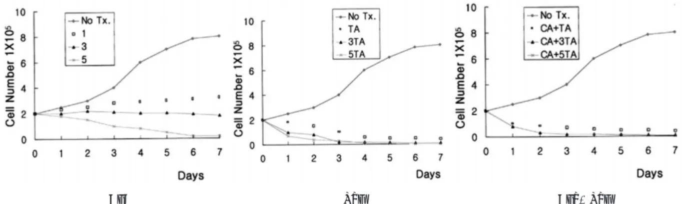
2. 성장곡선(Growth Curve)
약제를 투여하지 않은 대조군에서는 전형적인 S자형 성장 곡선을 나타냈으며, Cyclosporin A 1 ㎍/ml와 3 ㎍/ml의 농도로 투여한 실험군에서의 세포성장은 대조군에 비해 낮
은 성장을 나타냈고, 5 ㎍/ml의 농도로 투여한 군의 세포성 장은 3일째부터 감소하였다.
각 농도(1 ㎍/ml, 3 ㎍/ml, 5 ㎍/ml)에 따라 Taxol
�을 투여한 실험군과 1 ㎍/ml 농도의 Cyclosporin A에 각기 다른 농도(1 ㎍/ml, 3 ㎍/ml, 5 ㎍/ml)의 Taxol
�을 혼합 투여 한 실험군 모두에서 2일째부터 성장이 크게 감소하였 다(Fig. 3).
3. MTT assay(5-diphenyl dimethyltetrazolium bromide)
Cyclosporin A 1 ㎍/ml의 농도로 투여시 MTT는 거의 변화하지 않았으며 농도 증가시에도 크게 감소하지 않아 세 포내의 미토콘드리아에서 succinyl dehydrogenase의 활성 도가 유지됨을 알 수 있었고, cyclosporin A(1 ㎍/ml)와 Taxol
�(1 ㎍/ml, 3 ㎍/ml, 5 ㎍/ml)의 농도로 혼합투여 시에는 MTT의 급격한 감소를 나타내어 약제의 강한 효과 가 나타났다(Fig. 4).
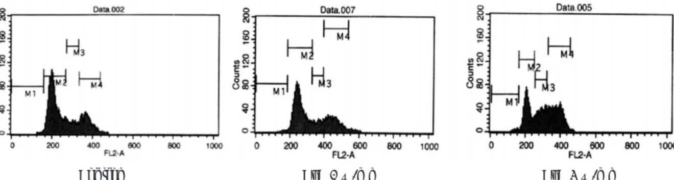
4. 유세포분석(Flow cytometry)
Cyclosporin A 투여시 3 ㎍/ml의 농도에서는 G
2/M phase 세포주기 억제가 일어났으며, Taxol
�투여시 저농도 (1 ㎍/ml)에서는 G
2/M phase, 고농도(3 ㎍/ml, 5 ㎍/ml) 에서는 S phase와 G
2/M phase 세포주기 억제가 나타 났다.
Cyclosporin A(1 ㎍/ml)와 Taxol
�(1 ㎍/ml, 3 ㎍/ml, 5 ㎍/ml)의 혼합투여시에는 Taxol
�단독투여시 보다 S phase와 G
2/M phase 세포주기 억제비율이 높게 나타났다 (Fig. 5-7).
Fig. 2. Drug Resistance Assay.
Fig. 3. Growth Curve.
CyA Tax Cya+Tax
5. 유전자 발현(cDNA Microarray)
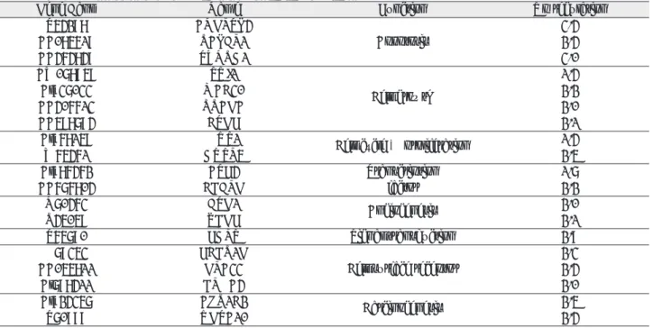
Cyclosporin A 투여후 CYR61(cysteine-rich, angio- genic inducer, 61)와 SERPINB2(serine proteinase inhibitor, Grade B, mem) SSR3 및 UPF3A 유전자 발 현이 대조군에 비해 증가하였으며 종양세포의 성장과 분화, 침투 및 전이 그리고 receptor activity와 전사조절인자에 관여하는 유전자들이 증가되었다(Table 2).
Taxol
�투여시 ANGPTL4, RALBP1, TXNRD1, SUI1, MAC30, RRAGA, HUS1, CEBPG, BTG1, FDPS, FCER1G, GPA33, UGTA10, ATF4, AKR1C2, CTGF, VEGF, DUSP5, EPHA4 등이 발현되었으며 이 들은 apoptosis, cell growth, cell cycle과 proliferation, Fig. 4. MTT Assay.
Fig. 5. Flow Cytometry (Control, Cyclosporin A 1 ㎍/ml, Cyclosporin A 3 ㎍/ml).
Fig. 6. Flow Cytometry (Taxol 1 ㎍/ml, Taxol 3 ㎍/ml, Taxol 5 ㎍/ml).
Fig. 7. Flow Cytometry.
Cyclosporin A+Taxol 1 ㎍/ml Cyclosporin A+Taxol 3 ㎍/ml Cyclosporin A+Taxol 5 ㎍/ml
Tax 1 ㎍/ml Tax 3 ㎍/ml Tax 5 ㎍/ml
Control CyA 1 ㎍/ml CyA 3 ㎍/ml
transcription 조절인자, angiogenesis, signal transduc- tion과 receptor activity 및 carcinogenesis에 관여하는 유전자로서 대부분 암세포의 억제효과가 있는 것으로 나타 났다(Table 3).
Cycolsporin A 단독 투여시와 Cyclosporin A와 Taxol
�(1 ㎍/ml)을 혼합투여시 공통으로 ALCAM 유전자가 발현 되었고, 이는 proteolytic cascades 의 기능을 증강시키는 인자로 transmembrane protein으로 알려져 있다.
Cyclosproin A와 Taxol
�(3 ㎍/ml) 고농도로 혼합투여시 세포주기를 조절하고 p53의 apoptosis를 억제하는 GTSE1유전자가 발현되었고 대조군에 비해 변화가 없었 다. 이는 고농도의(3 ㎍/ml) Taxol
�이 혼합 투여시 S
phase 초기부터 세포 정지가 나타나기 때문이라 추정되었 다(Table 4).
Ⅳ. 총괄 및 고찰
구강편평세포 암종의 치료법으로는 일반적으로 외과적 수 술, 방사선 치료, 항암요법 및 유전자 치료법 등이 사용되고 있다
19). 외과적 수술은 많은 구강악안면외과의사들의 노력 에 의해 다양한 수술기법의 개발로 높은 수준에 올라와 있 다. 그러나 항암요법 및 유전자 치료법의 경우 암세포에 대 한 높은 세포독성 및 세포분열 저해기능으로 높은 항암효과 를 나타내지만 신독성 및 간독성 또는 골수기능 의 저하 등 Table 2. Gene Expressions in HN22 Cell Line treated with Cyclosporin A
Gene Bank Name Function Up- regulation
A1014487 CYR61 Growth factor 2.1
T49159 SERPINB2 Carcinogenesis & metastasis 2.6
AI394435 SSR3 Receptor activity 2.0
AA931918 UPF3A Transcription factor 2.2
Table 3. Gene Expressions in HN22 Cell Line treated with Taxol
Gene Bank Name Function Up regulation
T54298 ANGPTL4 3.4
AA085619 RALBP1 Apoptosis 2.4
AA464849 TXNRD1 3.0
AW078959 SUI1 1.4
Al937033 MAC30 Cell growth 2.2
AA406613 RRAGA 2.0
AA598794 CTGF 2.1
Al968159 HUS1 Cell cycle & proliferation 1.4
W65461 DUSP5 2.5
Al986462 ATF4 Transcription 1.7
AA676804 CEBPG factor 2.2
N70463 BTG1 Angiogenesis 2.0
R45059 VEGF 2.1
T65790 FDPS Signal transduction 2.9
H79353 FCER1G 2.3
AA055811 GPA33 Cell surface receptor 2.4
Al798411 EPHA4 2.0
Al924357 AKR1C2 Carcinogenesis 2.5
T70999 UGTA10 2.4
Table 4. Gene Expressions in HN22 Cell Line treated with Taxol
�+Cyclosporin A
Gene Bank Name Function Up regulation
R39862 ALCAM Transmembrane protein 1.2
AI369284 GTSE1 cell cycle regulator 1.0
과 같은 면역능력이 떨어지는 2차적인 부작용을 유발하는 문제가 있다. 또한 유전자 발현 분석 및 각 인자들의 기능적 연구가 미미하여 임상적용에 어려움을 느끼고 있는 실정이 다. 최근에 다양한 천연물질의 항암효과에 대한 연구가 활 발히 진행되며 분자생물학 영역에서는 유전자 발현 분석법 의 첨단화에 노력을 경주하고 있다.
주목나무에서 추출된 천연물질인 Taxol은 미소관계 (microtubule system)을 안정화시키고 미소관의 합성을 억제하는 항미소관 약제(anti - microtubule agent)이다.
Taxol의 효능은 미소관에 결합하여 분해를 억제 안정화 시 키고 세포주기 G
2/M phase에서 세포분열을 억제하여 세포 사멸을 유도함으로써 항암작용을 나타낸다
1-7). 미소관을 안 정화 시키는 Taxol은 혈관형성 억제작용도 있는 것으로 알 려졌으며, 대식세포에서 TNF-α생성을 유도하고 미소관과 결합하여 방사능 감수성이 높게 나타나는 세포주기 G
2/M phase에 세포주기를 억제함으로써 radiosensitizer 역할을
한다
8-11). 미소관과 연관된 세포들의 기능들은 종양 치료에
있어 광범위한 미소관 억제인자들의 사용을 유도하게 되었 다. 즉, Taxol을 LNCaP 세포와 H460, RKO 세포에 처리 하여 나타내게 되는 G
2/M phase 세포주기억제 기간에 CDC2 Kinase의 활성도가 증가되며 Bcl-2의 인산화가 증 가 된다고 알려져 있다
23). 또한 G
1/S phase check point 기능을 하는 p21과
24), p53의
25)기능이 상실된 세포의 경우 Taxol에 의해 세포사멸이 증가된다고 보고하고 있다.
또한 신호전달 과정에서 세포 성장과 분화과정에 중요역 할을 담당하는 MAP kinase family가 세포사멸을 나타내 게 된다고 보고되고 있다
26). 즉 Taxol 처리시 MAP Kinase 와 전단계 신호전달 물질인 Raf, Ras 등이 활성화 되고 세포사멸 과정중에 protein Kinase C를 활성화시키게 된다
27).
Taxol은 미토콘드리아의 경로에 연관되어 있는데 대장암 의 경우는 caspase-8, caspase 3 등이 활성화 되어 미토콘 드리아의 활성막이 감소한다고 보고되고 있고, Taxol이 직 접 Cytochrome C의 방출을 유도하여 신경종양 세포로부 터 미토콘드리아의 분리에 작용한다고 발표되었다
28,29). 본 연구의 유세포 분석에서 taxol 투여시 저농도(1 ㎍/ml)에 서는 G
2/M phase 세포주기 억제가 나타났으며, 고농도(3
㎍/ml)투여시 S phase와 G
2/M phase에서 세포주기 억제 (cell arrest)가 나타나 세포사멸이 유도된다고 사료된다.
Cyclosporin은 Tolypocladium inflatum과 cylindro- carpum lucid에서 추출된 것으로 cyclophilin과 결합하여 유도된 lipophilic cyclic peptide이다
16). 주로 장기이식시 면역억제제로서 사용되며 자가면역 질환에도 사용되고 있 다. Cyclosporin A의 면역억제 기전은 B 임파구에는 영향 을 미치지 않으며 T 임파구에 선택적으로 작용하고 T- cytotoxic cell과 T-helper cell 기능을 억제한다. 즉 cal-
cineurin에 결합하여 활성을 저해하며 IL-2(interleukin - 2)와 같은 cytokine의 유전자 복제를 억제하여 T-cell 분화 를 차단시켜 동종항원(alloantigen)에 대한 세포 독성을 효 과적으로 차단한다
18).
Cyclosporin A는 면양 적혈구에 대한 과민반응, 이식편 대숙주(GVH), 피부거부반응, 알러지성 척수염 등에서 T임 파구의 기능을 억제하며 가슴샘 비의존형 I형인 lipopo- lysaccharide에 대한 plaque forming cell(PFC) 반응은 증진시키나, 가슴샘 의존형 II형 항원인 polyvinylpyroli- done(PVP)에 대한 체액성 면역반응은 억제하며 항원 과부 하에 의한 T세포의 활성을 억제한다는 보고도 있다
30).
Cyclosporin A는 림프구에서 신호전달과정에 영향을 줄 뿐 아니라 지방세포나 근육세포와 같은 다양한 세포에서 신 호 전달과정에 관여하여 각 세포의 세포사멸을 유도하는 것 으로 알려져 있다. Ciechomska 등은 신경종양세포에서 cyclosporin A에 의한 세포사멸을 소개하면서 이 과정에 fasL의 transcriptional activation과 AKT 신호전달과정 이 관련 되어 있다고 하였고
31), Bustamante 등은 lym- phoid leukemia에서 Cyclosporin A에 의해 미토콘드리아 의 탈분극이 나타나고 cytochrome c의 분비를 증강시켜 세 포사멸이 유도됨을 설명하였다
32).
또한 골수세포에서도 Cyclosporin A에 의해 세포사멸 과 정으로 핵분절, 다핵파골세포의 파괴 및 caspase-3의 활성 도가 증가하는 것으로 알려져 있다
33). Canudas 등은 가토 의 cerebellar granule neurone에서 Cyclosporin A가 colchicine에 매개된 cdk5와 cdk5/25의 발현을 증가시켜 세포사멸을 유도한다고 하였다
34). 본 연구 유세포 분석에서 도 cyclosporin A(3 ㎍/ml)투여시 G
2/M phase의 세포주 기 억제가 나타나 세포사멸을 유도한다고 추정된다.
Cyclosporin A와 Taxol을 병용하여 사용시 Cyclosporin A가 Taxol의 항암작용에 도움을 주는 것으로 알려져 있다.
즉 Cyclosporin A에 의해 multidrug resistance를 감소시 켜 p-glycoprotein이 억제됨으로 taxol의 장내 흡수를 증강 시키고 billiary track을 통한 소실을 막아주어 항암효과를 증강시킨다
35).
Kruijtzer 등
36)은 폐암에서 Cyclosporin A와 Taxol을 병 용 치료시 항암효과가 증가 한다고 보고하였고, Nomura 등
37)은 사람의 방광암 세포에서 Cyclosporin A에 의해 cal- cineurin activity의 억제와 Bcl-2의 항세포사멸 기능이 감 소되어 Taxol의 세포사멸 효과가 증가되었다고 보고하 였다.
Kemper 등
38)은 Cyclosporin A나 Psc833같은 P-
Glycoprotein inhibitor에 의해 Taxol의 brain uptake를
증가시킨다 하였고, Hepatoma에서 Taxol과 Cyclosporin
A를 병용 사용시 caspase9, caspase3 등의 활성도를 증가
시키고 HepG2 cell에서 미토콘드리아의 활성막이 저하되
어 세포사멸이 증가되며 이는 PI3 kinase-AKT 경로가 억 제되어 일어난다고 알려져 있다
39).
Bardelmeijer 등
40)도 마우스에서 p-Glycoprotein inhibitor에 의해 Taxol의 oral uptake를 상승시켜 장내흡 수가 증가한다 하였고, Koshkina 등
41)은 이종이식된 신장 암으로 유도된 마우스에 Cyclosporin투여시 Taxol의 항암 효과가 증가된다고 보고하였다. 본 연구에서도 Taxol 단독 투여 시 보다 Taxol과 Cyclosporin A를 혼합 투여시 S phase와 G
2/M phase 세포주기 억제 비율이 높게 나타 났다.
인간의 폐종양 세포(A549 cell)에 저농도 Taxol 투여에 의한 세포성장의 억제시 P53와 P21 발현의 증가가 나타난 다고 알려져 있으며 tumor suppressor gene인 P53은 세 포주기의 억제와 세포사멸을 유도함으로써 항암 효과가 나 타난다고 알려져 있다
42). 또한 고농도의 Taxol 투여시 mitosis에서 세포주기 억제가 일어나며 이때에도 세포주기 억제와 관련된 유전자가 발현이 되며 일반적 mitosis는 cyclin B와 복합체를 이루는 cdc2의 활성화가 억제되어 mitosis exit이 일어나게 된다
43). 또한 전사 과정에 관여하 는 유전자의 발현으로 cdc2의 down regulation과 p53의 upregulation으로 mitotic arrest를 유도한다고 알려져 있 다
44). Chen 등
45)은 미소관 안정 약제를 폐종양세포에 투여 시 BTG2, PTGF-B 등의 항종양증식 유전자 및 P21같은 세포사멸에 관여하는 유전자 발현을 통해 세포주기 억제를 보고하였고, mitotic exit는 G
2/M & spindle check point 유전자의 발현변화에 의해 유도된다고 설명하였으며, check point 유전자와 death receptor가 세포 사멸과 관계 있음을 보고하였다.
또한, philip 등
46)은 Taxol로 유도발현된 ATF-4, Krox- 24같은 transcription factor나 cox-2같은 효소가 세포사 멸을 유도한다고 하였다. 본 연구에서는 구강 편평세포암종 세포주에 Taxol 투여시 통계학적인 유의성이 있고 암세포 와 관련된 19가지의 유전자 발현이 증가됨을 확인할 수 있 었다. Apoptosis에 관여한 유전자로는 ANGPTL4, RALBP1 및 TXNRD1이 발현되었으며 세포성장인자와 관 련된 유전자로는 SUI1, MAC30, RRAGA와 CTGF 유전 자가 발현되었다. 세포주기와 증식에 관여한 유전자로는 HUS1, DUSP5 등이 발현되었으며 전사조절인자로는 ATF4, CEBPG 등이 비교적 높게 발현되었다. 또한 angiogenesis 에 관여한 유전자는 BTG1 VEGF 유전자 등 이 있었으며 신호전달이나 receptor activity 에 관여한 인 자로는 FDPS 와 FCER1G GPA33 및 EPHA4 등이 발현 되었다. 그 외 잘 알려져 있지 않으나 carcinogenesis와 연 관이 있는 유전자로 AKRIC2와 UGTA10 등도 발현되 었다.
구강편평세포 암종 세포주에 cyclosporin A 투여시 통계
학적인 유의성과 암세포와 관련된 유전자는 종양세포의 성 장과 연관된 CYR61, 종양세포의 침투전이에 관련된 SER- PINB2, receptor activity와 관련된 SSR3 및 전사조절인 자인 UPA3A 등이 발현되었다. 또한 Taxol과 cyclosporin A 혼합 투여 후 발현된 유전자는 Type I transmembrane protein으로 알려진 ALCAM과 세포주기 조절인자인 GTSE 등의 발현이 증가되었다.
Ⅴ. 결 론
구강편평세포 암종 세포주에 Taxol
�과 Cyclosporin A 투 여 후 나타난 세포독성 및 세포주기억제에 관련된 항암효과 와 cDNA microarray를 통해 발현된 유전자를 관찰한 후 다음과 같은 결과를 얻었다.
1. 구강편평세포 암종 세포주에 투여하는 Taxol
�과 Cyclosporin A의 농도는 각각 1 ㎍/ml, 3 ㎍/ml 이하에 서 시행하는 것이 적절하다고 판단되었다.
2. 대조군의 종양세포 성장은 전형적인 S자형을 보였으며, Cyclosporin A 1 ㎍/ml와 3 ㎍/ml의 농도로 투여한 실 험군에서의 세포성장은 대조군에 비해 낮은 성장을 나타 내었고, 5 ㎍/ml의 농도로 투여한 군의 세포성장은 3일 째부터 감소하였다. 각 농도(1 ㎍/ml, 3 ㎍/ml, 5 ㎍ /ml)에 따라 Taxol
�을 투여한 실험군과 1 ㎍/ml농도의 cyclosporin에 각기 다른 농도(1 ㎍/ml, 3 ㎍/ml, 5 ㎍ /ml)의 Taxol
�을 혼합투여한 실험군 모두에서 2일째부 터 성장이 크게 감소하였다.
3. Cyclosporin A 1 ㎍/ml의 농도로 투여시 세포내의 mitochondria에서 succinyl dehydrogenase의 활성도 가 유지되었고, Cyclosporin과 Taxol
�혼합투여시 MTT가 급격히 감소하여 약제의 강한 효과가 나타났다.
4. Cyclosporin A 투여시 3 ㎍/ml의 농도에서 G
2/M phase 세포주기 억제가 일어났으며 Taxol
�투여시 저농 도(1 ㎍/ml)에서는 G
2/M phase, 고농도(3 ㎍/ml, 5
㎍/ml)에서는 S phase, G
2/M phase 세포주기 억제가 나타났다. Cyclosporin(1 ㎍/ml)과 Taxol
�(1 ㎍/ml, 3
㎍/ml, 5 ㎍/ml)의 혼합투여시에는 Taxol
�단독투여시 보다 S phase와 G
2/M phase 세포주기 억제비율이 높 게 나타났다.
5. Cyclosporin A 투여후 CYR61(cysteine-rich, angio-
genic inducer, 61)와 SERPINB2(serine proteinase
inhibitor, Clade B, mem) SSR3 및 UPA3A 유전자가
발현되었고 대조군에 비해 증가하였으며 종양세포의 성
장과 분화, 침투 및 전이 그리고 receptor activity와 전
사조절인자에 관여하는 유전자가 발현되었다. Taxol
�투
여시 ANGPTL4, RALBP1, TXNRD1, SUI1,
MAC30, RRAGA, HUS1, CEBPG, BTG1, FDPS,
FCER1G, GPA33, UGTA10, ATF4, AKR1C2, CTGF, VEGF, DUSP5, EPHA4 등이 발현되었으며 이들은 Apoptosis, Cell growth, cell cycle과 prolifer- ation, transcription 조절인자, angiogenesis, signal transduction과 receptor activity 및 carcinogenesis 에 관여하는 유전자로 대부분 암세포 억제효과가 있는 것으로 나타났다. Cyclosporin A와 Taxol
�을 혼합투여 후에는 Transmembrane protein인 ALCAM과 세포주 기 조절인자인 GTSE1유전자가 발현되었다.
이상의 결과로 볼때 Taxol
�사용시 대부분의 유전자 발현 이 구강편평세포암종 세포주에 대한 세포사멸, 세포주기 조 절, 성장인자 및 전사인자 등에 관련되어 항암효과를 나타 내는 것으로 추정되며, Taxol
�과 cyclosporin A의 혼합투 여시에는 강한 종양세포성장 억제효과를 나타내어 추후 밀 도있는 임상적 적용과 관련된 연구를 통해 구강편평세포암 종의 치료에 기여할 수 있는 약제라 생각되었다.
참고문헌