• 검색 결과가 없습니다.

Pseudomonas aeruginosa Exotoxin A Induces Apoptosis in Chemoresistant YD-9 Human Oral Squamous Carcinoma Cell Line Via Accumulation of p53 and Activation of Caspases

N/A
N/A
Protected

Academic year: 2021

Share "Pseudomonas aeruginosa Exotoxin A Induces Apoptosis in Chemoresistant YD-9 Human Oral Squamous Carcinoma Cell Line Via Accumulation of p53 and Activation of Caspases"

Copied!
8
0
0

로드 중.... (전체 텍스트 보기)

전체 글

(1)

Pseudomonas aeruginosa Exotoxin A Induces Apoptosis in Chemoresistant YD-9 Human Oral Squamous Carcinoma Cell Line Via Accumulation of p53 and Activation of Caspases

Gyoo-Cheon Kim

1

and Young-Gi Gil*

Department of Anatomy College of Medicine, Kosin University, Busan 602-703, Korea

1

Department of Oral Anatomy School of Dentistry, Pusan National University, Busan 602-739, Korea, Received May 13 , 2009 /Accepted May 20, 2009

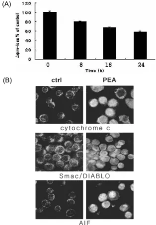
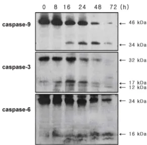
Oral squamous carcinoma (OSC) cells present resistance to chemotherapeutic agents-mediated apopto- sis in the late stages of malignancy. Advances in the understanding of bacterial toxins have produced new strategies for the treatment of cancers. It was demonstrated here that Pseudomonas aeruginosa exo- toxin A (PEA) significantly decreased the viability of chemoresistant YD-9 cells in the apoptosis mechanism. Apoptotic manifestations were evident through changes in nuclear morphology and gen- eration of DNA fragmentation. PEA treatment induced caspase-3, -6 and -9 cleavage, and activation.

These events preceded proteolysis of the caspase substrates poly (ADP-ribose) polymerase (PARP), DNA fragmentation factor 45 (DFF45), and lamin A in YD-9 cells. The reduction of mitochondrial membrane potential, release of cytochrome c and Smac/DIABLO from mitochondria to cytosol, and translocation of AIF into nucleus were shown. While p53, p21 and 14-3-3γ were upregulated, cyclin B and cdc2 were downregulated by PEA treatment. Taken together, PEA induces apoptosis in chemo- resistant YD-9 cells via activation of caspases, mitochondrial events and regulation of cell cycle genes.

Key words : Oral squamous carcinoma, apoptosis, P. aeruginosa exotoxin A, caspase

*Corresponding author

*Tel:+82-51-990-6412, Fax:+82-51-990-6438

*E-mail : [email protected]

Introduction

It has been reported that the mass size of mammalian can- cers was reduced when they were infected with microbial pathogens [6,25]. Furthermore, bacteria such as Shigella flex- neri, Salmonella typhimurium and Staphylococcus aureus were shown to induce apoptosis by the inhibition of survival pathways and the secretion of molecules activating the en- dogenous death machinery in the infected cell [11].

Considering that most tumors contain poorly vascularized areas, recent studies used anaerobic bacteria for treatment of cancer, resulting in significant regression of subcutaneous tumors [8]. However, the application of live bacteria pro- duces significant toxicity and side effects [8,17], limiting the use of bacteria in the treatment of human cancer [15].

Thus, bacterial toxins have emerged as powerful ther- apeutic agents with possible applications in cancer. Several bacterial toxins were used not only in the forms of im- muntoxin, which is composed of antibodies linked to bacte- rial toxins [26,4], but in purified forms [1,21,23]. Purified bac- terial toxins such as shiga toxin, cholera toxin and vacuolat-

ing cytotoxin showed apoptosis-inducing activity in cancer cells [5,23].

P. aeruginosa frequently causes pneumonia, septicemia, and other acute infections in immunocompromised patients.

This gram-negative bacterium produces several extracellular products such as proteases, hemolysins, exotoxin A, exoen- zyme S, elastase and pyocyanin [33]. Among them, P. aerugi- nosa exotoxin A (PEA) is known to be the most toxic factor secreted by P. aeruginosa. PEA enters into eukaryotic cells via receptor mediated endocytosis, and then is cleaved in the endocytic vesicles and translocated into the cytoplasm, where it catalyzes the transfer of the ADP-ribosyl moiety of NAD

+

to elongation factor 2 (EF-2). This inactivation of EF-2 in cells infected with P. aeruginosa is shown to lead inhibition of proteins synthesis, resulting in host cell death [16].

Human oral squamous carcinoma (OSC) is responsible for

the majority of malignancies in the oral cavity. Treatment

of OSC has primarily relied on classical modalities encom-

passing surgery, radiation and chemotherapy or a combina-

tion of these methods, but the result showed the lowest 5

years survival rate among the major cancers. At the late stage

of malignancy, OSC cells are highly resistant to cancer-ther-

apy-mediated apoptosis [7,32]. Chemoresistance in cancers is

(2)

a frequent impediment to successful chemotherapy. Thus, the selective removal of OSC cells by inducing apoptosis could be a promising strategy for treatment of OSC.

The present study was undertaken to investigate whether PEA has an anti-tumor activity in a highly chemoresistant OSC cell line YD-9 cells. I here demonstrate that PEA has a strong cytotoxic effect and induces apoptosis caspases- and mitochondria-dependently in YD-9 cells.

Materials and Methods

The following reagents were obtained commercially.

Antibodies

Mouse monoclonal anti-human caspase-9, cytochrome c, Lamin A and Smac/DIABLO and caspase-6 antibodies were from Upstate biotechnology (Lake Placid, NY, USA). Mouse monoclonal anti-human caspase-3 and PARP, p53, p21, cdc2 and cyclin B1, and rabbit polyclonal anti-human AIF and 14-3-3γ were from Santa Cruz Biotechnology (Santa Cruz, CA, USA). Mouse monoclonal anti-human DFF45 antibody was from MBL (Naka-ku, Nagoya, Japan).

Agents

PEA, Dimetyl sulfoxide (DMSO), Hoechst33258, RNase A, proteinase K, aprotinin, leupeptin, PMSF, thiazolyl blue tet- razolium bromide (MTT) were from Sigma Aldrich (St.

Louis, MO, USA). TUNEL reaction mixture was from Roche Molecular Biochemicals (Mannheim, Germany). ECL Western blotting detection reagents were from Amersham International (Buckinghamshire, UK) 5,5’,6,6’-tetrachloro- 1,1’,3,3’-tetraethylbenzimidazol carbocyanine iodide (JC-1) were from Biotium (Hayward, CA. USA).

Cell culture

YD-9 cell that established from OSC patient [22] were a generous gift of Professor Jong-In Yook (College of Dentistry, Yonsei University, Seoul, Korea). The cells were cultured at 37

o

C in a humidified atmosphere containing 5%

CO

2

in Dulbecco's modified Eagles medium: Nutrient Mixture F12 (DMEM/F12, 3:1) supplemented with 10%

heat-inactivated fetal bovine serum (FBS), 1% glutamine, and 100 μg/ml penicillin/streptomycin.

Cell viability assay

The viability of cultured cells was estimated by MTT

assay. In the MTT assay, cells were placed in a 96-well plate and incubated for 24 hr. Then cells were treated with PEA at 0, 1.5, 7.5, 15, 30 and 60 nM for 24 hr. And then, the cells were treated with 1 mg/ml of MTT in growth medium.

Cells were incubated at 37

o

C for 4 hr. The medium was aspi- rated and the formazan crystals, which are formed from MTT by NADH-generating dehydrogenases in metabolically active cell, were dissolved in 200 ml DMSO. Cell viability was evaluated in comparison to the control culture (taken as 100%) by measuring the intensity of the blue color (OD at 570 nm) by a multiwell reader Quant spectrophotometer (BIO-TEK Instruments Inc., Winooski, VT, USA).

Independent experiments were undertaken and each experi- ment was conducted in triplicates.

TUNEL assay

To detect DNA breaks in situ, the TUNEL assay was conducted with an TUNEL reaction mixture kit (Boehringer Mannheim, Germany). After PEA treatment, cells were washed twice with PBS, fixed in 4% paraf- ormaldehyde for 30 min, and applied permeabilisation sol- ution for 5 min at 4

o

C, and washed again with PBS. This was followed by in situ end labeling according to the man- ufacturer's instructions.

Hoechst staining

Cells treated with PEA for 24 hr and the cell suspension was centrifuged onto a clean fat-free glass slide with a cytocentrifuge. The samples were stained 4 μg/ml Hoechst 33258 at 37

o

C for 30 min and fixed for 10 min in 4%

paraformaldehyde.

DNA electrophoresis

2×10

6

cells were resuspended in 1.5 ml of lysis buffer [10 mM Tris (pH 7.5), 10 mM EDTA (pH 8.0), 10 mM NaCl and 0.5% SDS] into which proteinase K (200 μg/ml) was added.

After samples were incubated overnight at 48

o

C, 200 μl of

ice cold 5 M NaCl was added and the supernatant contain-

ing fragmented DNA was collected after centrifugation. The

DNA was then precipitated overnight at -20

o

C in 50% iso-

propanol RNase A-treated for 1 hr at 37

o

C. A loading buffer

containing 100 mM EDTA, 0.5% SDS, 40% sucrose, and

0.05% bromophenol blue were added at 1:5 (v/v). Separation

was achieved in 2% agarose gels in Tris-acetic acid/EDTA

buffer (containing 0.5 μg/ml ethidium bromide) using 50

mA for 1.5 hr.

(3)

Fig. 1. Effects of PEA treatment on the viability of chemoresistant YD-9 cells. After 24 h incubation with etoposide (2-10 μg/ml) and 5-FU (1-2000 μM), viabilities were determined by MTT assay. All of three cell lines showed resistance to etoposide and 5-FU. HOS, MG63 and YD-9 cells were treated with PEA (1.5-60 nM). PEA preferentially decreased the proliferation of YD-9 cells compared with other cell lines. Four independent assays were performed and data shown are the mean ±SD of the means obtained from triplicates of each assay.

Western blot analysis

Cells (2×10

6

) treated with PEA were washed twice with ice-cold PBS, resuspended in 200 μl ice-cold solubilizing buf- fer [300 mM NaCl, 50 mM Tris-Cl (pH 7.6), 0.5% TritonX-100, 2 mM PMSF, 2 μl /ml aprotinin and 2 μl /ml leupeptin]

and incubated at 4

o

C for 30 min. The lysates were centri- fuged at 14, 000 revolutions per min for 15 min at 4

o

C.

Protein concentrations of cell lysates were determined with Bradford protein assay (Bio-Rad, Richmond, CA, USA) and 50 μg of proteins were loaded onto 7.5-15% SDS/PAGE. The gels were transferred to nitrocellulose membrane and re- acted with each antibody. Immunostaining with antibodies was performed using Super Signal West Pico enhanced chemiluminescence substrate and detected with LAS- 3000PLUS (Fuji Photo Film Company, Kanagawa, Japan).

Immunofluorescent staining

Cells were cytocentrifuged and fixed for 10 min in 4%

paraformaldehyde, incubated with anti-cytochrome c, Smac/DIABLO and AIF antibodies for 1 hr, washed 3 times each for 5 min, and then incubated with goat-FITC-an- ti-mouse or rabbit IgG secondary antibody (Santa Cruz Biotechnology, Santa Cruz, CA, USA) for 1 hr at 37

o

C. Cells were mounted with PBS, and observed under Zeiss Axioscop (Germany).

Assay of mitochondrial membrane potential (MMP)

Changes in mitochondrial membrane potential after treat- ment with PEA for 24 hr were determined by staining cells with the indicator dye. JC-1 was added directly to the cell culture medium (1 μM final concentration) and incubated for 15 min. The medium was then replaced with PBS, and cells were quantitated for J-aggregated fluorescence intensity in a modular fluorimetric system (Spex Edison, NJ, USA)

using excitation and emission filters of 492 and 590 nm, respectively.

Results PEA reduced the viability of YD-9 cells

To investigate whether PEA has a cytotoxic effect on YD-9 cells, MTT assay was conducted. As well known che- moresistant osteosarcoma cell lines such as HOS and MG63, YD-9 cells did not significant decrease in cell viability after treatment with etoposide (2-10 μg/ml) and 5-FU (1-2000 μ M) at which concentration many cancer cells are responsive.

Furthermore, YD-9 cells proliferation were rarely affected by various concentrations of another antitumor agent genis- tein (data not shown). However, PEA at 1.5-60 nM sub- stantially reduced the viability of those cells. Noticeably, cell death inducing activity of PEA is significantly higher in YD-9 cells, compared with HOS and MG63 cells (Fig. 1).

Since PEA showed the highest cytotoxicity in YD-9 cells among three cells, the mechanism underlying its cytotox- icity was studied on this single cell. For this study, the sin- gle concentration 15 nM was utilized for further assessment of apoptosis, at which PEA showed half-maximal inhibition of viability.

Reduced viability of YD-9 cells induced by PEA resulted from apoptosis

I examined whether reduced viability of YD-9 cells were

caused by apoptosis. Hoechst staining showed nuclear con-

densation and fragmentation after treatment of 15 nM PEA

for 24 hr (Fig. 2A). In TUNEL assay, a large number of cells

treated with 15 nM PEA for 24 hr showed positive reaction

(Fig. 2B). Cells treated with 15 nM PEA showed DNA degra-

dation characteristic of apoptotic ladder (Fig. 2C).

(4)

Fig. 2. Demonstration of apoptosis in YD-9 cells treated with 15 nM PEA . (A) Nuclear condensation and fragmenta- tion were clearly shown by Hoechst staining. (B) TUNEL assay showed apoptotic cells in YD-9 cells treated with PEA. (C) DNA electrophoresis showed DNA ladder in YD-9 cells treated with PEA.

Mitochondrial events were involved in PEA-induced apoptosis of YD-9 cells

Loss of MMP (ΔΨm) is known to be a common event in many pathways of apoptosis. In this study, MMP decreased time-dependently after PEA treatment (Fig. 3A). I further ex- amined the subcellular location of mitochondrial apoptosis related factors after PEA treatment. Immunofluorescent mi- croscopy revealed that cytochrome c and Smac/DIABLO in control cells are shown in a punctuate pattern. However, it assumed a diffuse distribution in PEA-treated cells which supported the notion that PEA led to the release of those proteins from mitochondria into cytosol (Fig. 3B). On the other hand, the AIF protein which had been mainly dis- tributed out of the nucleus in the control cells was trans- located onto nucleus following treatment of PEA (Fig. 3B).

PEA-induced apoptosis of YD-9 cells was caspases-dependent

To examine whether the apoptosis was mediated via cas- pases, I carried out Western blotting experiments. Western blotting analysis showed that PEA activated an initiator cas- pase-9 and the two effector caspases caspase-3 and -6. Not only degradation of caspase-9, -3 and -6 were shown, but cleaved products of each caspase were also demonstrated (Fig. 4).

PEA treatment caused cleavage of various caspase target proteins

The effects of PEA on caspase target proteins such as DFF45, PARP and lamin A was investigated. As shown in Figure 5, degradation of PARP protein (116 kDa) and pro- duction of PARP 85 kDa cleavage products were shown. The

(A)

(B)

Fig. 3. Demonstration of mitochondrial events in YD-9 cells treated with 15 nM PEA. (A) MMP decreased after PEA treatment. (B) Immunofluorescence microscopy. In the control YD-9 cells, cytochome c, Smac/DIABLO and AIF are loacated in mitochondria. 24 hr after treatment with PEA, cytochrome c and Smac/ DIABLO are ob- served in the cytosol. Note that AIF was redistributed into nucleus following treatment of cells with PEA.

cleaved products of DFF45 (30 and 11 kDa) and lamin A (45 and 28 kDa) were observed.

PEA treatment induced upregulation or downregula- tion of cell cycle regulatory

I next asked whether PEA could modulate cell cycle regu- latory protein. I examined alterations in protein levels using antibodies against key players in cell cycle regulation (Fig.

6). Western blot analysis demonstrated that mutant type p53 increased at 8,16 and 24 hr, and then decreased. The ex- pression of 14-3-3γ and p21 were increased up to 24 hr, whereas a significant downregulation of cdc2 and cycline B was detected in time-dependent manner.

Discussion

I have shown that PEA in the form of purified protein

(5)

Fig. 4. Activation of caspases in YD-9 cells treated with15 nM PEA. Proform caspase-9 (46 kDa) was degraded, and 34 kDa cleaved product was produced after PEA treatment.

Proform of caspase-3 (32 kDa) was degraded, and 17 kDa and 12 kDa cleaved forms were produced after PEA treatment. Caspase-6 pro-enzyme (34 kDa) was de- graded, and 16 kDa cleaved forms were produced after PEA treatment.

Fig. 5. Degradation of caspase target proteins in YD-9 cells treat- ed with 15 nM PEA. Degradation of PARP protein (116 kDa) and production of 85 kDa cleavage product were demonstrated, which is prominent 24 hr after PEA treatment. Three cleaved products of DFF45 (30, 20 and 11 kDa) were demonstrated in YD-9 cells treated with PEA, which are evident in later time points (48 and 72 hr). Two cleaved products of lamin A (45 and 28 kDa) are demonstrated in YD-9 cells treated with PEA, which are shown at 24 hr after PEA treatment.

has a strong cytotocic effect on cheomresistant YD-9 OSC cells through inducing apoptosis. Chemoresistant tumor

Fig. 6. Western blot analysis of cell cycle regulation proteins in YD-9 cells treated with 15 nM PEA. The expression of p53 was increased at 8, 16 and 24 hr, and then decreased. The expression of p21 and 14-3-3γ were in- creased up to 24 hr, whereas the expression level of cdc2 and cyclin B were decreased time-dependently.

cells have acquired the ability to evade the action of multiple classes of anti-cancer drugs. One mechanism by which tu- mor cells survive in the presence of chemotherapy is the increase of anti-apoptotic activities. Because resistance to chemotherapy is a major failure in the treatment of OSC, there is a great interest in the search for new compounds that may destroy OSC cells, which eventually can be in- corporated into therapy for the treatment of OSC.

In the past two decades, bacterial toxins have been consid- ered as potential therapeutic agents for treatment of cancer.

The application of anthrax lethal factor to athymic nude mice carrying human melanoma xenograft induced complete tu- mor regression without side effects [21]. Verotoxin 1 has been proven to be highly effective in human colon cancer [3], myelogenous leukemia cells [23] and renal tumor xeno- grafts in SCID mice [14].

A number of extracellular toxic products, which P. aerugi- nosa synthesizes, are believed to be involved in the patho- genesis of its infection. Among those products, PEA is con- sidered to be the most toxic factor [12]. PEA is a 66 kDa protein which has three structurally and functionally differ- ent domains. Domain I is responsible for cell recognition, domain II for translocation, and domain III for inhibition of protein synthesis [27]. It is generally accepted that PEA is internalized by the cell surface receptor CD91 and exhibits its cytotoxicity by blocking protein synthesis through in- activation of EF-2 [16]. However, it was reported that protein synthesis inhibition by toxins is not sufficient to mediate tar- get cell death [24], suggesting that additional mechanisms such as apoptosis may be involved in PEA-induced cytotoxicity.

Previous studies elucidated that PEA induces cellular

(6)

death via apoptosis in normal cells including mouse hep- atocyte [30] and human mast cells [18]. Because of its apop- tosis-inducing activity, PEA has been widely used to kill tar- get cells. In most studies undertaken for the purpose of can- cer treatment, PEA has been employed in the form of immunotoxin. PEA-containing immunotoxin induced apop- tosis in breast cancer cells [2] and leukemia cells [9].

Although PEA-containing immunotoxin has been studied in various cancer cells, induction of apoptosis in cancer cells by PEA in the form of purified protein has not been demon- strated to date. I here demonstrated the induction of apopto- sis in chemoresistant cancer cells by purified PEA.

Etoposide and 5-FU are known to inhibit the proliferation of various cancer cells [13,29]. They have been widely used in treatment of OSC and reported inducing apoptosis in OSC cells. However, etoposide and 5-FU at usual doses did not inhibit substantially the proliferation of YD-9 cells.

Furthermore, genistein, at the doses inducing substantially apoptosis in human osteosarcoma cells, did not affect the viability in YD-9 cells [28]. My experimental results demon- strated that YD-9 cells are resistant to etoposide, 5-FU and genistein. Nevertheless, PEA at nanomolar concentrations strongly inhibited the growth of YD-9 cells dose-depend- ently, suggesting that PEA has a preferential cytotoxic effect on YD-9 chemoresistant OSC cells.

Caspases and mitochondria play an important role in the execution phase of apoptosis and are responsible for many of the biochemical and morphological changes associated with apoptosis. Caspases are known to cleave a number of proteins and the consequences of these cleavage events were suggested to be responsible for many of the phenotypic changes in the cell undergoing apoptosis [34]. Mitochondria play a critical role in control of apoptosis by acting as reser- voirs for death promoting factors such as cytochrome c, Smac/Diablo, AIF, endonuclease G and procaspases-2, -3, -8, -9 [10].

When bacteria toxins induce apoptosis in cancer cells, both caspase and mitochondria also play an important role.

Shiga toxin 1 induces apoptosis in the human myelogenous leukemia cell line THP-1 [23] and in Burkitt’s lymphoma cells [19] by a caspase-dependent mechanism. Diphtheria toxin contributes to apoptosis in human leukemia cells through caspase activation [31]. PTE-containing immunotox- in induced loss of mitochondrial membrane potential and activation of the caspase cascade in MA-11 breast cancer cells [2]. My data indicate that PEA also induces apoptosis on

YD-9 OSC via activation of caspases and mitochondrial events.

Usually p53 acts as an "emergency break" inducing either cell cycle arrest or apoptosis, protecting the genome from accumulating excess mutations. In response to stimuli, p53 is stabilized and activated by multiple mechanisms (including phosphorylation, dephosphorylation and acetyla- tion). Once activated, p53 either induces cell-cycle arrest at G1 or G2 by increasing the transcription of p21 gene or ini- tiate apoptosis [20]. Several of transcriptional targets of p53 including p21 and 14-3-3γ can inhibit cdc2. 14-3-3γ anchors cdc2 in the cytoplasm and inhibits the activation of cyclin B-cdc2, thereby deficiency of activated cyclin B-cdc2 induces cell cycle arrest at G2/M checkpoint. This study demon- strated that ETA treatment upregulated p21 and 14-3-3γ and downregulated cdc2 and cyclin B, which suggests that PEA induces cell cycle arrest and subsequent progress to apoptosis.

Taken together, PEA has a strong apoptotic effect on che- moresistant YD-9 OSC via activation of caspases , mitochon- drial events and regulation of cell cycle genes. It remains also an open question through which exact molecular mech- anism PEA induces apoptosis in chemoresistant OSC cells.

Acknowledgments

This study was supported by grant of Kosin University College of Medicine (2009) and this study was supported by for two years Pusan National University research grant.

References

1. Allam, M., R. Bertrand, G. Zhang-Sun, J. Pappas, and J.

Viallet. 1997. Cholera toxin triggers apoptosis in human lung cancer cell lines. Cancer Res. 57, 2615-2618.

2. Andersson, Y., S. Juell, and O. Fodstad. 2004. Downregula- tion of the antiapoptotic MCL-1 protein and apoptosis in MA-11 breast cancer cells induced by an anti-epidermal growth factor receptor-Pseudomonas exotoxin a immunotoxin. Int. J. Cancer 112, 475-483.

3. Bhattacharjee, R. N., K. S. Park, S. Uematsu, K. Okada, K.

Hoshino, K. Takeda, O. Takeuchi, S. Akira, T. Iida, and T.

Honda. 2005. Escherichia coli verotoxin 1 mediates apopto- sis in human HCT116 colon cancer cells by inducing over- expression of the GADD family of genes and S phase arrest.

FEBS Lett 579, 6604-6610.

4. Brinkmann, U. and I. Pastan. 1994. Immunotoxins against cancer. Biochim. Biophys . Acta 1198, 27-45.

5. Cho. S. J., N. S. Kang, S. Y. Park, B. O. Kim, D. K. Rhee,

(7)

and S. Pyo. 2003. Induction of apoptosis and expression of apoptosis related genes in human epithelial carcinoma cells by Helicobacter pylori VacA toxin. Toxicon. 42, 601-611.

6. Coley, W. B. 1991. The treatment of malignant tumors by repeated inoculations oferysipelas. With a report of ten orig- inal cases. 1893. Clin. Orthop. Relat. Res. 262, 3-11.

7. Crowe, D. L. and U. K. Sinha. 2006. p53 apoptotic response to DNA damage dependent on bcl2 but not bax in head and neck squamous cell carcinoma lines. Head Neck 28, 15-23.

8. Dang, L. H., C. Bettegowda, D. L. Huso, K. W. Kinzler, and B. Vogelstein. 2001. Combination bacteriolytic therapy for the treatment of experimental tumors. Proc. Natl. Acad. Sci.

USA 98, 15155-15160.

9. Decker, T., M. Oelsner, R. J. Kreitman, G. Salvatore, Q. C.

Wang, I. Pastan, C. Peschel, and T. Licht. 2004. Induction of caspase-dependent programmed cell death in B-cell chronic lymphocytic leukemia by anti-CD22 immunotoxins.

Blood 103, 2718-2726.

10. Du, C., M. Fang, Y. Li, L. Li, and X. Wang. 2000. Smac, a mitochondrial protein that promotes cytochrome c-de- pendent caspase activation by eliminating IAP inhibition.

Cell 102, 33-42.

11. Grassme, H., V. Jendrossek, and E. Gulbins. 2001. Molecular mechanisms of bacteria induced apoptosis. Apoptosis 6, 441-445.

12. Hamood, A. N., J. A. Griswold, and C. M. Duhan. 1996.

Production of extracellular virulence factors by Pseudomonas aeruginosa isolates obtained from tracheal, urinary tract, and wound infections. J. Surg. Res 61, 425-432.

13. Hande, K. R. 1998. Etoposide: four decades of development of a topoisomerase II inhibitor. Eur. J. Cancer 34, 1514-1521.

14. Heath-Engel, H. M. and C. A. Lingwood. 2003. Verotoxin sensitivity of ECV304 cells in vitro and in vivo in a xenograft tumour model: VT1 as a tumour neovascular marker.

Angiogenesis 6, 129-141.

15. Hunter, C. A., D. Yu, M. Gee, C. V. Ngo, C. Sevignani, M.

Goldschmidt, T. V. Golovkina, S. Evans, W.F. Lee, and A.

Thomas-Tikhonenko. 2001. Cutting edge: systemic in- hibition of angiogenesis underlies resistance to tumors dur- ing acute toxoplasmosis. J. Immunol. 166, 5878-5881.

16. Iglewki, B. H., P. V. Liu, and D. Kabat. 1977. Mechanism of action of Pseudomonas aeruginosa exotoxin Aiadenosine diphosphate-ribosylation of mammalian elongation factor 2 in vitro and in vivo . Infect. Immun 15, 138-144.

17. Jain, R. K. and N. S. Forbes, 2001. Can engineered bacteria help control cancer? Proc. Natl. Acad. Sci. USA 98, 14748-14750.

18. Jenkins, C. E., A. Swiatoniowski, A. C. Issekutz, and T. J.

Lin. 2004. Pseudomonas aeruginosa exotoxin A induces hu- man mast cell apoptosis by a caspase-8 and -3-dependent mechanism. J. Biol. Chem. 279, 37201-37207.

19. Kiyokawa, N., T. Mori, T. Taguchi, M. Saito, K. Mimori, T. Suzuki, T. Sekino, N. Sato, H. Nakajima, Y. U. Katagiri, T. Takeda, and J. Fujimoto. 2001. Activation of the caspase

cascade during Stx1-induced apoptosis in Burkitt's lympho- ma cells. J. Cell Biochem. 81, 128-142.

20. Ko, L. J. and C. Prives. 1996. p53: puzzle and paradigm.

Genes Dev. 10, 1054-1072.

21. Koo, H. M., M. VanBrocklin, M. J. McWilliams, S. H. Leppla, N. S. Duesbery, and G. F. Woude. 2002. Apoptosis and mel- anogenesis in human melanoma cells induced by anthrax lethal factor inactivation of mitogen-activated protein kinase kinase. Proc. Natl. Acad. Sci. USA 99, 3052-3057.

22. Lee, E. J., J. Kim, S. A. Lee, E. J. Kim, Y. C. Chun, M. H.

Ryu, and J. I. Yook. 2005. Characterization of newly estab- lished oral cancer cell lines derived from six squamous cell carcinoma and two mucoepidermoid carcinoma cells. Exp.

Mol. Med. 37, 379-390

23. Lee, S. Y., R. P. Cherla, I. Caliskan, and V. L. Tesh. 2005.

Shiga, toxin 1 induces apoptosis in the human myelogenous leukemia cell line THP-1 by a caspase-8-dependent, tumor necrosis factor receptor-independent mechanism. Infect.

Immun. 73, 5115-5126.

24. Morimoto, H., B. J. Bonavida. 1992. Diphtheria toxin- and Pseudomonas A toxin-mediated apoptosis. ADP ribosyla- tion of elongation factor-2 is required for DNA fragmenta- tion and cell lysis and synergy with tumor necrosis fac- tor-alpha. Immunol. 149, 2089-2094.

25. Nauts, H. C. and J. R. McLaren. 1990. Coley toxins--the first century. Adv. Exp. Med. Biol. 267, 483-500.

26. Pastan, I. 2003. Immunotoxins containing Pseudomonas exotoxin A: a short history. Cancer Immunol. Immunother. 52, 338-341.

27. Pastan, I. and D. FitzGerald. 1991. Recombinant toxins for cancer treatment. Science 254, 1173-1177.

28. Roh, M. S., C. W. Kim, B. S. Park, G. C. Kim, J. H. Jeong, H. C. Kwon, D. J. Suh, K. H. Cho, S. B. Yee, and Y. H.

Yoo. 2004. Mechanism of histone deacetylase inhibitor Trichostatin A induced apoptosis in human osteosarcoma cells. Apoptosis 9, 583-589.

29. Sarkar, F. H. and Y. Li. 2002. Mechanisms of cancer chemo- prevention by soy isoflavone genistein. Cancer Metastasis Rev. 21, 265-280.

30. Schumann, J., S. Angermuller, R. Bang, M. Lohoff, and G.

J. Tiegs. 1998. Acute hepatotoxicity of Pseudomonas aerugi- nosa exotoxin A in mice depends on T cells and TNF.

Immunol. 161, 5745-5754.

31. Thorburn, J., A. E. Frankel, and A. Thorburn. 2003.

Apoptosis by leukemia cell-targeted diphtheria toxin occurs via receptor-independent activation of Fas-associated death domain protein. Clin. Cancer Res. 9, 861-865.

32. Tong, D., M. Poot, D. Hu, and D. Oda. 2000. 5-Fluorouracil- induced apoptosis in cultured oral cancer cells. Oral. Oncol.

36, 236-241.

33. Van Delden, C., B. H. Iglewski. 1998. Cell-to-cell signaling and Pseudomonas aeruginosa infections. Emerg. Infect. Dis.

4, 551-560.

34. Yuan, J. 1996. Evolutionary conservation of a genetic path-

way of programmed cell death. J. Cell Biochem. 60, 4-11.

(8)

초록:항암제에 저항성을 가지는 YD-9 human oral squamous carcinoma cell line에서 Pseudomonas aeruginosa exotoxin A의 p53 단백질 누적과 caspase를 활성화 경로를 통 해 유도된 세포자멸사

김규천

1

․길영기*

(고신대학교 의과대학 해부학교실,

1

부산대학교 치의학전문대학원 구강해부학교실)

구강편평상피암종은 말기에서 종종 화학치료요법제들이 유도하는 세포자멸사에 저항성을 보인다. 박테리아의 독에 대한 진전된 이해는 암치료에 대한 새로운 치료전략으로 제기되어지고 있다. 본 연구는 Pseudomonas aeruginosa exotoxin A (PEA)가 세포자멸사 기작을 통해 항암제에 저항성을 보이는 YD-9 구강편평상피암종의 생존율을 현격 하게 떨어뜨림을 설명하고 있다. 세포자멸사현상은 핵의 형태학적 변화와 DNA 분절 생성을 통해 입증되었다.

PEA는 caspase-3, -6, -9의 분절과 활성화를 일으켰다. 그리고 이러한 반응들은 caspase의 기질에 해당하는 poly

(ADP-ribose) polymerase (PARP), DFF45, 그리고 lamin A의 단백질 분해를 야기했다. 사립체 막전위 감소, cyto-

chrome c와 Smac/DIABLO의 사립체로부터 세포질로의 유리, 그리고 AIF의 사립체에서 핵으로 이동 등이 관찰되

었다. p53, p21 그리고 14-3-3γ는 증가되는 반면 cyclin B와 cdc2는 감소되었다. 이상의 결과들을 종합해 보면 PEA는

caspase를 활성화시키고, 사립체에 변화를 야기시키고 더 나아가서 세포주기 유전자를 조절함으로써 항암제에

대한 강한 저항성을 보이는 YD-9 세포에서 세포자멸사를 유도한다.

수치

Fig. 1. Effects of PEA treatment on the viability of chemoresistant YD-9 cells. After 24 h incubation with etoposide (2-10 μg/ml) and 5-FU (1-2000 μM), viabilities were determined by MTT assay
Fig. 2. Demonstration of apoptosis in YD-9 cells treated with 15 nM PEA . (A) Nuclear condensation and  fragmenta-tion were clearly shown by Hoechst staining
Fig. 4. Activation of caspases in YD-9 cells treated with15 nM PEA. Proform caspase-9 (46 kDa) was degraded, and 34 kDa cleaved product was produced after PEA treatment.

참조

관련 문서

Concentration-Dependent inhibition of cell viability by adenosine in human oral fibroblast and FaDu human head and neck squamous cell carcinoma · · ·

It also suggest that cell proliferation inhibits by a novel signal transduction for adenosine effect in human FaDu hypopharynx squamous cell carcinoma cells....

Differential Expression of Desmoglein1, Desmoglein3, Epithelial Membrane Antigen, Ber-EP4 and CD10 in Basal Cell Carcinoma and Squamous Cell Carcinoma

Alginate appears to play a key role in the stabilization of biofilms formed by Pseudomonas aeruginosa and some other pseudomonas, with most Pseudomonas strains producing

In vitro cell migration in APE or JAG1 siRNA-treated M059K cell line Fig,11 Down-regulation of JAG1 induces S phase arrest in

Objective: The purpose of this study was to analyze recent trend in incidence of basal cell carcinoma and squamous cell carcinoma in patients from the Gwangju City

"ESKAPE" pathogens, consist of Enterococcus faecium, Staphylococcus aureus, Klebsiella pneumoniae, Acinetobacter baumannii, Pseudomonas aeruginosa,

Fig 4 : Inhibitory effects of classified methanol extracts of Acalypha australis the COX-2 protein expression of the human oral cavity carcinoma KB cells....