Printed in the Republic of Korea DOI 10.5012/jkcs.2010.54.5.573
요약. 본 연구에서는 염화제1철을 2,4-디히드록시살로펜과 조합하여 2,4-디히드록시살로펜-염화철을 합성하였다. 이 복합체 를 자외선-가시광선 분광법, IR 분광법으로 분석하였고, 천연의 송아지 흉선 DNA (ct-DNA)와 2,4-디히드록시살로펜-염화철 의 상호작용을 자외선-가시광선 분광법, 형광분광법, 열변성 분석법, 점성측정법을 이용하여 조사하였다. 분광학적 적정실험 으로 밝힌 2,4-디히드록시살로펜-염화철과 ct-DNA 사이의 결합상수는 (1.6 ± 0.2) × 103 M-1이었다. 형광분광분석을으로 브롬 화 에티디움의 DNA 결합이 2,4-디히드록시살로펜-염화철에 의하여 저해되는 것을 관찰하였다. 이 저해효과는 2,4-디히드록 시살로펜-염화철의 농도에 따라 선형의 Stern-Volmer 방정식을 따른다. 열변성 실험으로 2,4-디히드록시살로펜-염화철이 DNA의 녹는점을 약 4.3 oC 증가시킨다는 것을 관찰하였다. 이 결과들은 2,4-디히드록시살로펜-염화철이 대부분 ct-DNA의 큰고랑과 상호작용한다는 모델을 잘 설명하여 준다.
주제어: DNA, 쉬프염기, 상호작용, 형광, 자외선-가시광선 분광법
ABSTRACT. In this study, iron(III)-2,4-dihydroxysalophen chloride (Fe(2,4-DHSalophen)Cl), has been synthesized by combina- tion of 2,4-dihydroxysalophen (2,4-DHSalophen) with FeCl2 in a solvent system. This complex combination was characterized using UV-vis and IR spectroscopies. Subsequently, the interaction between native calf thymus deoxyribonucleic acid (ct-DNA) and Fe(2,4-DHSalophen)Cl, was investigated in 10 mM Tris/HCl buffer solution, pH 7.2, using UV-visible absorption and fluorescence spectroscopies, thermal denaturation technique and viscosity measurements. From spectrophotometric titration experiments, the binding constant of Fe(2,4-DHSalophen)Cl with ct-DNA was found to be (1.6 ± 0.2) × 103 M-1. The fluorescence study represents the quenching effect of Fe(2,4-DHSalophen)Cl on bound ethidium bromide to DNA. The quenching process obeys linear Stern- Volmer equation in extended range of Fe(2,4-DHSalophen)Cl concentration. Thermal denaturation experiments represent the increasing melting temperature of DNA (about 4.3 oC) due to binding of Fe(2,4-DHSalophen)Cl. These results are consistent with a binding mode dominated by interactions with the groove of ct-DNA.
Keywords: DNA, Schiff base, Interaction, Fluorescence, UV-vis spectroscopy
천연 DNA와 2,4-디히드록시살로펜-염화철(III)과 의 상호작용 연구
Mohammad-Reza Azani*, Azin Hassanpour, and Abdol-Khalegh Bordbar†
Departamento de Química Inorgánica, Facultad de Ciencias, Universidad Autónoma de Madrid, 28049 Madrid, Spain
†Laboratory of Biophysical Chemistry, Department of Chemistry, University of Isfahan, Isfahan, 81746-73441, I.R. Iran (접수 2010. 5. 10; 수정 2010. 6. 4; 게재확정 2010. 8. 18)
Study of Interaction of Native DNA with Iron(III)-(2,4-Dihydroxysalophen)chloride
Mohammad-Reza Azani*, Azin Hassanpour, and Abdol-Khalegh Bordbar†
Departamento de Química Inorgánica, Facultad de Ciencias, Universidad Autónoma de Madrid, 28049 Madrid, Spain
*E-mail: [email protected]
†Laboratory of Biophysical Chemistry, Department of Chemistry, University of Isfahan, Isfahan, 81746-73441, I.R. Iran (Received May 10, 2010; Revised June 4, 2010; Accepted August 18, 2010)
INTRODUCTION
The interaction of transition metal complexes, containing multidentate aromatic ligands, with DNA has recently gained much attention following the important biological and medi- cal roles played by potential metallointercalators.1 Schiff base metal complexes have been recognized as powerful catalysts in a great number of chemical reactions such as, electrochemical reduction of alkyl halides in aprotic sol- vents,2 oxygenation of indols, phenols, flavones, and others.3
In addition, some of these complexes are able to bind in a reversible form to molecular oxygen. These complexes have been also investigated as model compounds in the study of the natural oxygen carriers such as, hemoglobin, myoglobin hemocyanin4 and in the study of the catalytic properties of some cytochromes involved in the biological oxidative reactions. Finally, Schiff base tetradentate com- plexes of iron have been studied as mimetic models of pero- xidases, catalases and superoxide dismutase.5 In addition to these applications, the synthesis and further structural
C N N C Fe
O O
Cl
OH OH
Scheme 1. The chemical structure of Fe(2 ,4-DHsalophen)Cl and chemical characterization of new Schiff base tetradentate
ligands have been studied in many chemical and biological fields. Three major binding modes have been proposed for the binding of Schiff bases to DNA, intercalation, outside groove binding and outside binding with self-stacking in which the Schiff bases stacked along the DNA helix.6 The central metal ion strongly influences both the binding charac- teristics of the Schiff base complex to DNA and the DNA cleavage properties.7 The interaction of a number of iron(II) and iron(III) derivatives with nucleic acids has been widely investigated, but, no work has been published on the interac- tion of Fe(2,4-DHSalophen)Cl complexes with DNA. In this study, we have synthesized Fe(2,4-DHSalophen)Cl and in- vestigated its interaction with DNA in aqueous solutions by UV-vis, and fluorescence spectroscopes, thermal dena- turation and viscosity measurements.
EXPERIMENTAL Chemicals and solutions
1,2-Diaminobenzene and 2,4-dihydroxybenzaldehyde, were obtained from Aldrich Chemical Co. and were used as received. Anhydrous dimethylformamide (DMF) was purchased from Sigma. All other chemicals used in this work, were reagent quality, obtained from Sigma- Aldrich Co.
and used as received without further purification. 2,4-DHS has been synthesized by combination of 1,2-diaminobenzene and the 2,4-dihydroxybenzaldehyde. This ligand containing meta-quinone functional groups was characterized using UV-vis and IR spectroscopies, in non aqueous solvents, such as dimethylformamide (DMF). Fe(2,4-DHSalophen)Cl, has been synthesized by combination of 2,4-dihydroxy- salophen (2,4-DHSalophen) with FeCl2.4H2O in a solvent system. This complex was characterized using UV-vis and IR spectroscopies. Stock solutions of Fe(2,4-DHSalophen) Cl derivatives (typically 10 mM) were prepared just prior to use by dissolving the solids in DMF. All the solutions were extensively degassed under vacuum before measure- ments.
Water was purified with a Millipore Milli-Q system and all the experiments were carried out at room temperature.
Double stranded calf thymus DNA (ct-DNA, activated and lyophilized) was purchased from Sigma. ct-DNA stock solu- tions (2 mg/mL) were prepared in 10 mM Tris/HCl , pH 7.2 buffer. The DNA solutions gave a UV absorbance ratio (A260/ A280) of about 1.9, indicating that the DNA was sufficiently free from protein.8 The concentration in base pairs of DNA was determined using an extinction coefficient of 6600 cm-1 M-1 at 260 nm.9
Synthesis of 2,4-DHSalophen ligand
According to the traditional procedure of synthesis of tetradentate Schiff base ligands,10 the reaction of benzalde- hydes with 0.5 mol equivalent of diamines, in refluxing MeOH for a few hours, gives rise to the final products (75 - 85% yield) which were analytically pure solids after recry- stalization. To reflux the compounds, a 25 mL rounded bo- ttom flask 1,2-diaminobenzene (0.1080 g; 1.00 mmol) was mixed with 2,4-dihydroxybenzaldehyde (0.2762 g; 2.00 mmol) and 25 mL ethanol 95%. Afterwards the mixture was refluxed for 3 hours to obtain a fine yellow solid mix- ture. The obtained precipitate was filtered and washed several times with ethanol 96% and ether. The product was recry- stalized by MeOH to obtain yellow needles of pure 2,4-DHS (0.2819 g, 81%).
Synthesis of Fe(2,4-DHSalophen)Cl complex
The complex of Fe(2,4-DHSalophen)Cl Scheme 1, was synthesized by combination of 0.174 g (5.0 mmol) sample of 2,4-DHSalophen in 50 mL of ethanol and 0.099 g (5.0 mmol) of FeCl2.4H2O in 30ml of ethanol were added to the mixture drop by drop while stirring. Then the mixture was refluxed for 8 hours and cooled to room temperature. The synthesized complex was recrystallized from ethanol by diethyl ether and was filtered and dried under vacuum.
Spectroscopy
The IR measurements were done on Philips spectrophoto- meter. The absorbance measurements were carried out using UV-vis, Carry-500 double beam spectrophotometer, operat- ing from 200 to 800 nm in 1.0 cm quartz cells. The absor- bance titrations were performed at a fixed concentration of the specific Fe(2,4-DHSalophen)Cl complex while varying the concentration of double stranded ct-DNA.
Fluorescence spectra were recorded with a RF-5000 Shi- madzu spectrofluorimeter equipped with a Peltier system to control the temperature inside the cuvettes.
To achieve equilibration, samples of aqueous Fe(2,4- DHsalophen)Cl solutions were let for one week at room
[DNA]=0 µM 280 µM 900 µM 2110 µΜ 3600 µM 5400 µΜ 5880 µM
300 350 400 450 500 550 600
Wavelength (nm)
0.5 0.4 0.3 0.2
0.1 0.0
Abs.
[DNA] = 0 µM 280 µM 900 µM 2110 µM 3600µM 5400 µM 5880 µM
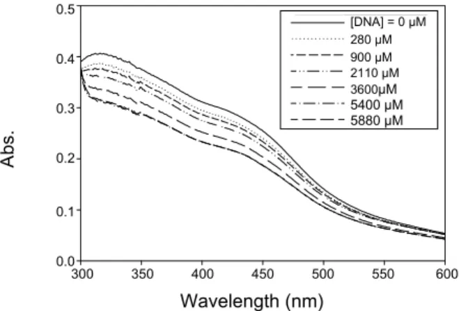
Fig. 1. The spectrum of Fe(2,4-DHsalophen)Cl in the presence of various concentration of ct-DNA. Measurements were done in 10 mM Tris/HCl buffer, pH 7.2 and at 25 ºC.
temperature. Stable and reproducible UV-vis absorption spectra were obtained at this equilibration. All the solu- tions were extensively degassed under vacuum before mea- surements.
Melting experiments
Melting curves were established using an UV-vis Carry- 500 double beam spectrophotometer in conjunction with a thermostated cell compartment. The measurements were carried out in 10 mM Tris/HCl, pH 7.2. The temperature inside the cuvette was measured with a platinum probe and was increased over the range 20 - 90 oC at a heating rate of 1 oC/min. The melting temperature, Tm, was obtained from the mid-point of the hyperchromic transition.
In all of the experiments, for the pH measurement, we used a potentiometer (Metrohm model, 744).
Viscosity measurements
The viscosity of ct-DNA solutions was measured at 25 ± 0.1 oC using an Ubbelohde viscometer. Typically, 10 mM Tris/HCl buffer solution, pH 7.2 was transferred to the visco- meter to obtain the reading of flow time. For determination of solution viscosity, 10.0 mL of buffered solution of 160 µM ct-DNA was taken to the viscometer and a flow time read- ing was obtained. An appropriate amount of Schiff base was then added to the viscometer to give a certain r (r = [Schiff base]/[DNA base pair]) value while keeping the ct-DNA con- centration constant and the flow time was read. The flow times of samples were measured after the achievement of thermal equilibrium (30 min). Each point measured was the average of at least five readings. The obtained data were presented as relative viscosity, η/η o, versus r, where η is the reduced specific viscosity of DNA in the presence of Schiff base and η o is the reduced specific viscosity of ct-DNA alone.11,12
RESULTS AND DISCUSSION
Structurals characterization of 2,4-DHS and Fe(2,4-DH- Salophen)Cl
IR spectra of tetradentate 2,4-DHSalophen ligand, syn- thesized base on the procedures described in Section 2. The IR information of this compound (KBr): νC=N 1614 cm-1; νAr-O-H 1300 cm-1; νC=C 1486 cm-1; νAr-H 730 cm-1. The signi- ficant frequencies were selected by comparing the IR spec- tra of the purified ligands with those were obtained for the commercially available salophen. This Schiff base ligand present a characteristic band in the range of 1605 - 1630 cm-1 attributable to the stretching vibration of the azomethine
(C=N) group. The presence of this band is concomitant with the absence of absorption bands in the range of 1680 - 1690 cm-1 corresponding to the starting carboxaldehyde gro- ups. The presence of phenolic hydroxylic groups can be ascer- tained by the presence of strong absorption bands in the range of 1270 - 1290 cm-1. The absorbance spectrum of 2,4-DH- Salophen showed the maximum wave length at 331 nm.13 IR spectra of tetra dentate Fe(2,4-DHSalophen)Cl com- plex, synthesized according with the procedures described in Section 3, contained this IR information (KBr): νC=N
1605 cm-1; νAr-O-H 1270 cm-1; νC=C 1440 cm-1 and νAr-H 720 cm-1. The decreasing of νC=N in Fe(2,4-DHSalophen)Cl (1605 cm-1) compared to 2,4-DHS (1614 cm-1)is a valuable sign for for- mation of Fe complex.The absorbance spectrum of Fe(2,4- DHSalophen)Cl also shows the maximum wave length at 316 nm.
UV-vis spectral studies
The binding of certain complex to DNA can be produced hypochromism, a broadening of the envelope, and a red shift of the complex absorption band. These effects are particu- larly pronounced for intercalators; with groove binders, a small changes observed on Soret band. A spectral change of Fe(2,4-DHSalophen)Cl due to addition of DNA is shown in Fig. 1. For obtaining these spectra, the fixed amount of Schiff base in Tric-HCl buffer pH 7.2 was titrated with a stock solution of ct-DNA. The changes in absorbance of the Soret band upon addition of ct-DNA were monitored at the maximum of the Soret band. It exhibited the low hypo- chromism and negligible red shift due to the incremental addition of ct-DNA indicating groove binding mode.
The apparent binding constant, Kapp, for the interaction between the Fe (2,4-DHsalophen)Cl complex and ct-DNA
0 20 40 60 80
[DNA]T × 10-6 M
60 50 40 30 20 10 0
Fig. 2. The plot of [DNA]T/(|εapp -εf|) versus [DNA]T.
[DHS] = 0.0 µΜ 1.03 µΜ 2.82 µΜ 5.30 µΜ 7.17 µΜ 9.81 µΜ
540 560 580 600 620 640
Wavelength (nm)
Relative Intensity
800
600
400
200
0
[DNA] = 0 µM 1.03 µM 2.82 µM 5.30 µM 7.17 µM 9.81 µM
Fig. 3. The emission spectrum of EB (5 µM) bound to DNA (12 µM) in the presence of various amounts of Fe(2,4-DHsalophen)Cl. Mea- surements were done in 10 mM Tris/HCl buffer, pH 7.2 and at 25 ºC.
The peak area is decreased with increasing of Fe(2,4-DHsalophen)Cl concentration in the direction of arrow. The excitation wavelength was 515 nm.
0 0.02 0.04 0.06 0.08 1.0
[Fe(2,4-DHSalophen)Cl]/[DNA]
Io/I
1.5
1.3
1.1
0.9
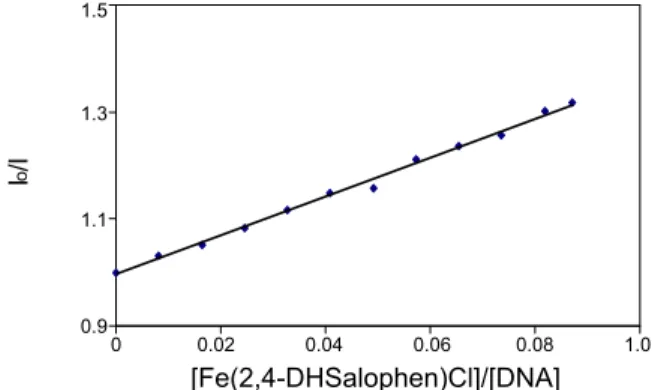
Fig. 4. The Stern-Volmer plot for quenching of EB bound to DNA by Fe(2 ,4-DHsalophen)Cl complex.
can be determined by analysis of absorption spectrophoto- metric titration data at room temperature using Eq. (1):14
) (
1 )
( ] [ ) (
] [
f b app f
b T f
app T
K DNA DNA
ε ε ε
ε ε
ε + −
= −
− (1)
Where [DNA]T,εapp,εf and εb correspond to total concen- tration of ct-DNA, Aobsd/[Schiff base], the extinction coeffici- ent for the free Schiff base and the extinction coefficient for the Schiff base in the fully bound form, respectively. In the plot of [DNA]T/(|εapp-εf|) versus [DNA]T that is shown in Fig. 2, Kapp is given by the ratio of the slope to the intercept.
The apparent binding constant of Fe(2,4-DHSalophen)Cl was determined by (1.6 ± 0.2) × 103 M-1.
Fluorescence spectroscopic studies
Ethidium bromide (EB) emits intense fluorescence light in the presence of DNA, due to its strong intercalation bet- ween the adjacent DNA base pairs. As it has been reported, the enhanced fluorescence can be quenched by the addition of a second molecule.15,16 In fluorescence titration experi- ment, the specified volumes of concentrated Fe(2,4-DHSalo- phen)Cl solution were added consecutively to the cell which contained 2000 µL DNA (12 µM) and EB (5 µM) solution.
Representative emission spectra of EB-DNA solution in the presence of various amounts of Fe(2 ,4-DHSalophen)Cl is shown in Fig. 3.
The pecks area was measured and analyzed using the classical Stern-Volmer equation Eq. (2):17
Io/I =1+Kr (2)
Where I0 and I are the fluorescence area peck in the ab- sence and the presence of complex, respectively, K is a linear Stern-Volmer quenching constant and r is the ratio of total concentration of complex to that of ct-DNA. The quen- ching extent of fluorescence of EB bound to ct-DNA was used to determine the extent of binding between the second molecule and ct-DNA. The Stern-Volmer plot for fluores- cence quenching of EB bound to ct-DNA by the Schiff base is shown in Fig. 4.The quenching plots illustrate that the quenching of EB bound to ct-DNA by the Schiff base is in good agreement with the linear Stern-Volmer equation which confirms the Schiff base bind to DNA. In the plot of I0/I versus [Schiff base]/[DNA], K is calculated by the ratio of the slope to intercept. The K value for Fe(2,4-DHSalophen)Cl was 6.2 The relative value of K compared with other known intercalators represents the non-intercalation mode for Fe (2,4-DHsalophen)Cl.13,18
20 40 60 80
T (oC)
Abs (259 nm)
2.2 2.0 1.8 1.6 1.4 1.2 1.0
Fig. 5. Thermal denaturation curves of DNA (10.0 × 10⁻⁵ M) in the various [Fe(2,4-DHsalophen)Cl]/[DNA] molar ratios of r1 = 0.0 (■), r2 = 0.2(▲) and r3 = 1.0(●). Measurements were done in 10 mM Tris/HCl buffer, pH 7.2 and at 25 ºC.
0 0.5 1 1.5 2
[Fe(2,4-DHSalophen)Cl]/[DNA]
Relative Viscosity
1.1 1.05
1
0.95
0.9
Fig. 6. Relative viscosity of calf thymus DNA (7.8 × 10⁻⁵ M) in Tris-HCl 10 mM in the presence of increasing amounts of Fe(2,4- DHSalophen)Cl at stoichiometric ratios r = [Fe(Salen)Cl]/[DNA- phosphate] = 0.0 - 2.0, plotted as (η/η o)1/3 vs. r.
Thermal denaturation experiments
When a molecule intercalates to ct-DNA, the stability of the helix increases and the temperature at which the helix denatures (Tm) goes up varies around 5 - 12 oC in r = 1.19,20 Thus, this parameter is most useful in analyzing the mode of interaction. The melting plot of ct-DNA was monitored by plotting the variation of maximum absorption of ct-DNA solution (100 µM) at 258 nm vs. temperature. The denatura- tion curves were measured at various [Fe(2,4-DHSalophen) Cl]/[DNA] molar ratios and is shown in Fig. 5.
The melting temperature of ct-DNA (Tm) was estimated from the midpoint of transition curves. With respecting to the results, the melting temperature of ct-DNA has been in- creased about 4.3 oC in the presence of Fe (2,4-DHSalophen) Cl. The observed small change in the Tm of ct-DNA in the presence of Fe (2,4-DHSalophen)Cl suggests that the interac- tion of these compounds with ct-DNA does not involve in- tercalation between the base pairs and could be ascribed to
interactions with the DNA grooves.21 Viscosity measurements
Optical or photo physical probes generally provide nece- ssary, but not sufficient, clues to support kind of binding model. Hydrodynamic measurements that are sensitive to length increases (i.e. viscosity, sedimentation, rotational diffusion as measured by transient electric diffusion) are the least ambiguous and most critical tests of the binding models in solution. The DNA helix lengthens as the base pairs are separated to accommodate the bound complex for the groove binding of the molecule, leading to lower increase in DNA viscosity. Hydrodynamic methods are thus suitable to detect such changes and, in the absence of crystallogra- phic structural data, provide essential evidence to support the intercalation model. In contrast, partial and/or nonclassi- cal intercalation of complex could bend (or kink) the DNA helix, reduce its effective length and in turn, its viscosity.
The effect of Fe(2,4-DHsalophen)Cl on the viscosity of DNA is shown in Fig. 6 The relative viscosity of DNA shows small rises with increase in the concentration of the Fe (2,4-DHsalophen)Cl, which is not similar to that of the classi- cal intercalation (i.e. ethidium bromide).22,23 The viscosity results unambiguously show that Fe(2,4-DHsalophen)Cl bind with DNA by groove binding mode, these results are in agreement with optical absorption experiments.
CONCLUSIONS
The results obtained from fluorescence, UV-vis, thermal denaturation and viscosity measurements exclude DNA out- side groove binding. From spectrophotometric titration ex- periments, the binding constants of Fe(2,4-DHSalophen)Cl with ct-DNA were found to be (1.6 ± 0.2) ×103 M-1. These results are consistent with a binding mode dominated by interactions with the groove of ct-DNA, analogously to what reported for a number of porphyrazines and metal-porphy- razine complexes interacting with ct-DNA.18
REFERENCES
1. Metcalfe, C.; Thomas, J. A. Chem. Soc. Rev. 2003, 32, 215.
2. Isse, A.; Gennaro, A.; Vianello, E. J. Electroanal. Chem. 1998, 444, 241.
3. Ramnath, R.; Al-Junaid, S.; Motevalli, M.; Parkin, B. C.; Sulli- van, A. C. Inorg. Chem. 2004, 43, 4072.
4. Chen, D.; Martell, A. E.; Sun, Y. Inorg. Chem. 1989, 28, 2647.
5. Haikarainen, A.; Sipila, J.; Pitkanen, P.; Pajunen, A.; Muti- kainen, I. Med. Chem. 2001, 9, 1633.
6. García, M.; Lorenzo, T.; Pariente, E. Biosensors and Bioelec- tronics 2007, 22, 2675.
7. Silvestri, A.; Barone, G.; Raisi, G.; Giudice, M.; Tumminello, S. Inorg. Biochem. 2004, 98, 589.
8. Marmur, J. J. Mol. Biol. 1961, 3, 208.
9. Duty, P.; Rice, S. A. Biochim. Biophys. Acta 1955, 16, 446.
10. Bailes, R. H.; Calvin, M. J. Am. Chem. Soc. 1947, 69, 1886.
11. Banville, D. L.; Marzilli, L. G.; Strickland, J. A.; Wilson, W.
D. J. Biopolymers. 1986, 25, 1837.
12. Gray, T. A.; Yue, K. T.; Marzilli, L. G. J. Inorg. Biochem. 1991, 41, 205.
13. Azani, M. R.; Hassanpour, A,; Bordbar, A, K,; Mirkhani, V, Bull. Korean Chem. Soc. 2009, 30, 1973.
14. Meehan, T.; Gamper, H.; Becker, J. F. J. Biol. Chem. 1982, 257, 10479.
15. Bagley, B. C.; Lebret, M. Biochemistry 1984, 23, 937.
16. Lowicz, J. R.; Webber, G. Biochemistry 1973, 12, 4161.
17. Cramb, D. T.; Beck, S. C. J. Photochem. Photobiol. A 2000, 134, 87.
18. Kang, J.; Wu, H.; Lu, X.; Wang, Y.; Zhou, L. Spectrochimica Acta A 2005, 61, 2041.
19. Kuswandi, B.; Tombelli, S.; Marazza, G.; Mancini, M. Chimia.
2005, 59, 236.
20. Wu, H. L.; Li, W. Y.; Miao, K.; He, X. W.; Liang, H. Spectrosc.
Lett. 2002, 35, 781.
21. Nair, R. B.; Teng, E. S.; Kirkland, S. L.; Murphy, C. J. Inorg.
Chem. 1998, 37, 139.
22. Cory, M.; Mckee, D. D.; Kagan, J.; Henry, D. W.; Miller, J. J.
Am.Chem. Soc. 1985, 107, 2528.
23. Waring, M. J. Mol. Biol. 1965, 13, 269.
![Fig. 5. Thermal denaturation curves of DNA (10.0 × 10 -5 M) in the various [Fe(2,4-DHsalophen)Cl]/[DNA] molar ratios of r 1 = 0.0 (■), r 2 = 0.2(▲) and r 3 = 1.0(●)](https://thumb-ap.123doks.com/thumbv2/123dokinfo/5299577.154676/5.892.95.420.145.366/thermal-denaturation-curves-various-dhsalophen-dna-molar-ratios.webp)
