대흉외지 2009;42:361-363 □ 증례보고 □
− 361 −
*조선대학교병원 흉부외과
Department of Thoracic and Cardiovascular Surgery, Chosun University Hospital 논문접수일:2008년 10월 6일, 심사통과일:2008년 10월 31일
책임저자:서홍주 (501-717) 광주시 동구 서석동 588번지, 조선대학교병원 흉부외과 (Tel) 062-220-3160, (Fax) 062-220-1444, E-mail: [email protected] 본 논문의 저작권 및 전자매체의 지적소유권은 대한흉부외과학회에 있다.
선천성 사엽성 반월형 판막
− 1예 보고 −
서 민 범*ㆍ서 홍 주*
Congenital Quadricuspid Semilunar Valve
A case report
Min Bum Seo, M.D.*, Hong-Joo Seo, M.D.*
A 17-year-old male patient was referred with symptoms of dyspnea. Multi-detector computerized tomography (MDCT) and echocardiography evaluation revealed quadricuspid aortic and pulmonary valves, an atrial septal defect (ASD), and pulmonary stenosis. We closed the ASD using a bovine patch and performed a commissurotomy of the pul- monary valve. Quadricuspid semilunar valves are very rare congenital abnormalities that are reported to occur nine times more frequently in the pulmonic valve than in the aortic valve. According to the Hurwitz and Roberts classi- fication, the aortic valve was type A, and the pulmonic valve was type B. The aortic valve had normal function, but the pulmonic valve was stenotic and had abnormal function.
(Korean J Thorac Cardiovasc Surg 2009;42:361-363) Key words: 1. Valve disease
2. Pulmonary valve, stenosis 3. Heart valve, abnormalities
CASE REPORT
A 17-year-old male patient was referred with dyspnea which was aggrevated 3 months ago. A past history of men- tal retardation and polydactyly of the right foot were notable without the presence of chromosomal abnormality such as Down syndrome. The patient was diagnosed as having con- genital heart disease, 14 years ago without proper treatment.
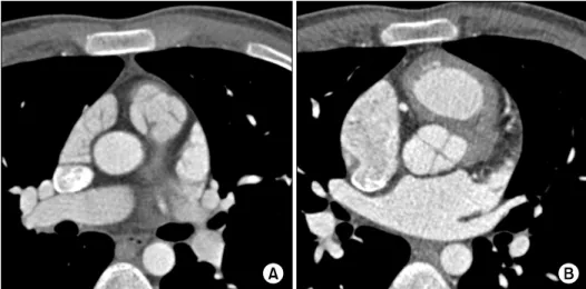
The patient first came to our hospital two years ago with symptoms of dyspnea and was diagnosed with ASD, thicken- ing of pulmonary valve, hypertrophy of the right ventricle, pulmonary hypertension and quadricuspid pulmonary and aortic valves through MDCT (Fig. 1).
On physical examination, systolic ejection murmur was auscultated on the left upper parasternal border, and there were no specific findings except right ventricle hypertrophy on electrocardiography. Mild hypoxemia was detected by ar- terial blood gas analysis; pH 7.423, pCO
244.0 mmHg, pO
270.5 mmHg, and sPO
294.4%. Right atrial and ventricular hy-
pertrophy, atrial septal defect, and pulmonary stenosis were
detected by echocardiography and maximal pressure gradient
between right ventricle and main pulmonary artery estimated
by Doppler echocardiography was 64 mmHg. Pulmonary val-
vular motion was restricted and the pressure gradient between
right ventricle and main pulmonary artery was 60 mmHg by
right atrial angiography. After diagnosing ASD and pulmo-
대흉외지
2009;42:361-363
− 362 −
Fig. 1. Multi-detector computerized tomogram (MDCT) image showing both pulmonary (A) and aortic (B) quadricuspid valves.
Fig. 2. Operative findings show quadricuspid pulmonary valve and 3-fused commissures. Commissurotomy of two site of the 3-fused commissures (arrow) was done.
nary stenosis, operation under routine cardiopulmonary bypass was performed.
After cardiopulmonary bypass, incisions to the pulmonary arteries were made. Quadricuspid pulmonary valve was con- firmed under direct surgeon’s visual field. Three parts of four commissures were fused. When performing commissurotomy on all three fused commissures, pulmonary regurgitation can occur. Therefore, commissurotomy was done on the left two commissures with 3 mm incision on each valve (Fig. 2).
Afterwards right atriotomy and ASD closure to the verified 12 mm-size ASD was done using the bovine pericardial
patch. The patient was discharged from the hospital on the seventh postoperative day without any complications. The aortic valve and pulmonary valve shows no abnormality on echocardiography at 11 months follow up after discharge.
DISCUSSION
Quadricuspid semilunar valves are very rare congenital malformations. To our knowledge, this is the first reported case of concomitant quadricuspid aortic and pulmonary valves.
This abnormality appears to be due to abnormal process of truncus arteriosus dividing into aorta and pulmonary trunk during embryological development of the heart. Incidence of Quadricuspid pulmonary valve is 0.02∼0.12% and that of quadricupid aortic valve is 0.008∼0.043%, the former occurs about 9 times more often[1-5].
Developmental mechanism of quadricuspid valves had not been understood exactly, but Fernandez et al. explained the occurrance of quadricuspid valves by a hypothesis using em- bryo of a Syrian hamster. Normal aortic valve originates from three mesodermal primordial cell differentiation and one of these three mesodermal cells is detached by invagination of endothelial cell at valve development period, so this process results in the formation of quadricuspid valves[4].
Quadricuspid semilunar valves were classified into 7 types
by Hurwitz and Roberts in 1973. Among the 121 cases, the
majority of quadricuspid pulmonary valve was type B in 72
cases (60%)[2]. In this case, 4 cusps of aortic valve were
same-sized, so called type A, pulmonary valve showed 3
서민범ㆍ서홍주