2003;9:1785-1791.
Clin Cancer Res
Jean-Charles Soria, Chulso Moon, Bonnie L. Kemp, et al.
Cancer
Outcome in Patients with Stage I Non-Small Cell Lung
Lack of Interleukin-10 Expression Could Predict Poor
Updated version
http://clincancerres.aacrjournals.org/content/9/5/1785
Access the most recent version of this article at:
Cited Articles
http://clincancerres.aacrjournals.org/content/9/5/1785.full.html#ref-list-1
This article cites by 28 articles, 18 of which you can access for free at:
Citing articles
http://clincancerres.aacrjournals.org/content/9/5/1785.full.html#related-urls
This article has been cited by 4 HighWire-hosted articles. Access the articles at:
E-mail alerts
Sign up to receive free email-alerts
related to this article or journal.
Subscriptions
Reprints and
.
[email protected]
Department at
To order reprints of this article or to subscribe to the journal, contact the AACR Publications
Permissions
.
[email protected]
Department at
Lack of Interleukin-10 Expression Could Predict Poor Outcome in
Patients with Stage I Non-Small Cell Lung Cancer
1
Jean-Charles Soria, Chulso Moon,
Bonnie L. Kemp, Diane D. Liu, Lei Feng,
Ximing Tang, Yoon-Soo Chang, Li Mao, and
Fadlo R. Khuri
2Departments of Thoracic/Head and Neck Medical Oncology [J-C. S., C. M., X. T., Y-S. C., L. M.], Pathology [B. L. K.], and Biostatistics [D. D. L., L. F.], The University of Texas M. D. Anderson Cancer Center, Houston, Texas 77030, and Winship Cancer Institute, Emory University, Atlanta, Georgia 30322 [F. R. K.]
ABSTRACT
Purpose: Interleukin-10 (IL-10) may play an important
role in controlling tumor growth and metastasis. Some re-ports have shown that IL-10 can be a potent inhibitor of tumor growth, but others suggest that IL-10 expression by the tumor is an adverse prognostic factor. Because normal bronchial epithelial cells constitutively produce IL-10, we decided to test the prognostic value of IL-10 in a well-defined population of patients with stage I non-small cell lung cancer (NSCLC) treated in a single institution.
Patients and Methods: Using immunohistochemical
analysis, we retrospectively analyzed IL-10 expression in specimens from 138 patients with completely resected clin-ical/radiographic stage I NSCLC for whom clinical follow-up data were available.
Results: IL-10 expression was retained (IL-10 labeling
index > 10%) in 94 patients (68.1%) and lost in 44 patients (31.9%). The duration of overall, disease-specific, and disease-free survival in the 44 patients lacking IL-10 expres-sion was worse than in the 94 patients with IL-10 expresexpres-sion (Pⴝ 0.08, 0.02, and 0.05, respectively; Log-rank test). In-terestingly, IL-10 expression was observed more frequently in tumors with squamous cell histology than in tumors of other histological subtypes (Pⴝ 0.04; 2
test). Multivariate analysis confirmed the independent prognostic value of IL-10 expression for disease-specific survival (Pⴝ 0.04).
Conclusion: Lack of IL-10 expression by the tumor was
associated with a significantly worse outcome of early stage NSCLC. The mechanisms underlying this clinically and bi-ologically important finding need to be further explored.
INTRODUCTION
Lung cancer is a major cause of mortality worldwide. Last year, in the United States alone, an estimated 169,400 new cases of lung cancer were diagnosed, and an estimated 154,900 deaths from lung cancer occurred (1). Improving the survival rate of patients with this disease requires a better understanding of tumor biology and the subsequent development of novel thera-peutic strategies. One area of intense lung cancer research has been in assessing the prognostic factors of NSCLC,3
focusing on stage I disease and molecular factors (2–5). This avenue of investigation may lead to the identification of patients with the highest risk stage I NSCLC or of those who are most likely to benefit from adjuvant or chemopreventive approaches.
IL-10 is thought to play a potential pathogenic or thera-peutic role in a number of human conditions, such as inflam-mation, autoimmunity, and cancer (6). The immunomodulatory effects of IL-10 have yielded mixed results in various tumor systems. On one hand, because many tumor types express IL-10, its role in helping tumors evade immunosurveillance has been suggested (7, 8). IL-10 inhibits the tumoricidal capacity of macrophages and the cytotoxicity and cytokine production of tumor-specific T cells and blocks the presentation of tumor antigens by antigen-presenting cells (9, 10). On the other hand, in vivo studies in different animal models have demonstrated that IL-10 is a potent inhibitor of tumor growth and metastasis (11–14). Additionally, systemic administration of IL-10 has inhibited tumor metastasis and stimulated antitumor immune responses in murine models (15). Nevertheless, recent data generated by analyzing human lung tissue samples suggest that IL-10 produced by NSCLC is a predictor of poor outcome (16). Because IL-10 is constitutively expressed in normal bron-chial epithelial cells, we hypothesized that loss of IL-10 expres-sion by lung tumors might be a prognostic factor for survival. Therefore, we decided to analyze the prognostic value of IL-10 expression in a homogeneous population of 138 patients with stage I NSCLC.
PATIENTS AND METHODS
Study Population. A total of 595 consecutive patients with stage I NSCLC underwent definitive surgical resection, defined as a lobectomy or a pneumonectomy, from 1975 to 1990
Received 8/12/02; revised 1/22/02; accepted 1/28/03.
The costs of publication of this article were defrayed in part by the payment of page charges. This article must therefore be hereby marked
advertisement in accordance with 18 U.S.C. Section 1734 solely to
indicate this fact.
1Supported by Biology, Education, Screening, Chemoprevention, and
Treatment Lung Cancer Program, Department of Defense Grant DAMD17-01-1-0689-1 (to F. R. K. and L. M.), Cancer Center Grant P30 CA 16620 (to M. D. Anderson Cancer Center), Tobacco Research Fund from the State of Texas (to M. D. Anderson Cancer Center), American Cancer Society Grant RPG-98-054 (to L. M.), and Fondation de France, AP-HP, and Lilly Foundation Grant (to J-C. S.).
2To whom requests for reprints should be addressed, at Winship Cancer
Institute, 1365 Clifton Road, Suite B4100, Atlanta, GA 30322. Phone: (404) 778-4250; Fax: (404) 778-5016; E-mail: [email protected].
3The abbreviations used are: NSCLC, non-small cell lung cancer; SCC,
squamous cell carcinoma; TNM, Tumor-Node-Metastasis; CI, confi-dence interval; IL, interleukin; TIMP, tissue inhibitor of metallopro-tease; NK, natural killer; MMP, matrix metalloprotease.
at The University of Texas M. D. Anderson Cancer Center. We retrospectively examined 138 cases for which both tissue sam-ples and a median follow-up period of⬎5 years were available at the time of this study. The patient population was identified through a search of the Tumor Registry database maintained by the Department of Medical Informatics at The University of Texas M. D. Anderson Cancer Center. Survival status was ver-ified and updated from Tumor Registry records as of December 1, 2000.
The study population consisted of 106 men and 32 women; 120 patients were Caucasian, and 18 patients were of other ethnicities (Table 1). The mean age of patients at surgery was 63 years. Histological subtypes included 58 cases of SCC, 60 cases of adenocarcinoma, 10 cases of bronchioalveolar carcinoma, 5 cases of large cell carcinoma, 3 cases of adenosquamous carci-noma, and 2 cases unclassified.
Immunohistochemical Staining for IL-10 Protein. All available tissue blocks from each patient were reviewed for the presence of tumor by a thoracic pathologist (B. L. K.). Paraffin-embedded, 4-m-thick tissue sections from all 154 primary tumors were stained for IL-10 protein using a primary goat polyclonal antihuman IL-10 antibody (AF-217-NA; R&D Sys-tems, Minneapolis, MN). Slides were deparaffinized through a series of xylene baths. Rehydration was performed using graded alcohol. The sections were then immersed in methanol contain-ing 0.3% hydrogen peroxidase for 20 min to block the endog-enous peroxidase activity and incubated in 2.5% blocking serum to reduce nonspecific binding. Sections were incubated over-night at 4°C with primary anti-IL-10 antibody at a dilution of 1:75 (1.33 g/ml). The sections were then processed using a standard avidin-biotin immunohistochemical assay according to the manufacturer’s recommendations (Vector Laboratories,
Bur-lingame, CA). Diaminobenzidine was used as a chromogen, and commercial hematoxylin was used for counterstaining. Rou-tinely processed tissue sections of normal lymph nodes and tonsils were used as positive staining controls and also stained with the primary antibody omitted to confirm staining specific-ity. Normal bronchial epithelial cells that constitutively produce IL-10 were also used as internal positive controls (17).
The IL-10 labeling index was defined as the percentage of tumor cells displaying cytoplasmic immunoreactivity and cal-culated by counting IL-10-stained tumor cells among ⱖ1000 tumor cells for each section. Representative areas of each tissue section were selected, and cells were counted in at least four fields (magnified 400 times) in these areas. On the basis of previous reports, ifⱖ10% of the tumor cells were positive for IL-10, the case was considered to be IL-10 positive (18, 19). Tumors with⬍10% of the cells stained were counted as nega-tive. All slides were scored concomitantly by a pathologist (X. T.) and another investigator (J-C. S.). Immunohistochemical analysis was performed in a blinded manner with respect to clinical information about the subjects.
Statistical Analysis. Overall, disease-specific, and disease-free survival were analyzed in this study. Survival curves were estimated by the Kaplan-Meier method. The Log-rank test was used to compare patient’s survival time between groups. The Fisher exact and2
tests were used to analyze the association between two categorical variables. The Wilcoxon rank-sum test was used for differences in median of age. The Cox proportional hazards model was used for univariate analy-sis to evaluate the association between survival time and risk factors and for multivariate analysis to model the risks of IL-10 expression on survival time, with adjustment for clinical and histopathological parameters (age, sex, race, tumor histology,
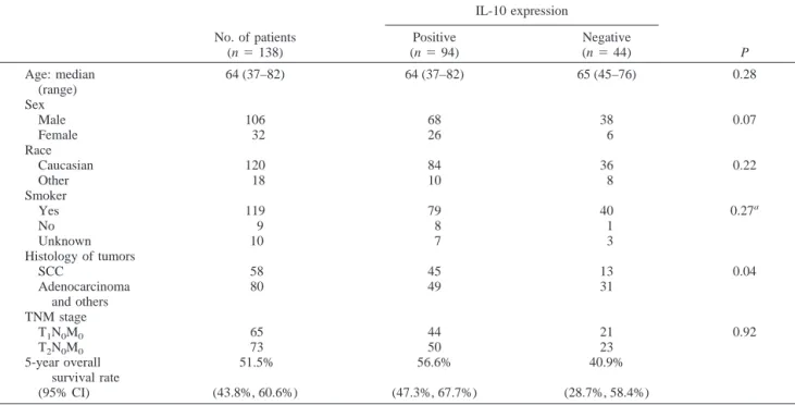
Table 1 IL-10 status in stage I NSCLC tumors according to clinicopathological features of patients
No. of patients (n⫽ 138) IL-10 expression P Positive (n⫽ 94) Negative (n⫽ 44) Age: median (range) 64 (37–82) 64 (37–82) 65 (45–76) 0.28 Sex Male 106 68 38 0.07 Female 32 26 6 Race Caucasian 120 84 36 0.22 Other 18 10 8 Smoker Yes 119 79 40 0.27a No 9 8 1 Unknown 10 7 3 Histology of tumors SCC 58 45 13 0.04 Adenocarcinoma and others 80 49 31 TNM stage T1N0M0 65 44 21 0.92 T2N0M0 73 50 23 5-year overall survival rate 51.5% 56.6% 40.9% (95% CI) (43.8%, 60.6%) (47.3%, 67.7%) (28.7%, 58.4%)
aP calculated comparing smoking vs. nonsmoking patients. 1786 IL-10 Expression As a Prognostic Marker for NSCLC
and tumor size). All statistical tests are two sided, and a P ⬍ 0.05 was considered to be statistically significant.
All survival curves were calculated from the date of sur-gery. Overall survival took all deaths (cancer related or not) into account. Disease-specific survival time was calculated from the date of surgery to death from cancer-related causes. Disease-free survival time was calculated from the date of surgery to relapse or death from cancer-related causes.
RESULTS
A total of 138 formalin-fixed, paraffin-embedded NSCLC tumor specimens was stained using a standard immunohisto-chemical technique reported previously for the identification of IL-10 expression (18, 19). The usual pattern of positive staining for IL-10 in NSCLC was cytoplasmic and not nuclear (Fig. 1, A–C). Even if tumors cells were negative for IL-10 staining, normal bronchial epithelial cells in the section were positive and used as an internal positive control of the staining for IL-10. In peribronchial gland cells or alveolar pneumocytes, IL-10 ex-pression was not detectable. Lymphoid cells of tumor areas were occasionally immunostained. Only 20 of 138 samples displayed tumor-infiltrating lymphocytes, therefore hindering any relevant analysis of IL-10 production by infiltrating immune cells. In the positive control tissues (tonsil), the normal stratified squamous
epithelium displayed IL-10-positive cells. In the adjacent lymph nodes, IL-10-positive cells were localized predominantly in the germinal centers (data not shown). IL-10 immunohistochemical staining showed a wide heterogeneity from rare scattered cells to a homogeneous pattern for the vast majority of cells exam-ined, suggesting that phenotypic heterogeneity is a major feature in NSCLC (Fig. 1).
IL-10 expression was observed in 94 (68.1%) of the 138 stage I NSCLC specimens. Lack of staining was observed in 44 tumors (31.9%). Table 1 shows the relationships between the expression of IL-10 and clinicopathological factors. There were no statistically significant differences in TNM stage, sex, smok-ing status, age, and race between the groups with IL-10-positive and -negative staining. Interestingly, IL-10 expression was more prevalent in the SCC subtype than it was in other histological subtypes. Forty-five (77.6%) of the 58 cases of SCC exhibited IL-10 expression, whereas 49 (61.3%) of 80 patients with non-SCC tumors (mainly adenocarcinoma) showed IL-10 expression (P⫽ 0.04; 2
test).
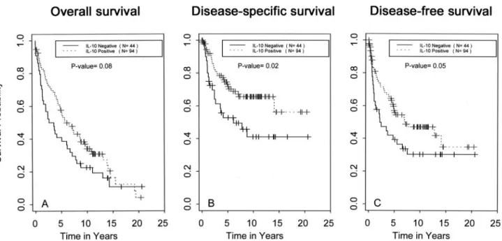
We subsequently analyzed the relationship between IL-10 expression and length of survival. The median follow-up time for the patient population was 10.6 years. Fig. 2A shows the Kaplan-Meier overall survival curves for patients whose tumors were IL-10 positive and negative. Patients with tumors that were
Fig. 1 Immunohistochemical staining patterns of IL-10 in stage I NSCLC. A, a well-differentiated adenocarcinoma with most cancer cells expressing
IL-10 in the cytoplasm. B, a SCC with most carcinoma cells positive for IL-10. C, a bronchioalveolar carcinoma tumor with IL-10 expression. D, a SCC tumor negative for IL-10 expression (original magnification,⫻400).
IL-10 negative had a shorter survival time than did patients with tumors that were IL-10 positive (P ⫽ 0.08; Log-rank test). Five-year overall survival rate for patients whose tumors were IL-10 positive was 56.6% (95% CI⫽ 47.3–67.7%) and 40.9% (95% CI⫽ 28.7–58.4%) for patients whose tumors were IL-10 negative (Table 1). Fig. 2B shows that patients with negative IL-10 expression had significantly shorter disease-specific sur-vival times than did patients with positive IL-10 expression (P⫽ 0.02; Log-rank test). A comparison of disease-free sur-vival curves in IL-10-negative and -positive patients yielded similar results (P⫽ 0.05; Log-rank test; Fig. 2C). The prognos-tic significance of IL-10 expression on disease-specific survival
was further explored in each major histological subtype. IL-10 negativity was a borderline significant adverse prognostic factor among patients with non-SCC tumors (P⫽ 0.06; Log-rank test; Fig. 3A). A similar trend was observed for patients with SCC of the lung, but this trend was not significant (P⫽ 0.25; Log-rank test; Fig. 3B). Univariate Cox proportional hazards model was used to evaluate the association between IL-10, clinicopatho-logical variables (age, sex, race, histoclinicopatho-logical subtype, and TNM), and survival time. Table 2 shows the results on disease-specific survival. In multivariate Cox proportional hazards model, among all clinicopathological variables, IL-10 expres-sion was the only significant independent prognostic indicator
Fig. 2 Survival curves of patients with IL-10-positive and -negative NSCLC. The patients lacking IL-10 expression (solid line, n⫽ 44) had worst
outcomes than the patients with IL-10 expression (broken line, n⫽ 94) for overall (A), disease-specific (B), and disease-free survival (C).
Fig. 3 A, disease-specific
sur-vival curve of patients with stage I SCC according to IL-10 expression; B, disease-specific survival curve of patients with adenocarcinoma or other histo-logical subtypes according to IL-10 expression.
for disease-specific survival. The hazard of cancer death for patients whose tumor was IL-10 positive was only 55% of the hazard for patients whose tumor was IL-10 negative (P⫽ 0.04, Cox model).
DISCUSSION
Human lung cancer displays an extremely aggressive clin-ical course and represents the leading cause of malignancy-related mortality in the United States (1). This behavior may reflect an increased capacity to evade detection and containment by host immune response. Because IL-10 demonstrates in vitro immunosuppressive activities (9, 10), some groups have hypoth-esized that IL-10 production by cancer cells may help the tumor evade immunosurveillance (7, 8). Nevertheless, IL-10 is also able to inhibit tumor growth and metastasis in various tumor models (11–14). These conflicting results imply that it is all about “fine tuning” in case of IL-10-mediated immunosuppres-sion or immunostimulation. Because normal bronchial epithelial cells constitutively express IL-10 (17), loss of IL-10 expression by lung cancer cells would represent a specific change in the tumor as compared with its normal epithelial counterpart. In the present study, we explored the prognostic value of IL-10 ex-pression by lung cancer cells in a large and homogeneous population of 138 completely resected clinical/radiographic stage I NSCLC for whom a median follow-up of 10.6 years was available. We have demonstrated that IL-10 is retained in a significant percentage of stage I NSCLCs. Overall, 94 (68.1%) of 138 tumors expressed IL-10 inⱖ10% tumor cells, whereas loss of IL-10 expression was observed in 44 patients (31.9%). Our data show that lack of IL-10 expression is a poor prognostic factor in patients with stage I NSCLC. The poor prognostic value of lack of IL-10 expression was observed for disease-specific and -free survival, with a trend for overall survival. Furthermore, multivariate analysis confirmed the independent prognostic value of lack of IL-10 expression. The prognostic value of IL-10 was retained even when we changed the cutoff level of positivity from 10% (18) to 5 or 15%. Our results are in contrast to a previous report by Hatanaka et al. (16), who suggested that IL-10 expression by the tumor was an indicator of poor prognosis. We have analyzed IL-10’s prognostic value in a large and homogeneous population of patients with early stage lung cancer (n⫽ 138), whereas Hatanaka et al. performed their analysis using a smaller and more heterogeneous popula-tion that included 82 patients with stage I–IIIb disease. Further-more, the fact that all of the patients in our study were treated at a single institution and received lengthy follow-up care after
surgery helps to increase the credibility of our survival analysis. Finally, Hatanake et al. (16) evaluated IL-10 expression by RT-PCR as opposed to immunohistochemistry in this study. Thus, our different results may be related in part to differences in patient population and the technique used to evaluate IL-10 expression. Indeed, we evaluated IL-10 expression at the protein level as compared with the mRNA level for Hatanake et al. Furthermore, both studies were retrospectively conducted and therefore potentially subject to some degree of selection bias.
We have analyzed IL-10 expression by performing immu-nohistochemical analysis with a polyclonal antihuman IL-10 antibody reported previously (18, 19). Other anti-IL-10 antibod-ies have also been used to evaluate IL-10 expression in paraffin-embedded tissue sections (17, 20). A good concordance between reverse transcription-PCR analysis and immunohistochemical analysis for IL-10 has been suggested in different reports (7, 17). We have used internal and external positive controls to assess the specificity of the staining. We found that even if tumor cells were negative for IL-10 staining, normal bronchial epithelial cells in the section (when present) were positive (17), thus ruling out a false negative result.
Although the mechanisms underlying the current data are not clear, there are several potential explanations for the poor outcome of patients with IL-10-negative tumors. Several labo-ratories have demonstrated that IL-10 is a potent inhibitor of tumor growth and metastasis in multiple animal models and tumor types, including melanoma, breast and prostate cancers, and Burkitt’s lymphoma (11, 13, 20, 21). In vivo, the effects of IL-10 may be multifold. They can be related to direct inhibition of IL-10 on the angiogenic process per se or indirectly by affecting the angiogenic capacity or signals from tumor and/or tumor-infiltrating cells. Compelling evidence indicates that the antiangiogenic effect of IL-10 results from the inhibition of angiogenic factor release and production by the tumor and/or stromal cells. IL-10 induces production of TIMP-1, an inhibitor of angiogenesis, and inhibits MMP-2 and MMP-9 secretion by cancer cell lines, blocking the induction of microvessel forma-tion in vitro (20, 22). It has also been suggested that IL-10 can directly inhibit endothelial cell response to angiogenic factors (21). Moreover, in murine mammary tumors, the antimetastatic and antitumor activity resulting from IL-10 gene transfer is related to enhanced production of nitric oxide (23).
One of the major roles of IL-10 in the regulation of im-mune responses involves its deactivating effect on macrophages (6). From the many cells and cell products within a tumor that serve as inducers or modulators of angiogenesis, macrophages
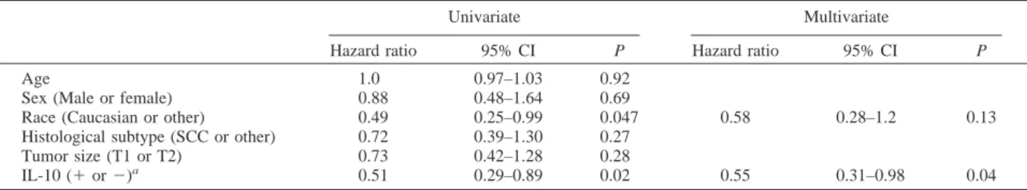
Table 2 Univariate and multivariate Cox proportional hazards model on disease-specific survival
Univariate Multivariate
Hazard ratio 95% CI P Hazard ratio 95% CI P
Age 1.0 0.97–1.03 0.92
Sex (Male or female) 0.88 0.48–1.64 0.69
Race (Caucasian or other) 0.49 0.25–0.99 0.047 0.58 0.28–1.2 0.13
Histological subtype (SCC or other) 0.72 0.39–1.30 0.27
Tumor size (T1 or T2) 0.73 0.42–1.28 0.28
IL-10 (⫹ or ⫺)a 0.51 0.29–0.89 0.02 0.55 0.31–0.98 0.04
have emerged as a major component. IL-10 secreted by the tumor cells may prevent the migration of macrophages from the periphery into the tumor tissue, thus preventing macrophage infiltration (24). IL-10 also inhibits the expression of angiogenic factors (vascular endothelial growth factor, IL-1, tumor necro-sis factor-␣, IL-6, and MMP-9) in tumor-associated macro-phages (25). These changes correlate with decreased neovascu-larization of the tumors. Alternatively, the inhibitory effect of IL-10 on tumor metastasis has been suggested to be mediated through a NK cell-dependent mechanism (6, 12). IL-10 is able to affect the activities of NK cells, and NK cells were recently shown to contribute to the antiangiogenic effects of IL-12 through the killing of endothelial cells (26).
The observation that IL-10 expression differs among his-tological subtypes highlights the biological differences among different subtypes of NSCLC. Different abnormalities in onco-genes and tumor suppressor onco-genes among histological subtypes of NSCLC are well known. Indeed, K-ras mutations are much more common in adenocarcinomas than in SCCs, whereas cy-clin B1 overexpression or the p53 mutant immunophenotype is more frequent in SCCs than in adenocarcinoma (2, 27, 28).
In conclusion, we found that a lack of IL-10 expression is a prognostic factor of poor outcome in stage I NSCLC. This result may be explained by the antitumor effects of IL-10, which contrast with the immunomodulatory effects that this cytokine displays in vitro. The mechanisms behind IL-10 antitumor ef-fects might include inhibition of angiogenesis, stimulation of TIMPs, inhibition of MMP secretion, and inhibition of macro-phage activity (20 –25). Nevertheless, this result needs to be interpreted with caution because of potential limitations in the present study: (a) IL-10 production by tumor-infiltrating lym-phocytes was not addressed because only a small fraction of our tissue samples displayed immune infiltrating cells; and (b) the role of IL-10 in cancer progression or regression might be very different according to the level of cytokine produced by the tumor and infiltrating immune cells. Additional studies are clearly required to confirm the present data and resolve the role of IL-10 in tumor growth and metastasis. We plan to conduct additional studies that will help in assessing the clinical impor-tance of the present IL-10 findings and in understanding their possible mechanisms. These studies will be conducted using resected tissue from patients with stage I NSCLC, analyze the expression of MMPs and TIMPs, microvessel density, and the presence of tumor-infiltrating lymphocytes and their phenotype, and relate these factors to IL-10 expression by the tumor and to overall prognosis.
ACKNOWLEDGMENTS
We thank Christophe Borg for helpful discussions, as well as Susan Cweren and Lakshmi Kakarala for technical assistance with immuno-histochemistry. We also thank Dawn Chalaire for editing this manu-script and Sandra Ideker for the artwork.
REFERENCES
1. Jemal, A., Thomas, A., Murray, T., and Thun, M. Cancer statistics, 2002. CA Cancer J. Clin., 52: 23– 47, 2002.
2. Soria, J. C., Jang, S. J., Khuri, F. R., Hassan, K., Liu, D., Hong, W. K., and Mao, L. Overexpression of cyclin B1 in early-stage
non-small cell lung cancer and its clinical implication. Cancer Res., 60: 4000 – 4004, 2000.
3. Zhou, X., Kemp, B. L., Khuri, F. R., Liu, D., Lee, J. J., Wu, W., Hong, W. K., and Mao, L. Prognostic implication of microsatellite alteration profiles in early-stage non-small cell lung cancer. Clin. Cancer Res., 6: 559 –565, 2000.
4. Khuri, F. R., Lotan, R., Kemp, B. L., Lippman, S. M., Wu, H., Feng, L., Lee, J. J., Cooksley, C. S., Parr, B., Chang, E., Walsh, G. L., Lee, J. S., Hong, W. K., and Xu, X. C. Retinoic acid receptor-beta as a prognostic indicator in stage I non-small cell lung cancer. J. Clin. Oncol., 18: 2798 –2804, 2000.
5. Khuri, F. R., Wu, H., Lee, J. J., Kemp, B. L., Lotan, R., Lippman, S. M., Feng, L., Hong, W. K., and Xu, X. C. Cyclooxygenase-2 overexpression is a marker of poor prognosis in stage I non-small cell lung cancer. Clin. Cancer Res., 7: 861– 867, 2001.
6. Howard, M., and O’Garra, A. Biological properties of interleukin 10. Immunol. Today, 13: 198 –200, 1992.
7. Smith, D. R., Kunkel, S. L., Burdick, M. D., Wilke, C. A., Orringer, M. B., Whyte, R. I., and Strieter, R. M. Production of interleukin-10 by human bronchogenic carcinoma. Am. J. Pathol., 145: 18 –25, 1994. 8. Young, M. R., Wright, M. A., Lozano, Y., Matthews, J. P., Bene-field, J., and Prechel, M. M. Mechanisms of immune suppression in patients with head and neck cancer: influence on the immune infiltrate of the cancer. Int. J. Cancer, 67: 333–338, 1996.
9. Rohrer, J. W., and Coggin, J. H. CD81 T cell clones inhibit antitumor T cell function by secreting IL-10. J. Immunol., 155: 5719 –5727, 1995. 10. Beissert, S., Hosoi, J., Grabbe, S., Asahina, A., and Granstein, R. D. IL-10 inhibits tumor antigen presentation by epidermal antigen present-ing cells. J. Immunol., 154: 1280 –1286, 1995.
11. Kundu, N., Beaty, T. L., Jackson, M. J., and Fulton, A. M. Anti-metastasic and antitumor activities of interleukin10 in a murine model of breast cancer. J. Natl. Cancer Inst. (Bethesda), 88: 536 –541, 1996. 12. Zheng, L. M., Ojcius, D. M., Garaud, F., Roth, C., Maxwell, E., Li, Z., Rong, H., Chen, J., Wang, X. Y., Catino, J. J., and King I. Interleu-kin-10 inhibits tumor metastasis through an NK cell-dependent mech-anism. J. Exp. Med., 184: 579 –584, 1996.
13. Huang, S., Xie, K., Bucana, C. D., Ullrich, S. E., and Bar-Eli, M. Interleukin 10 suppresses tumor growth and metastasis of human mel-anoma cells: potential inhibition of angiogenesis. Clin. Cancer Res., 2: 1969 –1979, 1996.
14. Giovarelli, M., Musiani, P., Modesti, A., Dellabona, P., Casorati, G., Allione, A., Consalvo, M., Cavallo, F., di Pierro, F., De Giovanni, C., Musso, T., and Forni, G. Local release of IL-10 by transfected mouse mammary adenocarcinoma cells does not suppress but enhances antitu-mor reaction and elicits a strong cytotoxic lymphocyte and antibody-dependent immune memory. J. Immunol., 155: 3112–3123, 1995. 15. Berman, R. M., Susuki, T., Tahara, H., Robbins, P. D., Narula, S. K., and Lotze, M. T. Systemic administration of cellular IL-10 induces an effective, specific, and long-lived immune response against established tumors in mice. J. Immunol., 157: 231–238, 1996. 16. Hatanaka, H., Abe, Y., Kamiya, T., Morino, F., Nagata, J., Toku-naga, T., Oshika, Y., Suemizu, H., Kijima, H., Tsuchida, T., Yamazaki, H., Inoue, H., Nakamura, M., and Ueyama, Y. Clinical implications of interleukin (IL)-10 induced by non-small cell lung cancer. Ann. Oncol.,
11: 815– 819, 2000.
17. Bonfield, T. L., Konstan, M. W., Burfeind, P., Panuska, J. R., Hilliard, J. B., and Berger, M. Normal bronchial epithelial cells consti-tutively produce the anti-inflammatory cytokine interleukin-10, which is downregulated in cystic fibrosis. Am. J. Respir. Cell Mol. Biol., 13: 257–261, 1995.
18. Huang, M., Wang, J., Lee, P., Sharma, S., Mao, J. T., Meissner, H., Uyemura, K., Modlin, R., Wollman, J., and Dubinett, S. M. Human non-small cell lung cancer cells express a type 2 cytokine pattern. Cancer Res., 55: 3847–3853, 1995.
19. Fujieda, S., Lee, K., Sunaga, H., Tsuzuki, H., Ikawa, H., Fan, G. K., Imanaka, M., Takenaka, H., and Saito, H. Staining of interleukin-10 predicts clinical outcome in patients with nasopharyngeal carcinoma. Cancer, 85: 1439 –1445, 1999.
20. Stearns, M. E., Garcia, F. U., Fudge, K., Rhim, J., and Wang, M. Role of interleukin 10 and transforming growth factor  1 in the angiogenesis and metastasis of prostate primary tumor lines from or-thotopic implants in severe combined immunodeficiency mice. Clin. Cancer Res., 5: 711–720, 1999.
21. Cervenak, L., Morbidelli, L., Donati, D., Donnini, S., Kambayashi, T., Wilson, J. L., Axelson, H., Castanos-Velez, E., Ljunggren, H. G., Malefyt, R. D., Granger, H. J., Ziche, M., and Bejarano, M. T. Abol-ished angiogenicity and tumorigenicity of Burkitt lymphoma by inter-leukin-10. Blood, 96: 2568 –2573, 2000.
22. Stearns, M. E., Fudge, K., Garcia, F., and Wang, M. IL-10 inhibi-tion of human prostate PC-3 ML cell metastases in SCID mice: IL-10 stimulation of TIMP-1 and inhibition of MMP-2/MMP-9 expression. Invasion Metastasis, 17: 62–74, 1997.
23. Kundu, N., Dorsey, R., Jackson, M. J., Guiterrez, P., Wilson, K., Fu, S., Ramanujam, K., Thomas, E., and Fulton, A. M. Interleukin-10 gene transfer inhibits murine mammary tumors and elevates nitric oxide. Int. J. Cancer, 76: 713–719, 1998.
24. Richter, G., Kruger-Kasagakes, S., Hein, G., Huls, C., Schmitt, E., Diamantstein, T., and Blankenstein, T. Interleukin-10 transfected into Chinese hamster ovary cells prevents tumor growth and macrophage infiltration. Cancer Res., 53: 4134 – 4137, 1993.
25. Di Carlo, E., Coletti, A., Modesti, A., Giovarelli, M., Forni, G., and Musiani, P. Local release of interleukin-10 by transfected mouse ade-nocarcinoma cells exhibit pro- and anti-inflammatory activity and re-sults in a delayed tumor rejection. Eur. Cytokine Netw., 9: 61– 68, 1998. 26. Yao, L., Sagadari, C., Furuke, K., Bloom, E. T., Teruya-Feldstein, J., and Tosato, G. Contribution of natural killer cells to inhibition of angiogenesis by IL-12. Blood, 93: 1612–1621, 1999.
27. Rossell, R., Li, S., Skacel, Z., Mate, J. L., Maestre, J., Canela, M., Tolosa, M., Tolosa, E., Armengol, P., Barnadas, A., and Ariza, A. Prognostic impact of mutated K-ras gene in surgically resected non-small cell lung cancer patients. Oncogene, 8: 2407–2412, 1993 28. Kishimoto, Y., Murakami, Y., Shiraishi, M., Hayashi, K., and Sekiya, T. Aberrations of the p53 tumor suppressor gene in human non-small cell carcinomas of the lung. Cancer Res., 52: 4799 – 4804, 1992.