https://doi.org/10.20307/nps.2021.27.1.18
18
Cytotoxic Constituents from the Stem Bark of Chisocheton pentandrus
Rurini Retnowati1,*, Hermin Sulistyarti1, Nikmatus Zahro Wahidah1, Anisa Lailatusy Syarifah2, Suprianto Salam3, Nurlelasari3, Agus Safari3, Desi Harneti3, Mulyadi Tanjung4, Ace Tatang Hidayat3,5, Rani Maharani3,5,
Unang Supratman3,5, and Yoshihito Shiono6
1Department of Chemistry, Faculty of Mathematics and Natural Sciences, Universitas Brawijaya, Malang 65145, Indonesia 2Academy of Pharmacy of Putra Indonesia, Malang 65123, Indonesia
3Department of Chemistry, Faculty of Mathematics and Natural Sciences,
Universitas Padjadjaran, Jatinangor 45363, Indonesia
4Department of Chemistry, Faculty of Science and Technology, Universitas Airlangga, Surabaya 60286, Indonesia 5Central Laboratory, Universitas Padjadjaran, Jatinangor 45363, Indonesia
6Department of Food, Life, and Environmental Science, Faculty of Agriculture,
Yamagata University, Tsuruoka, Yamagata 997-8555, Japan.
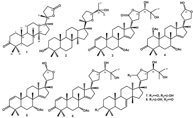
Abstract Eight cytotoxic constituents, consisting of six triterpenoids, cabralealactone (1), cabraleadiol (2), prototiamin A (3), 23-desmethyllimocin B (5), melianodiol (7) and indicalilacol (8) along with one limonoid, neemfruitins A (4) and one protolimonoid, protoxylocarpin G (6), were isolated from the extract of n-hexane of the stembark of Chisocheton pentandrus. The chemical structures were identified on the basis of spectroscopic evidence and compared to previously reported spectra. These isolated compounds appear for the first time in the plant. Compounds 1 - 8 were evaluated for their cytotoxic effect against MCF-7 breast cancer lines in vitro. Among the isolated compounds, melianodiol (7) showed the strongest cytotoxic activity with IC50 values of 16.8 M.
Keywords Chisocheton pentandrus, MCF-7 cell lines, Meliaceae, limonoid, triterpenoids
Introduction
The genus Chisocheton has approximately 150 species that are widely distributed throughout the tropical regions such as Thailand, Malaysia, and Indonesia.1,2 Many Chisocheton plants have been traditionally used by local people to treat various diseases,3,4 and previous studies on the chemical constituents of the this genus have led to the isolation and determination of a number of compounds, such as triterpenoids5-7 and limonoids.8-12 In addition, biological effects of the constituents have been reported, such as cytotoxic, anti-inflammatory, antifungal, antimalarial and antimycobacterial effects.2,8,13-15 During the course of our continuing search for anticancer substances from Indonesia Chisocheton plants,1,7,16,17 the methanol extract of the stem bark of Chisocheton pentandrus was found to inhibit significant cytotoxic against MCF-7 breast cancer
lines.
C. pentandrus is distributed in Sulawesi and Sumatera islands of Indonesia and previous investigation have led to the isolation of cytotoxic triterpenoids and limonoids.17,18 In the further search for cytotoxic constituent from this plant, we investigated the n-hexane extract of the bark of C. pentandrus and obtained six triterpenoids and two limonoids (Fig. 1). The cytotoxic effects of these compounds against MCF-7 breast cancer cells were evaluated. Here, we described the structural elucidation of the isolates and their cytotoxic activity.
Experimental
General experimental procedures The IR spectra were obtained in KBr on a SHIMADZU IR Prestige-2. Mass spectra were measured with a Water QTOF-HR-TOFMS-XEVotm mass spectrometer. NMR spectra was recorded using a JEOL ECZ-500 and ECZ-600 spectro-meter using tetramethyl silane (TMS) as an internal standard. Furthermore, chromatographic separations was carried out on silica gel 60 (70-230 mesh and 230-400 mesh, Merck). The PTLC glass plates were precoated
*Author for correspondence
Rurini Retnowati, Department of Chemistry, Faculty of Mathemat-ics and Natural Sciences, Universitas Brawijaya, Malang 65145, Indonesia
Tel: +62-341-361876; E-mail: [email protected]; [email protected]
with silica gel GF254 (Merck, 0.25 mm). Similarly, the TLC plates were precoated with silica gel GF254 (Merck, 0.25 mm), and the detection was carried out using 10% H2SO4 in ethanol, followed by heating.
Plant materials The stem bark of C. pentandrus was collected at Bogor Botanical Garden, West Java Province, Indonesia in June 2016. The plant was identified by the staff of the Herbarium Bogoriense, Bogor and an exhibition specimen (No. Bo-104) was deposited in the herbarium.
Extraction and isolation The dried stem bark of C. pentandrus (1.8 kg) was grounded to powder and extracted with methanol (4 × 4 L, 4 days each) at room temperature, and concentrated using a rotary evaporator, yielding a concentrated extract (340 g). Furthermore, about 300 g of the methanol extract was suspended in water (600 mL) and successively partitioned with n-hexane, ethyl acetate and n-butanol. The mixture was then evaporated under reduced pressure, resulting in 10.9, 25.2, and 228.6 g of crude extracts, respectively. In addition, 10.0 g of the n-hexane fraction was column chromatographed (CC) using silica gel (70-230 mesh), and eluted with n-hexane-ethyl acetate-methanol (10% stepwise), resulting to eight fraction groups (A-H).
Fraction B (439 mg) was subjected to silica gel CC (50 g, 230-400 mesh) and eluted with
n-hexane-dichloro-methane-ethyl acetate (5% stepwise) to give seven sub-fraction groups (B1-B7). Subsequently, part of the group, B3 (230 mg) was purified by crystallization in methanol to yield 1 (86.2 mg), while subfraction B7 (132.3 mg) was chromatographed using silica gel CC (10 g, 230-400 mesh) with gradient elution of n-hexane- dichloromethane-ethyl acetate (5% stepwise) to produce seven subfractions (B7A-B7F). Therefore, subfraction B7B (13.2 mg) was separated over silica gel CC (5 g, 230-400 mesh), and eluted with n-hexane: dichloromethane (1:1) to yield 2 (2.3 mg). Similarly, subfraction B7C (43.2 mg) was separated over silica gel CC (6 g, 230-400 mesh), and eluted with n-hexane-dichloromethane-ethyl acetate (5: 4.5:0.5) to produce 3 (14.2 mg), while Fr. B7D (20.1 mg) was purified by silica gel CC (5 g, 230-400 mesh), and eluted with n-hexane: dichloromethane (6:4) to generate 4 (8.1 mg).
Fraction C (700 mg) was separated over silica gel CC (70 g, 70-230 mesh) with a gradient eluent of n-hexane-dichloromethane-ethyl acetate (5% stepwise) to produce five subfractions (C1-C5). Therefore, subfraction C3 (300 mg) was further separated over silica gel CC (30 g, 230-400 mesh), and eluted with n-hexane: dichloromethane (4:1) to yield four subfractions (C3A-C3D). Subsequently, subfraction C3C (110 mg) was purified by silica gel CC (15 g, 230-400 mesh), and eluted with dichloromethane :
ethyl acetate (7.5:2.5) to generate 5 (14.2 mg), similarly, subfraction C3D (14.5 mg) was separated over silica gel CC (5 g, 230-400 mesh), and eluted with chloroform: ethyl acetate (6:4) for the production of 6 (1.9 mg).
Fraction D (725 mg) was separated on CC silica gel (80 g, 70-230 mesh) using an elution gradient consisting of n-hexane-chloroform-ethyl acetate (5% stepwise) to generate seven fractions (D1-D7). Furthermore, sub-fraction D4 (141.5 mg) was separated with CC (15 g, 230-400 mesh), and eluted with n-hexane: chloroform : ethyl acetate (7:1.5:1.5), to yield five subfractions (D4A-D4E), while subfraction D4C (41.2 mg) was purified on CC (5 g, 230-400 mesh), and eluted with dichloromethane-chloroform-ethyl acetate (6.0:1.5:2.5) to produce 7 (11.2 mg) and eluted with dichloromethane: ethyl acetate (7.5:2.5) to obtain 8 (2.1 mg).
Cabralealactone (1) – colorless crystals; m.p. 138-140 oC; IR (KBr) max cm1: 2937, 2870, 1754, 1720, 1464, 1379, 1280, 1056; 1H-NMR (CDCl 3, 500 MHz): H 2.52 (1H, d, J = 10 Hz, H-23a), 2.01 (1H, m, H-22b), 1.95 (1H, m, H-5), 1.91 (1H, m, H-12b), 1.90 (1H, m, H-15a), 1.62 (1H, d, J = 10 Hz, H-23b), 1.56 (1H, m, H-7b), 1.53 (1H, m, 13), 1.52 (1H, m, 16a), 1.51 (1H, m, H-11b), 1.50 (1H, m, H-1b), 1.49 (1H, m, H-12a), 1.47 (1H, m, H-22a), 1.47 (1H, m, H-2b), 1.41 (1H, dd, J = 2.4, 13.2 Hz, H-9), 1.40 (1H, m, H-2a), 1.39 (1H, m, H-7a), 1.37 (1H, m, H-6b), 1.33 (3H, s, CH3-21), 1.32 (1H, m, H-6a), 1.23 (1H, m, H-17), 1.20 (1H, m, H-11a), 1.17 (1H, m, 1a), 1.15 (1H, m, 16b), 1.10 (1H, m, H-15b), 0.92 (3H, s, CH3-18), 0.91 (3H, s, CH3-28), 0.87 (3H, s, CH3-30), 0.82 (3H, s, CH3-19), 0.81 (3H, s, CH3 -29); 13C-NMR (CDCl 3, 125 MHz), Table 1; HR-TOFMS, m/z 415.3311 [M+H] +, (calcd. C 27H42O3 m/z 414.3134). Cabraleadiol (2) – colorless needle crystals; m.p. 178-180oC; max cm1: 3450, 2942, 1471, 1387, 1075; 1 H-NMR (CDCl3, 500 MHz): H 4.33 (1H, ddd, J = 2.4, 6.8, 9.6 Hz, H-3), 3.62 (1H, dd, J = 4.8, 10.2 Hz, H-24), 2.62 (1H, m, 23b), 2.01 (1H, m, 22b), 1.90 (1H, m, H-15b), 1.85 (1H, m, H-23a), 1.83 (1H, m, H-17), 1.75 (1H, m, H-12b), 1.62 (1H, m, H-13), 1.55 (1H, m, H-2b), 1.53 (1H, m, 11b), 1.51 (1H, m, 16b), 1.50 (1H, m, H-7b), 1.49 (1H, m, H-12a), 1.46 (1H, m, H-16a), 1.44 (1H, dd, J = 2.4, 13.2 Hz, H-9), 1.42 (1H, m, H-1b), 1.39 (1H, m, H-6b), 1.38 (1H, m, H-2a), 1.34 (1H, m, H-7a), 1.32 (1H, m, H-6a), 1.24 (1H, m, H-5), 1.22 (1H, m, H-22a), 1.20 (1H, m, H-11a), 1.17 (1H, m, H-1a), 1.17 (3H, s, CH3-26), 1.13 (3H, s, CH3-21), 1.09 (3H, s, CH3-27), 1.04 (1H, m, H-15a), 0.95 (3H, s, CH3-18), 0.92 (3H, s, CH3 -28), 0.87 (3H, s, CH3-30), 0.84 (3H, s, CH3-19), 0.82 (3H, s, CH3-29); 13C-NMR (CDCl3, 125 MHz), Table 1; HR-TOFMS m/z 461.3271 [M+H] +, (calcd. C 30H52O3 m/z 460.3916).
Prototiamin A (3) – White amorphous powder; IR (KBr) max cm1: 3430, 2945, 1720, 1639, 1456, 1387, 1074, 847; 1H-NMR (CDCl 3, 600 MHz): H 7.14 (1H, d, J = 10.2 Hz, H-1), 5.86 (1H, d, J = 10.2 Hz, H-2), 5.28 (1H, br.s, H-15a), 5.05 (1H, m, H-7), 4.47 (1H, dd, J = 9.6, 1.8 Hz, H-23), 3.92 (1H, d, J = 9.6 Hz, H-24), 2.71 (1H, m, 20), 2.53 (1H, m, 6b), 2.25 (1H, m, H-6a), 2.20 (1H, m, H-5), 2.19 (1H, m, H-17), 2.19 (1H, m, H-9), 2.05 (1H, m, H-22b), 1.94 (3H, s, CH3-2), 1.82 (1H, m, 22a), 1.82 (1H, m, 16b), 1.76 (1H, m, H-12b), 1.63 (1H, m, H-13), 1.57 (1H, m, H-12a), 1.50 (1H, m, H-11b), 1.44 (3H, s, CH3-27), 1.35 (3H, s, CH3-26), 1.22 (1H, m, H-11a), 1.17 (1H, m, H-16a), 1.16 (3H, s, CH3-30), 1.16 (3H, s, CH3-18), 1.07 (3H, s, CH3-19), 1.06 (3H, s, CH3-28), 1.06 (3H, s, CH3-29); 13C-NMR (CDCl3, 150 MHz), Table 1. HR-TOFMS, m/z 543.1260 [M+H] +, (calcd. C32H46O7 m/z 542.1244).
Neemfruitin A (4) – Colorless solid; IR (KBr) max cm1: 3360, 2939, 1725, 1710, 1458, 1380, 1109; 1 H-NMR (CDCl3, 600 MHz): H 5.34 (1H, t, J = 2.4 Hz, H-1), 5.33 (1H, t, J = 2.4 Hz, H-7), 5.28 (1H, dd, J = 1.8, 3.6 Hz, H-15), 4.57 (1H, dd, J = 9.6, 1.8 Hz, H-23), 4.04 (2H, d, J = 9.6 Hz, 21), 2.70 (1H, m, 20), 2.53 (1H, m, 6b), 2.50 (1H, dd, J = 8.0, 10.4 Hz, 5), 2.25 (1H, m, H-6a), 2.20 (1H, m, H-9), 2.17 (1H, m, H-22a), 2.16 (1H, m, H-22b), 2.04 (1H, m, H-16a), 1.97 (3H, s, CH3-2), 1.93 (1H, m, H-2a), 1.93 (1H, m, H-11a), 1.91 (3H, s, CH3-2), 1.73 (1H, m, 12b), 1.70 (1H, dt, J = 3.0, 10.2 Hz, H-17), 1.61 (1H, m, H-2b), 1.56 (1H, m, H-11b), 1.54 (1H, m, H-12a), 1.45 (1H, m, H-16b), 1.16 (3H, s, CH3-30), 1.16 (3H, s, CH3-19), 1.09 (3H, s, CH3-29), 1.06 (3H, s, CH3-28), 1.00 (3H, s, CH3-18); 13C-NMR (CDCl3, 150 MHz), Table 1. HR-TOFMS, m/z 517.4498 [M+H] +, (calcd. C30H44O7 m/z 516.4087).
23-Desmethyllimocin B (5) white solid; IR (KBr) max cm1: 3300, 2949, 1705, 1620, 1457, 1380, 1080; 1 H-NMR (CDCl3, 600 MHz): H 7.14 (1H, d, J = 10.2 Hz, H-1), 5.86 (1H, d, J = 10.2 Hz, H-2), 5.33 (1H, t, J = 2.4 Hz, H-7), 5.28 (1H, dd, J = 1.8, 3.6 Hz, H-15), 4.57 (1H, dd, J = 9.6, 1.8 Hz, H-23), 4.04 (2H, d, J = 9.6 Hz, H-21), 2.70 (1H, m, H-20), 2.53 (1H, m, H-6b), 2.50 (1H, dd, J = 8.0, 12.4 Hz, H-5), 2.25 (1H, m, H-6a), 2.20 (1H, m, H-10), 2.17 (1H, m, H-22a), 2.16 (3H, s, CH3-2), 2.16 (1H, m, 22b), 2.04 (1H, m, 16a), 1.93 (1H, m, H-11a), 1.92 (1H, m, H-16b), 1.73 (1H, m, H-12b), 1.70 (1H, dt, J = 3.0, 10.2 Hz, H-17), 1.58 (1H, m, H-11b), 1.54 (1H, m, H-12a), 1.16 (3H, s, CH3-19), 1.06 (3H, s, CH3-29), 1.06 (3H, s, CH3-30), 1.00 (3H, s, CH3-18); 13
C-NMR (CD3OD, 150 MHz), Table 1. HR-TOFMS, m/z 455.0712 [M+H] +, (calcd. C
28H40O5 m/z 456.2876). Protoxylocarpin G (6) White solid; IR (KBr) max cm1: 3420, 2949, 1715, 1645, 1457, 1380, 1080; 1 H-NMR (CD3OD, 600 MHz): H 7.14 (1H, d, J = 10.2 Hz, 1), 5.86 (1H, d, J = 10.2 Hz, 2), 5.28 (1H, br.s, H-15), 4.16 (2H, d, J = 9.6 Hz, H-23), 3.92 (1H, d, J = 9.6 Hz, H-24), 2.71 (H, m, H-20), 2.53 (1H, m, H-6b), 2.25 (1H, m, H-6a), 2.20 (1H, m, H-5), 2.19 (1H, m, H-17), 2.19 (1H, m, H-9), 1.94 (3H, s, CH3-2), 1.87 (1H, m, H-16a), 1.78 (1H, m, H-12a), 1.69 (1H, m, H-7b), 1.65 (m, H-7a), 1.57 (1H, m, H-12b), 1.52 (1H, m, H-11a), 1.50 (1H, m, H-11b), 1.44 (3H, s, CH3-27),1.35 (3H, s, CH3 -26), 1.17 (1H, m, H-16b), 1.16 (3H, s, CH3-30), 1.16 (3H, s, CH3-18), 1.01 (3H, s, CH3-19), 1.06 (3H, s, CH3-28), 1.06 (3H, s, CH3-29); 13C-NMR (CD3OD, 150 MHz)
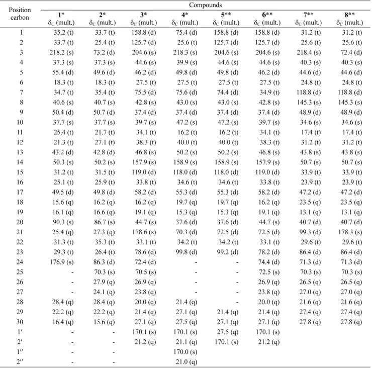
Table 1. 13C-NMR data for compounds 1 - 8
Position carbon Compounds 1* C (mult.) 2* C (mult.) 3* C (mult.) 4* C (mult.) 5** C (mult.) 6** C (mult.) 7** C (mult.) 8** C (mult.) 91 35.2 (t) 33.7 (t) 158.8 (d) 75.4 (d) 158.8 (d) 158.8 (d) 31.2 (t) 31.2 (t) 92 33.7 (t) 25.4 (t) 125.7 (d) 25.6 (t) 125.7 (d) 125.7 (d) 25.6 (t) 25.6 (t) 93218.2 (s) 73.2 (d) 204.6 (s) 218.3 (s) 204.6 (s) 204.6 (s) 218.4 (s) 72.4 (d) 94 37.3 (s) 37.3 (s) 44.6 (s) 39.9 (s) 44.6 (s) 44.6 (s) 40.3 (s) 40.3 (s) 95 55.4 (d) 49.6 (d) 46.2 (d) 49.8 (d) 49.8 (d) 46.2 (d) 44.6 (d) 44.6 (d) 96 18.3 (t) 18.3 (t) 27.5 (t) 27.5 (t) 27.5 (t) 27.5 (t) 24.8 (t) 24.8 (t) 97 34.7 (t) 35.4 (t) 75.5 (d) 75.6 (d) 74.4 (d) 34.9 (t) 118.8 (d) 118.8 (d) 98 40.6 (s) 40.7 (s) 42.8 (s) 43.0 (s) 43.0 (s) 42.8 (s) 145.3 (s) 145.3 (s) 99 50.4 (d) 50.7 (d) 37.4 (d) 37.4 (d) 37.4 (d) 37.4 (d) 48.9 (d) 48.9 (d) 10 37.7 (s) 37.7 (s) 39.7 (s) 47.2 (s) 47.2 (s) 39.7 (s) 34.6 (s) 34.6 (s) 11 25.4 (t) 21.7 (t) 34.1 (t) 16.2 (t) 16.2 (t) 34.1 (t) 17.4 (t) 17.4 (t) 12 21.3 (t) 27.1 (t) 38.3 (t) 40.0 (t) 40.0 (t) 38.3 (t) 31.2 (t) 31.2 (t) 13 43.2 (d) 42.8 (d) 46.8 (s) 50.2 (s) 50.2 (s) 46.8 (s) 43.8 (s) 43.8 (s) 14 50.3 (s) 50.2 (s) 157.9 (s) 158.9 (s) 158.9 (s) 157.9 (s) 50.7 (s) 50.7 (s) 15 31.2 (t) 31.5 (t) 119.0 (d) 118.0 (d) 118.0 (d) 119.0 (d) 33.9 (t) 33.9 (t) 16 25.1 (t) 25.9 (t) 33.8 (t) 34.6 (t) 34.6 (t) 33.8 (t) 23.9 (t) 23.9 (t) 17 49.5 (d) 49.8 (d) 58.2 (d) 55.3 (d) 55.3 (d) 58.2 (d) 47.2 (d) 47.2 (d) 18 15.6 (q) 16.2 (q) 16.2 (q) 19.7 (q) 19.7 (q) 16.2 (q) 23.5 (q) 23.5 (q) 19 16.1 (q) 16.6 (q) 19.1 (q) 15.3 (q) 15.3 (q) 19.1 (q) 13.1 (q) 13.1 (q) 20 90.3 (s) 86.7 (s) 44.7 (s) 37.6 (d) 37.6 (d) 44.7 (s) 40.7 (d) 40.7 (d) 21 25.4 (q) 27.3 (q) 178.6 (s) 70.3 (d) 72.5 (d) 72.5 (d) 99.3 (d) 178.3 (s) 22 31.3 (t) 35.3 (t) 33.1 (t) 34.2 (t) 34.2 (t) 33.1 (t) 29.6 (t) 29.6 (t) 2329.3 (t) 26.4 (t) 78.6 (d) 99.8 (d) 99.2 (d) 78.2 (d) 86.4 (d) 86.4 (d) 24 176.9 (s) 86.3 (d) 72.4 (d) - - 74.4 (d) 71.3 (d) 71.3 (d) 25 - 70.3 (s) 70.5 (s) - - 72.5 (s) 70.3 (s) 70.3 (s) 26 - 27.9 (q) 26.9 (q) - - 26.9 (q) 26.5 (q) 26.5 (q) 27 - 24.1 (q) 23.8 (q) - - 23.8 (q) 27.0 (q) 27.0 (q) 28 28.4 (q) 28.4 (q) 20.0 (q) 21.4 (q) - 20.0 (q) 21.6 (q) 21.6 (q) 29 22.2 (q) 22.2 (q) 21.4 (q) 27.1 (q) 21.4 (q) 21.4 (q) 27.4 (q) 27.4 (q) 30 16.4 (q) 15.6 (q) 27.1 (q) 27.5 (q) 27.1 (q) 27.1 (q) 27.8 (q) 27.8 (q) 1 - - 170.1 (s) 170.1 (s) 27.5 (q) 170.1 (s) 2 - - 21.2 (q) 21.1 (q) 170.1 (s) 21.2 (q) 1 - - 170.0 (s) 2 - - 21.0 (q)
*Measured in 500 MHz for 1H and 125 MHz for 13C
Table 1. HR-TOFMS, m/z 527.2984 [M+H] +, (calcd. C32H48O6 m/z 528.2951).
Melianodiol (7) – Colorless needle crystal; m.p. 216-218oC; IR (KBr) max cm1 : 3350, 2935, 1725, 1645, 1457, 1380, 1055; 1H-NMR (CDCl 3, 600 MHz): δH 5.26 (1H, dd, J = 2.6, 5.8 Hz, 7), 4.63 (1H, d, J = 5.6 Hz, H-21), 4.05 (1H, br.s, H-24), 3.64 (1H, ddd, J = 1.3, 6.2, 8.7 Hz, H-23), 2.67 (1H, ddd, J = 5.7, 8.4, 11.7 Hz, H-20), 2.38 (1H, m, 22a), 2.34 (1H, m, 17), 2.24 (1H, m, H-9), 2.20 (1H, ddd, J = 6.2, 8.4, 11.7 Hz, H-22b), 2.09 (1H, d, J = 14.7, H-6a), 1.93 (1H, m, H-2a), 1.78 (1H, m, H-5), 1.76 (1H, m, H-12a), 1.73 (1H, m, H-16b), 1.63 (1H, m, H-11a), 1.61 (1H, m, H-2b), 1.61 (1H, m, H-12b), 1.57 (1H, m, 16a), 1.53 (1H, m, 11b), 1.52 (1H, m, H-15b), 1.49 (1H, m, H-1a), 1.41 (1H, m, H-6b), 1.39 (3H, s, CH3-27), 1.38 (1H, dt, J = 3.2, 12.2 Hz, H-1b), 1.32 (3H, s, CH3-26), 1.06 (1H, m, H-15a), 1.01 (3H, s, CH3 -30), 0.93 (3H, s, CH3-18), 0.93 (3H, s, CH3-28), 0.92 (3H, s, CH3-29), 0.78 (3H, s, CH3-19); 13C-NMR (CDCl3, 150 MHz) Table 1. HR-TOFMS, m/z 487.4535 [M+H] +, (calcd. C32H50O7 m/z 488.4502).
Indicalilacol B (8) – White solid; IR (KBr) max cm1: 3430, 2930, 1760, 1655, 1420, 1378, 1210, 1110; 1 H-NMR (CDCl3, 600 MHz): δH 5.26 (1H, dd, J = 2.6, 5.8 Hz, H-7), 4.63 (1H, d, J = 5.6 Hz, H-21), 4.32 (1H, d, J = 5.4 Hz, H-3), 4.05 (1H, br.s, H-24), 3.64 (1H, ddd, J = 1.3, 6.2, 8.7 Hz, H-23), 2.67 (1H, ddd, J = 5.7, 8.4, 11.7 Hz, H-20), 2.38 (1H, m, H-22a), 2.34 (1H, m, H-17), 2.24 (1H, m, H-9), 2.20 (1H, ddd, J = 6.2, 8.4, 11.7 Hz, H-22b), 2.09 (1H, d, J = 14.7, H-6a), 1.93 (1H, m, H-2a), 1.78 (1H, m, 5), 1.76 (1H, m, 12a), 1.73 (1H, m, H-16b), 1.63 (1H, m, H-11a), 1.61 (1H, m, H-12b), 1.61 (1H, m, 2b), 1.57 (1H, m, 16a), 1.53 (1H, m, H-11b), 1.52 (1H, m, H-15b), 1.49 (1H, m, H-1a), 1.41 (1H, m, H-6b), 1.39 (3H, s, CH3-27), 1.38 (1H, dt, J = 3.2, 12.2 Hz, H-1b), 1.32 (3H, s, CH3-26), 1.06 (1H, m, H-15a), 1.01 (3H, s, CH3-30), 0.93 (3H, s, CH3-28), 0.92 (3H, s, CH3-29), 0.84 (3H, s, CH3-18), 0.78 (3H, s, CH3-19); 13 C-NMR (CDCl3, 160 MHz) see Table 1. HR-TOFMS, m/z 487.4535 [M+H] +, (calcd. C
32H50O7 m/z 488.4502). Cytotoxicity assay – The MCF-7 Cells were plated at a density of 5 × 10³cells/well in 96-well plate, incubated at 37oC overnight and treated in triplicate with decreasing concentrations isolated compound from C. pentandrus extract. Furthermore, all the extracted were dissolved in DMSO and Cis-platin (Sigma) was used as a positive control. After 48 hours of treatment, cell viability was evaluated using the MTT reagent 3-(4,5-dimethylthiazol-2-yl)-2,5-diphenyl tetrazolium bromide was added accor-ding to the manufactured protocols, followed by a 4-hour
incubation. The results were expressed as relative viable cells compared to the controls (untreated cells). The 50% inhibitory concentration (IC50) values of antiproliferative activity were calculated and converted into the IC50 Dose.
Results and Discussion
The methanol extract of the of C. pentandrus stem bark was partitioned successfully with n-hexane, ethyl acetate and n-butanol. Column chromatography was repeated using silica gel of the soluble fraction in n-hexane led to the isolation of eight isolated compounds (Fig. 1). The structures of the isolated compounds were identified by spectroscopic methods including 1D, 2D NMR and HR-TOFMS. To the best our knowledge, compounds 1 - 8, were isolated from C. pentandrus for the first time.
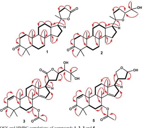
Compound 1 was obtained as colorless crystals (MeOH). The molecular composition was determined was C27H42O3 based on HR-TOFMS ([M+H]+ m/z 415.3311, calcd. for C27H42O3 m/z 414.3334) and NMR analysis (Table 1), and seven degrees of unsaturation is required. Furthermore, IR spectra showed absorption peaks at 2937 and 2870 cm1 (C-H sp3), 1754 cm1 (C=O), 1720 (C=O, lactone) 1464 cm1 (C=C), 1379-1280 cm1 (gem-dimethyl), and 1056 cm1 (C-O groups). The 1H-NMR spectrum showed the presence of six tertiary methyl groups, resonating at δH 1.33 (CH3-21), 0.92 (CH3-18), 0.91 (CH3-28), 0.87 (CH3 -30), 0.82 (CH3-19) and 0.81 (CH3-29). The aliphatic protons in the upfield region were observed also in the 1H-NMR spectra. The 13C-NMR, HMQC and DEPT 135° spectra of 1 showed the presence of six methyl groups, exhibiting the characteristics of dammarane-type triter-penoid, one oxygenated quaternary carbon at δC 90.3 (C-20), one carbonyl lactone at δC 176.9 (C-24) and one carbonyl ketone at δC 218.2 (C-3). These functions represented two of the total of seven degrees of unsaturation, and the remaining five degrees of unsatura-tion matched the dammarane-type triterpenoid framework with an addition of a five-membered lactone ring.19,20 The 1H-1H correlations (Fig. 2) from H-1/H-2, H-5/H-6/H-7, H-9/H-11/H-12/H-13, H-15/H-16/H-17 and H-22/H-23 were aided by the presence of the dammarane-type in compound 1. The HMBC cross peaks (Fig. 2) from H-1 (δH 1.17 and 1.50) and H-2 (δH 1.40 and 1.47) to the carbonyl ketone at C-3 (δC 218.2) indicated the presence of a ketone group at C-3. Protons signal H-17 (δH 1.23) and CH3-21 (δH 1.33) were correlated to C-20 (δC 90.3), whereas methylene protons at C-23 (H 2.52 and 2.62) was correlated to C-24 (C 176.9) indicated the presence of five-membered lactone ring at C-17. The relative
configuration of 1 was determined by NOESY experi-ments, which is supported by the presence of dammarane-type triterpenoids in Chisocheton species.19,20 The NOESY correlations (Fig. 3) from H-17/CH3-30 identified the -lactone ring at C-17 as -oriented, while the cross peak observed between CH3-30/H-17/CH3-21, indicates the CH3-21 as -oriented. A comparison of the NMR data of 1 with cabralealactone obtained from the whole plant of Cleome africana,19 revealed that the structures of the two compounds were very similar, consequently, compound 1 was identified as cabralealactone.
Compound 2 was obtained as a colorless needle crystal. Its molecular composition C30H52O3, was determined from HR-TOFMS spectra (m/z 461.3271 [M+H] +, calculated, C30H52O3 m/z 460.3274) and NMR data (Table 1). The IR spectra showed absorption peaks at 3450 cm1 (OH), 2942 cm1 (C-H sp3), 1471 and 1387 cm1 (gem-dimethyl), and 1075 cm1 (C-O) groups. The 1H-NMR spectrum showed the presence of eight tertiary methyl groups at δH 1.17 (CH3-26), 1.13 (CH3-21), 1.09 (CH3 -27), 0.95 (CH3-18), 0.92 (CH3-28), 0.87 (CH3-30), 0.84 (CH3-19) and 0.82 (CH3-29) resonances. Two oxygenated
Fig. 2. Selected 1H-1H COSY and HMBC correlations of compounds 1, 2, 3 and 5.
methines resonances at H 4.33 (1H, ddd, J = 2.4, 6.8, 9.6 Hz, H-3) and H 3.62 (1H, dd, J = 4.8, 10.2 Hz, H-24) were also observed in the 1H-NMR spectra. The 13 C-NMR spectra showed 30 carbons and were classified by DEPT 135o and HMQC experiments as eight methyl groups, exhibiting the characteristics of dammarane-type triterpenoid,19,20 an oxymethine group at δ
C δC 86.3 (C-24) and 73.2 (C-3) as well as an oxygenated quaternary carbon at C 70.3 (C-25). The 1H-1H correlations (Fig. 2) from H-1/H-2/H-3, H-5/H-6/H-7, H-9/H-11/H-12/H-13, H-15/H-16/H-17 and H-22/H-23 were aided by the presence of the dammarane-type in compound 219,20. The HMBC cross peaks (Fig. 2) from CH3-28 (δH 0.92), CH3 -29 (δH 0.82), and the methylene protons at H-2 (δH 1.55 and 1.38) to the oxymethine carbon at C-3 (δC 73.2) indicated the presence of a hydroxy group at C-3. The correlation of H-22 (δH 1.22 and 2.01) and H-23 (δH 2.52 and 2.62) to C-24 (δC 86.3) and C-20 (δC 86.7) indicates that the position of furan ring at C-20/C-24. Furthermore, methyl protons at H 1.17 (CH3-26) and 1.09 (CH3-27) as well as the oxygenated proton at H 3.62 correlated with oxygenated quaternary carbon at C 86.3 (C-24), indicating that an isopropyl alcohol moiety was located at C-24. In addition, the key NOESY correlation (Fig. 3) between CH3-21 and CH3-26, CH3-27 specifies the isopropyl group at C-24 as a -orientation, which is supported by the presence of dammarane-type triterpenoids in Chisocheton species.19,20 The NMR spectra of 2 were in good agreement with that reported cabraleadiol isolated from Chisocheton penduliflorus,20 therefore, compound 2 was identified as a cabraleadiol.
Compound 3 was isolated as a white amorphous powder. Its molecular composition was assigned as C32H46O7 on the basis of the HRTOFMS (m/z 543.1260 [M+H] +, (calcd. C
32H46O7 m/z 542.1244 NMR data (Table 1), therefore required ten double-bond equivalents. IR spectra showed absorption peaks at 3430 cm1 (OH), 2945 cm1 (C-H sp3), 1720 (C=O), 1639 cm1 (C=C), 1456 and 1378 cm1 (gem-dimethyl) and 1076 cm1 (C-O) groups. The 1H-NMR spectrum showed the presence of seven tertiary methyl groups, resonance at δH 1.44 (CH3-27), 1.35 (CH3-26), 1.16 (CH3-30), 1.16 (CH3-18), 1.07 (CH3-19), 1.06 (CH3-28) and 1.06 (CH3-29). The signal of one acetyl at δH 1.94 (CH3-2), two oxygenated methines at δH 4.47 (1H, dd, J = 9.6, 1.8 Hz, H-23), 3.92 (1H, d, J = 9.6 Hz, H-24), three methine sp2 at δ
H 7.14 (1H, d, J = 10.2 Hz, H-1), 5.86 (1H, d, J = 10.2 Hz, H-2) and 5.28 (1H, br.s, H-15) were observed also in the 1 H-NMR spectrum. The 13C-NMR spectrum (Table 1) showed the presence 32 carbons, which were assigned to
one ketone (C 204.6), one ester (C 170.1), one lactone (C 178.6), four sp2 carbons (C 158.8, 157.9, 125.7 and 119.0), eight methyls (C 27.1, 26.9, 23.8, 21.4, 21.2, 20.0, 19.1 and 16.2), six methylenes and seven methines. These properties represented a total of five or ten degrees of unsaturation, and the remaining five degrees of unsaturation corresponded to the pentacyclic framework of the apotirucallan-type triterpenoid.21 The proton pairing was also confirmed with the 1H-1H COSY spectrum (Fig. 2) and supported the presence of apotirucallan-type triterpenoid in 3. The HMBC crosspeaks (Fig. 2) from H-2 (δH 5.86) to C-1 (δC 158.8) and carbonyl at C-3 (δC 204.6), indicated the presence of ,-unsaturated ketone at C-1, C-2 and C-3, respectively. Correlation which was arising from H-7 (δH 5.33) to C-1' (δC 170.1) and C-6 (δC 27.5) as well as from CH3-2'' (δH 1.94) to to C-1' (δC 170.1), indicate that acetyl group was attached at C-7. The correlation from H-15 (δH 5.28) to C-16 (δC 33.8) and C-14 (δC 157.9), indicated that another double-bond was located at C-14 and C-15, respectively. An oxygenated proton at H-23 (δH 4.47) and methine proton at H-17 (δH 2.19) was correlated to carbonyl at C-21 (δC 178.6), indicate the lactone ring was attached at C-17. Furthermore, the correlation from CH3-26 (δH 1.35) to C-24 (δC 72.4) and C-25 (δC 70.5), suggesting that 1,2-diol side chain was attached at C-23. The detailed examination of the NMR data and comparison with those reported for prototiamin A, an apotirucallane-type triterpenoid isolated from the bark of Entandrophragma congoense,21 showed that the structures of the two compounds were very similar. Therefore, compound 3 was identified as proto-tiamin A.
Compound 4 was isolated as colorless solid, with the molecular formula determined as C30H44O7 based on HRTOFMS (m/z 517.4498 [M+H] +, (calcd. C
30H44O7 m/z 516.4087) and NMR spectra (Table 1). In addition, 13C NMR analysis show that nine degrees of unsaturation is required, while, the IR absorption bands at 3560, 2939, 1725 and 1109 cm1 imply the presence of hydroxyl, aliphatic, carbonyl ester and ether functionalities. The 1 H-NMR spectrum showed the presence of five methyl tertiary signals at δH 1.16 (CH3-19), 1.16 (CH3-30), 1.06 (CH3-28), 1.06 (CH3-29), 1.00 (CH3-18) and two acetyl signals at δH 1.91 and 1.97. One olefinic proton at δH 5.28 (1H, dd, J = 1.8, 3.6 Hz, H-15) and three oxygenated methane signals at δH [5.39 (1H, m, 1), 5.33 (1H, m, H-7) and 4.57 (1H, dd, J=9.6, 1.8 Hz, H-23)] were also observed in the 1H-NMR spectrum. Moreover, the 13C NMR spectrum (Table 1) recognized 30 nonequivalent carbon signals, which are characterized by one carbonyl
ketone (C 218.2), two acetyls (C 170.1 and 170.0), two sp2 carbon (
C 158.9 and 118.0), seven methyl signals at (C 27.5, 27.1, 21.4, 21.1, 21.0, 19.7 and 15.3), seven sp3 methylenes (including one oxygenated methylene type at C 70.3), seven sp3 methynes (including two oxygenated types at δC 75.4, 75.6 and a hemiacetal at δC 99.8) and four quaternary carbons. These properties represented a total of four or nine degrees of unsaturation, and the remaining five degrees of unsaturation corresponded to limonoid framework.16,17,22 Comprehensive analyses of the above mentioned 1H and 13C NMR data exhibited that the structure of 4 was similar to that of neemfruitin,22 a limonoid isolated from Azadirachta indica. Consequently, compound 4 was identified as neemfruitin A.
Compound 5 was obtained as a white solid with a proposed molecular formula of C28H40O5 based on HRTOFMS (m/z 455.0712 [M+H] +, (calcd. C
28H40O5 m/z 456.2876) and NMR data (Table 1), therefore nine indices of hydrogen deficiency were required. Furthermore, the IR absorption bands at 3300, 1705 and 1080 cm1 show the presence of hydroxyl, carbonyl and ether properties, while the NMR data was highly similar to 4. However, differences were observed in the disappearance of one of acetyl group at [H 1.91 (3H, s), C 21.7, 1701.1], oxygenated methyne at [H 5.39 (1H, m), C 75.4] and the appearance of olefinic signals at [H 7.14 (1H, d, J = 10.2 Hz), 5.86 (1H, d, J = 10.2 Hz); C 158.8, 125.7), suggesting 5 as a 1-deacetyl derivative of 4. The proton pairing was also confirmed with the 1H-1H COSY spectrum (Fig. 2) and supported the presence of limonoid structure in 5.22,23 The HMBC crosspeaks (Fig. 2) from H-2 (δH 5.86) to C-1 (δC 158.8) and carbonyl at C-3 (δC 204.6), indicated the presence of ,-unsaturated ketone at C-1, C-2 and C-3, respectively. Correlation which was arising from H-7 (δH 5.33) to C-1' (δC 170.1) and C-6 (δC 27.5) as well as from CH3-2'' (δH 1.94) to to C-1' (δC 170.1), indicate that acetyl group was attached at C-7. The above-mentioned NMR data suggested that compound 5 is a limonoid, for which NMR data (Table 1) showed many similarities to those of 23-desmethyllimocin-B, a limonoid isolated from the seed of Azadirachta indica.23 Therefore, compound 5 was identified as 23-desmethyllimocin-B.
Compound 6 was obtained as a white solid with a molecular composition of C32H48O6, based on HRTOFMS (m/z 527.2984 [M+H] +, (calcd. C
32H48O6 m/z 528.2951) and NMR analysis, therefore nine degrees of unsaturation are required. Furthermore, the IR spectrum showed the absorption bands for hydroxyl (3420 cm1), aliphatic (2949 and 2860 cm1), carbonyl (1715 cm1) and ether (1080 cm1) moieties, while the NMR data observed in
Table 1 was highly similar to 5. However, the difference was identified in the absence of a hemiacetal group at [H 4.57 (1H, dd, J = 9.6, 1.8 Hz), C 99.2], and also the appearance of a newly oxygenated methynes at [H 4.47 (1H, dd, J = 9.6, 1.8 Hz), 3.92 (1H, d, J = 9.6 Hz), C 78.2, 74.4], two methyls at [H 1.35 (3H, s), 1.44 (3H, s), C 26.9, 23.8] and one oxygenated quaternary carbon at C 72.5. These observations suggest that 6 is a 2-methylbutane-2,3-diol derivative of 5 and is attached to C-23. The aforementioned data as well as biogenetic considerations suggested that compound 6 is protoli-monoid, with a structure similar to that of protoxylocarpin G, a protolimonoid isolated from Xylocarpus granatum.24 Consequently, compound 6 was identified as protoxylocarpin G.
Compound 7 was obtained as a colorless crystal. Its molecular composition C30H48O5, was determined from HRTOFMS (m/z 487.4535 [M+H] +, (calcd. C
32H50O7 m/z 488.4502) and NMR data (Table 1), therefore seven unsaturated degrees were required. The IR spectra showed absorption peaks at 3350 cm1 (OH), 2935 cm1 (C-H sp3), 1725 (C=O), 1645 (C=C), 1457 and 1380 cm1 (gem-dimethyl groups), and 1055 cm1 (C-O) groups. The 1H-NMR spectrum showed the presence of seven tertiary methyl groups at H 1.39 (CH3-27), 1.32 (CH3-26), 1.01 (CH3-30), 0.93 (CH3-28), 0.92 (CH3-29), 0.84 (CH3-18) and 0.78 (CH3-19). One olefinic proton at H 5.26 (1H, dd, J = 2.6, 5.8 Hz, H-7), two oxygenated methyne signals at H 3.64 (1H, ddd, J = 1.3, 6.2, 8.7 Hz, H-23), 4.06 (1H, br.s, H-24) and one hemiacetal signal at H 4.63 (1H, d, J = 5.6 Hz) were also observed in the 1H-NMR spectrum. The 13C-NMR spectrum of 7 showed 30 carbons and were classified as seven methyl groups at C 27.8, 27.4, 27.0, 26.5, 23.5, 21.6 and 13.1, one carbonyl ketone at C 218.4 (C-3), two oxymethine signals at C 86.4 (C-23) and 71.3 (C-24), one oxygenated quaternary
Table 2. IC50 inhibition values of compounds 1 - 8 against
MCF-7 breast cancer cell line
Compounds IC50 (µM) Cabrealeolactone (1) 61.18 Cabreadiol (2) 3 3 .12 Prototiamin A (3) 76.08 Neemfruitins A (4) 181.12 Desmethyllimocin B (5) 98.18 Protoxylocarpin G (6) 90.24 Melianodiol (7) 16.84 Indicalilacol B (8) 20.23 Cisplatin* 13.20 *positive control
carbon at C 70.3 (C-24), hemiacetal signal at C 99.3 (C-21) and two sp2 carbon at
C 118.8 (C-7) and 145.3 (C-8), respectively. These properties represented two of seven total degrees of unsaturation, and the remaining five degrees of unsaturation were consistent with the limonoid structure.24,25 The above data showed general features similar to those of melianodiol, a limonoid isolated from the leaves of Aglaia andamanica.25 Therefore, compound 7 was identified as melianodiol.
Compound 8 was obtained as a white solid having a molecular composition of C30H48O5, based on HRTOFMS (m/z 487.4535 [M+H] +, (calcd. C
32H50O7 m/z 488.4502) along with NMR analysis, therefore seven degrees of unsaturation are required. Furthermore, the IR spectrum showed the absorption bands for hydroxyl (3430 cm1), aliphatic (2930), carbonyl (1760 cm1), gem-dimethyl (1420 and 1378 cm1) and ether (1080 cm1) moieties, while the NMR data observed in Table 1 was highly similar to 7. However, the difference was identified in the absence of a hemiacetal signal at [H 4.63 (1H, d, J = 5.6 Hz), C 99.3] and ketone signal at C 218.4, and appearance of a newly carbonyl lactone at C 178.3 (C-21) and hydroxyl signal at [H 4.32 (1H, d, J = 5.4 Hz), C 72.4]. These observations indicate that 8 is 3-hydroxy and lactone derivative of 7. The above data revealed that 8 is a tirucallane-type triterpenoid similar to indicalilacol B, isolated from Azadiracta indica.26 Consequently, compound 8 was identified as indicalilacol B.
The isolated compounds, 1 - 8, were evaluated for their cytotoxic against MCF-7 breast cancer lines using a previously described method,17,18,27 using cis-Platin as a positive control.28,29 Among the compounds tested, melia-nodiol (7), exhibited the strongest cytotoxicity with an IC50 value of 16.84 M. Interestingly, Compounds 7 and 8 share the same structure and only differed at the functional of the cyclopentane ring, the cytotoxic activities of compounds 7 and 8 differed greatly, suggested the presence of hemiacetal group increase the cytotoxic activity. These results were supported from previously studies that the presence of a hemiacetal group in triter-penoids structure can increase cytotoxic activity.17,24,25 In addition, despite close structural similarities to 7, a low activity was observed in 6, which indicates the possible contribution of an acetyl group to cytotoxicity.25 This results was supported from previously studies that the presence of a hemiacetal group in triterpenoids structure can increase cytotoxic activity.25 Therefore, the structural comparison of compound 7 with the other compounds indicates that carbonyl, hydroxyl and acetyl groups present contribute to the cytotoxic activity.
Acknowledgments
This investigation was financially supported by Universitas Brawijaya, Indonesia (PPKI Grant, No: 455.5/ UN10.C10/PN/2020 by RR); (Universitas Padjadjaran, Indonesia (PPKI Grant, No: 1427/UN6.3.1/LT/2020 by US) and Universitas Airlangga, Indonesia (PPKI Grant, No: 305/UN3.14/PT/2020 by MT).
References
(1) Katja, D. G.; Farabi, K.; Nurlelasari.; Harneti, D.; Mayanti, T.; Supratman, U.; Awang, K.; Hayashi, H. J. Asian Nat. Prod. Res. 2017, 19, 194-200.
(2) Shilpi, J. A.; Saha, S.; Chong, S. L.; Nahar, L.; Sarker, S. D.; Awang, K. Chem. Biodivers. 2016, 13, 483-503.
(3) Awang, K.; Lim, C. S.; Mohamad, K.; Hiroshi, M.; Hirasawa, Y.; Takeya, K.; Thoison, O.; Hadi, A. H. A. Bioorg. Med. Chem. 2007, 15, 5997-6002.
(4) Tan, Q. G.; Luo, X. D. Chem. Rev. 2011, 111, 7437-7522.
(5) Yang, M. H.; Wang, J. S.; Luo, J. G.; Wang, X. B.; Kong, L. Y. J. Nat. Prod. 2009, 72, 2014-2018.
(6) Mohamad, K.; Hirasawa, Y.; Litaudon, M.; Awang, K.; Hadi, A. H. A.; Takeya, K.; Ekasari, W.; Widyawaruyanti, A.; Zaini, N. C.; Morita, H. Bioorg. Med. Chem. 2009, 17, 727-730.
(7) Supratman, U.; Naibaho, W.; Salam, S.; Maharani, R.; Hidayat, A. T.; Harneti, D.; Nurlelasari.; Shiono, Y. Phytochem. Lett. 2019, 30, 81-87. (8) Chong, S. L.; Hematpoor, A.; Hazni, H.; Sofian-Azirun, M.; Litaudon, M.; Supratman, U.; Murata, M.; Awang, K. Phytochem. Lett. 2019, 30, 69-73.
(9) Najmuldeen, I. A.; Hadi, A. H. A.; Mohamad, K.; Awang, K.; Ketuly, K. A.; Mukhtar, M. R.; Taha, H.; Nordin, N.; Litaudon, M.; Guéritte, F.; Nugroho, A. E.; Morita, H. Heterocycles 2012, 84, 1265-1270.
(10) Fang, X.; Di, Y. T.; Hao, X. J. Current Organic Chemistry 2011, 15, 1363-1391.
(11) Haldar, S.; Kolet, S. P.; Thulasiram, H. V. Green Chemistry 2013, 15, 1311-1317.
(12) Nagoor, N. H.; Muttiah, N. S. J.; Lim, C. S.; In, L. L. A.; Mohamad, K.; Awang, K. PLoS One. 2011, 6, e23661.
(13) Wong, C. P.; Shimada, M.; Nagakura, Y.; Nugroho, A. E.; Hirasawa, Y.; Kaneda, T.; Awang, K.; Hadi, A. H. A.; Mohamad, K.; Shiro, M.; Morita, H. Chem. Pharm. Bull. 2011, 59, 407-411.
(14) Yang, M. H.; Wang, J. S.; Luo, J. G.; Wang, X. B.; Kong, L. Y. Bioorg. Med. Chem. 2011, 19, 1409-1417.
(15) Bordoloi, M.; Saikia, B.; Mathur, R. K.; Goswami, B. N. Phytochemistry 1993, 34, 583-584.
(16) Nurlelasari.; Katja, D. G.; Harneti, D.; Wardayo, M. M.; Supratman, U.; Awang, K. Chem. Nat. Compd. 2017, 53, 83-87.
(17) Supriatno.; Nurlelasari.; Herlina, T.; Harneti, D.; Maharani, R.; Hidayat, A. T.; Mayanti, T.; Supratman, U.; Azmi, M. N.; Shiono, Y. Nat. Prod. Res. 2018, 25, 1-7.
(18) Supratman, U.; Salam, S.; Naibaho, W.; Fajar, M.; Nurlelasari.; Katja, D. G.; Harneti, D.; Maharani, R.; Hidayat, A. T.; Lesmana, R.; Nafiah, M. A.; Shiono, Y. Phytochem. Lett. 2020, 35, 63-67.
(19) Nagaya, H.; Tobita, Y.; Nagae, T.; Itokawa, H.; Takeya, K.; Halim, A. F.; Abdel-Halim, O. B. Phytochemistry 1997, 44, 1115-1119.
(20) Phongmaykin, J.; Kumamoto, T.; Ishikawa, T.; Suttisri, R.; Saifah, E. Arch. Pharm. Res. 2008, 31, 21-27.
S.; Bauer, J. O.; Strohmann, C.; Spiteller, M. J. Nat. Prod. 2015, 78, 604-614.
(22) Chianese, G.; Yerbanga, S. R.; Lucantoni, L.; Habluetzel, A.; Basilico, N.; Taramelli, D.; Fattorusso, E.; Taglialatela-Scafati, O. J. Nat. Prod. 2010, 73, 1448-1452.
(23) Kumar, C. S. S. R.; Srinivas, M.; Yakkundi, S. Phytochemistry 1996, 43, 451-455.
(24) Pudhom, K.; Sommit, D.; Nuclear, P.; Ngamrojanavanich, N.; Petsom, A. J. Nat. Prod. 2009, 72, 2188-2192.
(25) Puripattanavong, J.; Weber, S.; Brecht, V.; Frahm, A. W. Planta Med. 2000, 66, 740-745.
(26) Kurimoto, S.; Takaishi, Y.; Ahmed, F. A.; Kashiwada, Y. Fitoterapia 2014, 92, 200-205.
(27) Skehan, P.; Storeng, R.; Scudiero, D.; Monks, A.; McMahon, J.; Vistica, D.; Warren, J. T.; Bokesch, H.; Kenney, S.; Boyd, M. R. J. Natl. Cancer Inst. 1990, 82, 1107-1112.
(28) Hadisaputri, Y. E.; Miyazaki, T.; Suzuki, S.; Yokobori, T.; Kobayashi, T.; Tanaka, N.; Inose, T.; Sohda, M.; Kuwano, H. Ann. Surg. Oncol. 2012, 19, S589-S596.
(29) Chavoshi, H.; Vahedian, V.; Saghaei, S.; Pirouzpanah, M. B.; Raeisi, M.; Samadi, N. Asian Pac. J. Cancer Prev. 2017, 18, 2243-2247.
Received November 17, 2020 Revised February 3, 2021 Accepted February 9, 2021
![Table 1 was highly similar to 5. However, the difference was identified in the absence of a hemiacetal group at [ H 4.57 (1H, dd, J = 9.6, 1.8 Hz), C 99.2], and also the appearance of a newly oxygenated methynes at [ H 4.47 (1H, dd, J = 9](https://thumb-ap.123doks.com/thumbv2/123dokinfo/4983472.55385/8.892.459.817.150.353/similar-difference-identified-absence-hemiacetal-appearance-oxygenated-methynes.webp)
