서
론
이두건 장두(long head of biceps tendon)의 병 변은 견관절의 전방부 통증의 여러 원인 중에 하나 에 해당한다.1)이두건 장두의 병변이 의심되는 경우 에는 신체 검사로서 Speed 검사, Yergason 검사, 또는 이두구(bicipital groove)의 압통 등을 확인해 볼 수 있으나 특이도 및 민감도에서 다른 병변들에 비하여 낮아서,2-5) 신체 검사 만으로 확진을 하기는 쉽지 않다. 단순 방사선 검사에서도 이두건 장두의 병변을 의심해 볼 수 있는 특징적인 소견을 찾기가 쉽지 않다. 이두건 장두의 병변에 대한 자기공명영 상 검사(MRI)에서도 관절경으로 확진한 소견과 낮 은 일치도를 보였다.6) 최근에는 정형외과 외래 진료 시에 초음파 검사 장비를 많이 활용하고 있으며 다양한 근골격계 질환 에 대한 진단 및 치료에 대하여 접근하고 있다. 이에 초음파 검사에서 이두건 장두 병변과 관련된 소견을 알아보고자 하였다.
검사 자세
환자는 주관절을 90도 구부린 상태에서 전완부를 회외전(supination)시킨 상태에서 초음파 검사를 시작한다. 주관절을 굴곡 상태로 검사하는 이유는 견관절의 회전 각도를 쉽게 조절하기 위한 것이다. 환자의 해부학적인 차이 또는 견관절의 운동 범위의 차이에 따라서 초음파 기기의 변환기(transducer) 를 피부에 대는 위치를 이동시킬 필요가 있다.이두건 장두의 정상 해부학
이두근의 근위부는 장두와 단두(short head of biceps tendon)로 나뉘어서 기시한다. 장두는 상부 관절와에서 기시하며, 단두는 오구돌기(coracoids process)에서 기시한다. 이두건 장두는 상완골의 대 결절과 소결절 사이에 형성된 이두구를 통과하며, 이 두구의 근위부는 횡 상완 인대(transverse humeral ligament)와 falciform ligament가 터널을 형성한 Hyun Seok Song, M.D.Department of Orthopedic Surgery, St. Paul’s Hospital, The Catholic University of Korea, Seoul, Korea Lesion of long head of biceps tendon is one of the causes making an anterior shoulder pain. According to the anthro-pometric study of the bicipital groove the meanings of the width, depth and the medial wall angle were suggested. Confirming the images and location of the long head of biceps tendon itself would be helpful in diagnosis.
Key Words: Rupture, Subluxation, Dislocation, Long head of biceps tendon, Ultrasonography
통신저자: 송 현 석
서울특별시 동대문구 왕산로 180
가톨릭대학교 의과대학 성바오로병원 정형외과 Tel: 02-958-2159 Fax: 02-965-1456
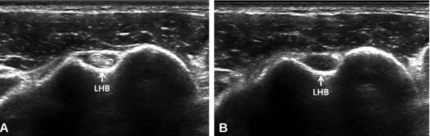
다. 이두구는 관절와-상완 관절과 통하며 이두구 내면은 활액막으로 되어 있다. 단축(sort axis) 초음파 영상으로 이두건 장두를 확인하는 것으로 검사를 시작하게 된다. 이는 단축 초음파 영상에서 이두구를 찾기가 훨씬 용이하기 때 문이다. 이두구 내에서 둥글고 고 에코로 보이는 구 조물이 이두건 장두이다(Fig. 1A). 그러나 이두건 장두를 이루는 건 섬유에 수직 각도로 초음파 영상 을 얻지 못한다면 비등방성(anisotrophy) 허상 (artifact)에 의해서 저 에코로 관찰되어서 구조물 을 확인하지 못하게 된다(Fig. 1B). 이러한 비등방 성 허상을 이용하면 오히려 정상적인 이두건 장두를 쉽게 확인하는데 도움을 받을 수 있다. 단축 초음파 영상을 원위부로 이동시켜서, 이두건 장두의 전체 주행을 확인하는 것이 중요하다. 대흉근
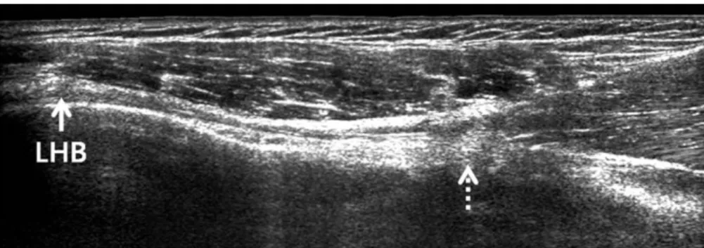
(pectoralis major) 부착부(Fig. 2)를 거쳐서, 근육 부위에서 이두건 장두와 단두를 확인할 수도 있다. 장축(long axis) 초음파 영상으로 이두건 장두를 확인해 볼 수도 있다(Fig. 3).
이두구의 해부학
이두건 장두가 지나는 이두구의 단면은 반원 형태 를 이루며(Fig. 4), 그 크기나 위치는 개인적인 차 이를 보인다. 이두구의 형태에 대한 여러 해부학적 인 연구가 진행되었다. 해부학적인 연구에서 이두구 를 폭, 깊이, 내측벽 각(medial wall angle)의 골성 지표로 구분하여서 분석하였다. 이두구의 폭, 깊이 등은 남녀의 차이가 존재한다.7)이두구의 폭이 이두 건 장두의 단면적보다 지나치게 좁은 경우에는 이두 건 장두의 건염 및 부분 파열을 일으키기 쉽다고 한 다(Fig. 5).7) 이두구의 깊이가 얕은 경우에는, 이두 건의 불안정성을 유발하게 된다. 이두구의 내측벽 각도가 작은 경우에도 이두건 장두의 내측 탈구를 막지 못하여서 불안정성을 보이는 경우가 많다고 한 다. 대부분의 이러한 해부학 연구는 컴퓨터 단층촬 영 검사(CT) 혹은 자기공명영상 검사8) 를 이용한 것 이며 이러한 이두구의 골성 지표를 초음파 검사에서 명확히 측정하기가 어려울 수도 있다. 그러나 초음 파 검사에서 연부 조직과 골 조직 모두를 명확히 구 분할 수 있다는 장점이 존재하므로 이두건 장두의 단면과 이두구의 형태와의 관계는 다른 영상 검사법 에 비하여 초음파 검사에서 더 쉽게 확인할 수 있다. 초음파 영상에서 이두구 주변에 형성된 골극을 확 Fig. 1. Short axis images at the bicipital groove level showing the long head of the biceps tendon (arrow).According to the angle of the transducer, the echo of the tendon can be changed from the normal image (A) to the anisotrophy artifact (B). (LHB: long head of the biceps tendon)
A
B
Fig. 2. Short axis image at the insertion of the pectoralis
Fig. 3. Long axis image of the long head of biceps tendon (arrow) showing the musculotendinous junction (dotted
arrow). (LHB: long head of the biceps tendon)
Fig. 4. Short axis image at the bicipital groove level
showing the smooth groove wall and the long head of the biceps tendon (arrow) (LHB: long head of the biceps tendon)
Fig. 5. Short axis image at the bicipital groove level
showing the relationship between the narrow groove and the long head of the biceps tendon (arrow) (LHB: long head of the biceps tendon)
Fig. 6. Short axis image at the bicipital groove level
showing the spur formation (arrows) at the medi-al wmedi-all (arrow) and floor (two crosses and arrow).
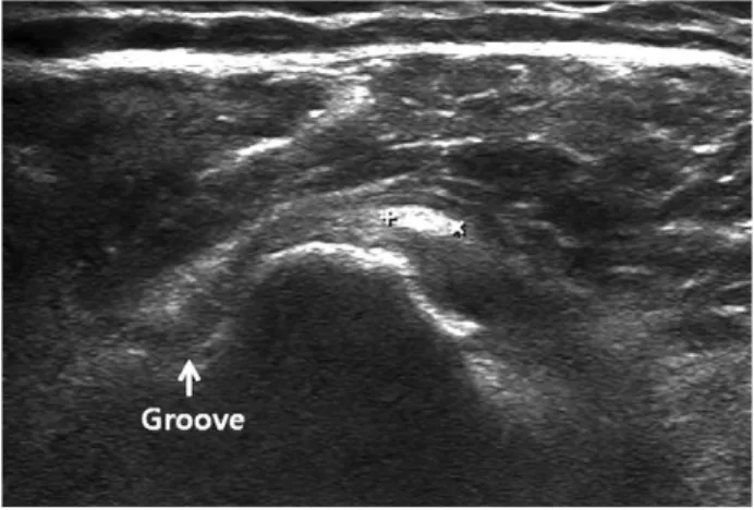
Fig. 7. Short axis image of the long head of the biceps
tendon (LHB) (arrow) showing a hypo-echoic effusion.
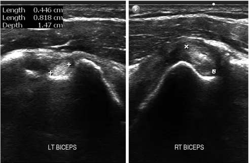
되었다. 그러나 초음파를 비롯하여서 컴퓨터 단층촬 영 검사 및 자기공명영상 검사와 같은 영상 검사에 서 이두건 부분 파열을 진단하기는 쉽지 않다. 이두건에 대한 초음파 검사에서 이두구 내에서 체 액의 증가를 쉽게 확인해 볼 수 있다(Fig. 7). 그러 나 이두구가 관절와-상완 관절과 교통하기 때문에 관절내 체액의 증가에 의해서 이두구 내까지 체액이 관찰될 수 있다. 관절내 체액의 증가가 일어나는 경 우로는 활액막염, 관절연골 손상에 따른 관절염, 회 전근 개 손상 등을 의심해 볼 수 있다.10) 따라서 이 두구 내에서 관찰되는 체액의 증가는 이두건 장두의 병변에 대한 진단적 의미가 높지 않다. 이두건 장두의 건염 및 부분 파열의 일부 형태에서 이두건 장두의 단면적(굵기)이 증가하게 된다. 정상적 인 이두건 장두의 단면적은 개인적인 차이를 보이며 한 개인에서도 해부학적인 위치에 따라서 단면적의 차 이를 보인다. 그러므로 이두구의 형태와 비교하여서 이두건 장두의 단면적의 상대적인 차이에 의미를 두는 것이 낫다고 판단된다. 또는 증상이 없는 반대편과 비 교하여 보는 것도 도움이 될 수 있다(Fig. 8). 이두건 장두의 부분 파열이 진행되면서, 둥근 단 면을 유지하지 못하고 이두구의 내측연을 넘어가는 형태를 관찰할 수 있다(Fig. 9). 이는 이두건 장두 의 병변이 발생하는 요인 중에 이두구 내측 활차 (medial pulley)의 병변에 의하여 이두건 장두의 불안정성이 발생하고 이로 인한 부분 파열로 진행된 것일 수도 있다. 그러나 Amstrong 등11) 에 의하면 초음파 검사에 서 이두건 장두의 부분 파열은 한 예도 진단하지 못 하였다고 보고하였다.
이두건 완전 파열
이두건 장두의 부분 파열이 진행되거나 혹은 갑자 기 무리한 근육 수축 중에 이두건 장두의 완전 파열 이 발생하게 된다. 파열되는 해부학적인 위치는 차 이를 보일 수 있다. 원위부가 후퇴(retraction)하는 정도는 개인적인 차이를 보일 수 있다. 흔히 이두건Fig. 8. Short axis image of the long head of the biceps tendon showing a difference between both shoulders. The
right (RT) biceps tendon was larger than the left (LT) one.
Fig. 9. Short axis image at the bicipital groove level
showing the deformed long head of biceps ten-don (arrows).
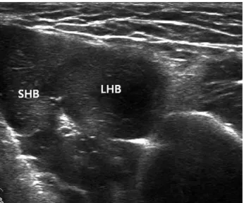
조직 혹은 다른 조직 영상이 관찰될 수도 있어서 오 진한 경우를 경험하였다. 초음파 검사는 이러한 Popeye 변형의 진단에 유용 하다고 생각된다. 정상적인 이두근의 단축 영상은 이 두건 단두 및 장두 부분의 근육 영상이 동일한 에코 및 양상을 보인다(Fig. 10). 그러나 이두건 장두의 완전 파열이 발견된 경우에는, 원위부의 후퇴에 의하 여서 이두건 단두의 근육이 관찰되는 지점에서 장두 의 건 부분이 확인된다(Fig. 11). 이두건 장두 주변 에 급성 파열인 경우에는 혈액, 진구성 파열인 경우 에는 체액이 고여 있는 것을 흔히 발견할 수 있다. 이 러한 소견은 앞서 언급한 피하 지방이 두꺼운 경우, 여성의 경우에서도 진단율을 높일 수 있을 것이다. 로 탈구되기도 한다. 자기공명영상 검사에서는 완전 탈구된 경우에 이 두구 내에서 저신호 강도의 이두건 장두가 보이지 않 고 관절내 혹은 견갑하건 내에서 저신호 강도의 구조 물을 확인함으로써 비교적 쉽게 진단할 수 있다. 초음파 검사에서는 소결절의 전방, 즉 견갑하건의 바깥, 건 내, 관절내에서 이두건 장두 자체의 영상을 확인하는 것이 중요하다. 이두구 내에 이두건 장두 가 없으나, 반흔 조직 등의 섬유가 고 에코로 보이면 서 이두건 장두로 오인하게 만들 수도 있다. 이두구 에서 탈구된 이두건 장두를 직접 확인하는 것이 진 단에 도움이 되며 이두건의 단축 영상에서 비등방성 허상을 이용하면 변환기의 각도를 변화시키면서 고 에코와 저 에코로 바뀌는 둥근 구조물을 찾으면 된
Fig. 10. Short axis image at the biceps muscle level
showing the short head and long head of biceps tendon. (SHB: short head of the biceps tendon / LHB: long head of the biceps tendon)
Fig. 11. Short axis image at the biceps muscle level
showing the retracted long head of biceps tendon (dotted arrow) and effusion (arrows). (LHB: long head of the biceps tendon)
다. 이는 단축 영상으로 확인할 때, 견갑하건의 건 섬유 방향과 탈구된 이두건 장두의 건 섬유 방향이 다르기 때문에 가능하다(Fig. 12). Amstrong 등11) 에 의하면 초음파 검사에서 이두건 장두의 아탈구/ 탈구에 대한 진단은 100%의 특이도 96%의 민감도 를 보고하였다.
결
론
이두건 장두에 대한 초음파 영상은 비교적 쉽게 얻 을 수 있다. 이두구 및 이두건 장두의 정상 해부학에 있어서 개인적인 차이가 있음을 알고 있어야 한다. 이두구의 형태, 이두건 장두 자체의 영상 소견 및 위 치를 확인함으로써 진단에 도움을 받을 수 있다.참고문헌
01. Krupp RJ, Kevern MA, Gaines MD, Kotara S, Singleton SB. Long head of the biceps tendon
pain: differential diagnosis and treatment. J Orthop Sports Phys Ther. 2009;39:55-70.
02. Ben Kibler W, Sciascia AD, Hester P, Dome
D, Jacobs C. Clinical utility of traditional and
new tests in the diagnosis of biceps tendon injuries and superior labrum anterior and poste-rior lesions in the shoulder. Am J Sports Med.
2009;37:1840-7.
03. Holtby R, Razmjou H. Accuracy of the Speed's
and Yergason’s tests in detecting biceps patholo-gy and SLAP lesions: comparison with arthro-scopic findings. Arthroscopy. 2004;20:231-6.
04. Chen HS, Lin SH, Hsu YH, Chen SC, Kang
JH. A comparison of physical examinations with
musculoskeletal ultrasound in the diagnosis of biceps long head tendinitis. Ultrasound Med Biol. 2011;37:1392-8.
05. Calis M, Akgun K, Birtane M, Karacan I,
Calis H, Tuzun F. Diagnostic values of clinical
diagnostic tests in subacromial impingement syn-drome. Ann Rheum Dis. 2000;59:44-7.
06. Mohtadi NG, Vellet AD, Clark ML et al. A
prospective, double-blind comparison of magnet-ic resonance imaging and arthroscopy in the evaluation of patients presenting with shoulder pain. J Shoulder Elbow Surg. 2004;13:258-65.
07. Pfahler M, Branner S, Refior HJ. The role of
the bicipital groove in tendopathy of the long biceps tendon. J Shoulder Elbow Surg. 1999;8:419-24.
08. Abboud JA, Bartolozzi AR, Widmer BJ,
DeMola PM. Bicipital groove morphology on
MRI has no correlation to intra-articular biceps tendon pathology. J Shoulder Elbow Surg. 2010; 19:790-4.
09. Cone RO, Danzig L, Resnick D, Goldman
AB. The bicipital groove: radiographic,
anatom-ic, and pathologic study. AJR Am J Roentgenol. 1983;141:781-8.
10. Chang KV, Chen WS, Wang TG, Hung CY,
Chien KL. Associations of sonographic
abnor-malities of the shoulder with various grades of biceps peritendinous effusion (BPE). Ultrasound Med Biol. 2014;40:313-21.
11. Armstrong A, Teefey SA, Wu T et al. The
effi-cacy of ultrasound in the diagnosis of long head of the biceps tendon pathology. J Shoulder Elbow Surg. 2006;15:7-11.
12. Conrad MR, Nelms BA. Empty bicipital
groove due to rupture and retraction of the biceps tendon. J Ultrasound Med. 1990;9:231-3.
Fig. 12. Short axis image at the bicipital groove level
showing the dislocated long head of biceps tendon (two crosses). Arrow indicates the bicipital groove.