서 론
부신(adrenal gland)은 신장(kidney) 위에 양쪽 좌우에 삼 각형 모양으로 내분비계통의 호르몬을 관장하고 있다. 이 러한 부신에서 발생하는 부신 종양은 복부 CT(computed tomography) 검사의 최대 5%에서 확인되고 있으며(Dun
nick and Korobkin 2002), 부신 선종(adrenal adenoma)은 흔 한 양성 종양으로서, 세포질에 다량의 지방 성분을 함유한 조직이다. 이와 관련하여 CT는 HU(Hounsfield Unit)의 CT 감약계수로 부신 선종을 진단하고 있다(Boland et al. 1998). 조영전의 CT에서 부신 선종은 -2~16HU의 범위의 평균 감약을 보이고, 지질(lipid)이 풍부한 경우는 20~25HU이고 부족한 경우에도 HU로 나타나고 있다(Johnson et al. 2009).
부신 CT는 기능적 부신 종양 및 부신 암 병기를 암시하는
호르몬 이상을 포함한 다양한 증상에 대해 수행되고 있다.
부신
CT
검사에서
HU
의 조영증강 및 선량 평가
권 대 철1· 최 지 원2,*
1신한대학교 바이오생태보건대학 방사선학과, 2전주대학교 방사선학과
Evaluation of Contrast Enhancement of HU and
Effective Dose for Adrenal MDCT
Dae Cheol Kweon
1and Jiwon Choi
2,*
1Department of Radiological Science, College of Bioecological Health, Shinhan University,
Uijeongbu 11644, Republic of Korea
2Department of Radiological Sciences, Jeonju University, Jeonju 56079, Republic of Korea
Abstract - The purpose is to improve the accuracy of dose reduction and adrenal gland diagnosis by analyzing the HU and evaluating the effective dose of contrast CT scan of the adrenal gland in order to evaluate the accuracy of contrast enhancement of adrenal adenomas. We analyzed the dose information of patient DICOM file that underwent CT scan of adrenal gland examination. CT scan of the adrenal gland performed pre contrast, portal phase, a 3 minute delay and a 15 minute delay. The HU measurement selects the largest region of adrenal lesion. And, mean, standard deviation, and area of the adrenal lesion were analyzed by ROI. Absolute percentage
washout(APW) and relative percentage washout(RPW) calculated and the adrenal adenomas
diagnosed by threshold value of the percentage washout rate were above APW 60% and RPW 40%. Effective dose was calculated based on DLP of DICOM dose report for adrenal CT scan. HU analyzed of the adrenal gland by calculating APW and RPW. The APW and RPW were analyzed by 52% and 41.8% at 3 minute delay, 73.5% and 59% at 15 minute delay, respectively.
The diagnosis of adrenal adenomas was consistent with APW(73.5%) and RPW(59%) above the
threshold at 15 minute delay. The effective dose of adrenal CT scan was 16.08 mSv. Therefore, it is necessary for radiation dose reduction.
Key words : Adrenal CT, Absolute percentage washout, HU, Relative percentage washout
─ 157 ─
Technical Paper
* Corresponding author: Jiwon Choi, Tel. +82632203260, Fax. +82632202054, Email. [email protected]
CT 검사의 프로토콜 설계 및 영상의 데이터를 진단하는 정 보를 이해하는 것이 중요하다. 이러한 CT를 이용하여 조영 제를 주입하여 영상의 조영증강의 정도를 이용하여 병변을 진단하기 위한 명확한 전략을 수립하기 위한 목적으로 다양 한 부신 선종의 특성이 있다. 부신병변 감별 진단을 위한 CT는 많은 연구를 하고 있으 며, CT 스캐너를 사용하여 조영제 주입 전과 후에 CT 스캔 의 정확성이 보고되었다. 조영증강되지 않은 CT의 경우, 10 HU 미만의 병변 내 밀도 값은 높은 정확도의 선종을 나타 낸다. 대조적으로 10HU보다 큰 병기 밀도 값은 비 선종에 서 더 흔하지만 세포질 내 지방 함량이 낮은 선종의 가능성 을 배제할 수는 없다. 지연은 대조 물질의 정맥 주사 후 15 분으로 표현되고 다른 경험에 따르면 5분에 수행된 초기 CT 스캔이 분야에서 사용될 수 있다(Korobkin et al. 1996; Choyke 2006). 연구의 목적은 부신의 CT 검사에서 조영제의 증강에 따 른 배출율의 정확성을 평가하기 위해 조영제를 주입 후에 문맥기, 3분 지연기, 15분 지연기에 부신을 CT 스캔하여 HU를 분석하고 유효선량을 평가하여 선량 감소 및 부신진 단의 정확성을 높이는 데 목적이 있다.
대상 및 방법
1. Scanning Protocols 부신 CT 검사 사례 환자는 42세 남자로 MDCT(Brilliance CT 64channel, Philips, The Netherlands)를 이용하여 CT 검 사를 실시한 환자 DICOM 파일의 선량 정보를 분석하였다. 부신 검사 환자를 3ml·sec-1의 속도로 주입되는 자동 주 입기를 이용하여 80ml 조영제(Ultravist, Schering, Berlin, Germany)를 정맥 주사하고 두께 5mm, pitch 1:1로 촬영 을 하였다. CT 검사의 매개 변수는 120kVp, 180mAs, FOV (350mm), 5mm thickness로 부신을 조영제 주입 전과 문맥 기, 3분 지연기, 15분 지연기를 스캔하였다. 연구 대상 환자 의 동의 하에 CT 검사를 진행하여 영상을 획득하여 연구를 진행하였다. 2. Analysis of HU부신의 CT 스캔 영상은 PACS(picture archiving and communication system)으로 전송하여 영상을 분석하였다. CT 감약계수인 HU 측정은 부신병변이 가장 크게 보이는 부위를 선택하여 부신 선종병변의 가운데를 ROI(region of interest)를 65.42mm2 설정하지만 주변 조직과의 부분 체적 평균을 피하기 위해 부신병변의 경계 내에 완전히 포함된 병변의 평균(HU), 표준편차, 면적을 분석하였다(Fig. 1). CT의 조영제 주입의 시기별 스캔에서 감약계수(HU)를 측정한 후 다음과 같은 공식에 따라 절대조영증강배출율 인 APW(absolute percentage washout)과 상대조영증강배출 율 RPW(relative percentage washout)을 계산하였다. 조영
증강배출율의 문턱값은 APW는 60%를 기준으로 하였고,
RPW는 40% 이상은 부신 선종으로 진단하였다(Blake et al. 2006; Johnson et al. 2009).
APW(absolute percentage washout)는 공식 1과 같이 계 산하였다.
Absolute percentage washout=
(HUportal venous phase)-(HUdelayed) 100×
[
---]
(1) (HUportal venous phase)-(HUnonenhanced) RPW(relative percentage washout)는 공식 2와 같이 계산 하였다.Relative percentage washout=
(HUportal venous phase)-(HUdelayed) 100×
[
---]
(2) (HUnonenhanced)3. Analysis of effective dose
부신 CT 검사에 대한 DICOM의 선량보고서의 DLP(dose length product)를 기준으로 하여 유효선량을 산출하였다. 유효선량을 산출하기 위해 DLP(mGy·cm)에 conversion factor를 곱하여 선량을 산출하였다. DLP에 변환계수(k)인 0.0153mSv·mGy-1×cm를 곱하여 유효선량을 계산하였다 (Christner et al. 2010; Deak et al. 2010).
부신의 CT 검사에서 유효선량(E)은 다음과 같이 변환계 수(k)에 DLP를 곱하여 계산하였다.
E=k×DLP (3) 부신의 조영증강에 따른 문맥기, 3분 지연기, 15분 지연기 스캔에 따른 조영증강의 회귀분석을 Microsoft Excel 2010 프로그램(MS Excel; Microsoft, Redmond, MA, USA)을 이 용하였다.
결 과
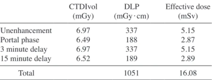
부신 선종의 CT 검사의 ROI에 대한 조영증가의 HU의 분석은 APW, RPW를 계산하여 3분 지연기에서 APW 52%, RPW 41.8%이었고, 15분 지연기는 APW 73.5%, RPW 59% 로 분석되었다(Table 1). 부신 선종에 대한 ROI 설정의 직선성은 회귀분석은 R2= 1로 직선성을 보였고, 문맥기에서 가장 높은 조영증강을 보 였고, 3분 지연기, 15분 지연기, 조영증강 순으로 조영증강을 나타냈다(Fig. 2). 부신 CT의 CTDIvol과 DLP는 PACS의 선량보고서를 기 반으로 하여 유효선량을 산출하였다. CT 검사에서 CTDIvol 은 조영제 주입 전 6.97mGy, 문맥기 6.49mGy, 3분 지연 기 6.97mGy, 15분 지연기 6.52mGy로 산출되었다. 선량보 고서의 DLP는 조영제 주입 전 337mGy·cm, 문맥기 188 mGy·cm, 3분 지연기 337mGy·cm, 15분 지연기 189 mGy·cm로 산출되었다(Table 2). 부신 CT 검사의 조영증강에 따른 유효선량의 산출은 DLP를 기반으로 하여 조영제 주입 전 5.15mSv, 문맥기 2.87mSv, 3분 지연기 5.15mSv, 15분 지연기 2.89mSv로 분 석되었고, 총 유효선량은 16.08mSv로 계산되었다(Fig. 3).
고 찰
부신 종양의 진단 및 검사는 최근에 초음파검사, CT, MR 검사의 영향으로 부신 종양에 대한 발견율이 증가하고 있고 진단은 대부분 양성으로 보고되어 있다(Kloos et al. 1995). 이러한 부신을 검사하는 CT의 부신 선종은 조영증강 전에 CT에서 감약계수 10HU 이상의 감약계수를 보이는 특징이 있으며 크기는 3cm 미만으로 작은 특징이 있다(Sohn et al. 2003). 또한, 조영되지 않은 CT 스캔의 경우 병변 내 밀도 10HU 미만의 값은 높은 선종을 나타낸다는 보고가 있다 (Choyke 2006). CT 검사에서 조영제의 비증강, 동적 및 지연된 CT 검사 에 대한 부신 종양의 특성에 대한 많은 연구가 보고되었Fig. 1. 42year old man with adrenal CT. Unenhancement CT scan shows left adrenal that measured 24HU(a). Arterial phase of enhanced
CT scan shows left adrenal that measured 172HU(b). 3 minute phase of enhanced CT scan shows left adrenal that measured 71HU(c). 15 minute phase of enhanced CT scan shows left adrenal that measured 50HU(d).
(a) (b)
(c) (d)
Table 1. Results of CT dose for adrenal CT scanning
Unenhancement Portal phase 3 minute delay 15 minute delay
SD(HU) 13.11 16.46 14.03 14.72
Mean(HU) 24 122 71 50
Absolute percentage washout(%) 52 73.5
고, 연구 결과에 따라 부신 결절을 특징짓는 데 있어 조영 증강되지 않은 CT에서 HU 값으로 확인되고 있다(Szolar and Kammerhuber 1998; Pena et al. 2000). CT 선량의 표시 는 CTDI(computed tomography dose index) 및 DLP(dose length product)가 있다(Kweon et al. 2009).
부신 선종 검사에서 조영제를 주입한 후 지연시기에 조 영증강 문맥시기에 비해 얼마나 감소하는지를 나타내는 지
연시기 조영증강 배출율에는 APW와 RPW로 나누고 있다
(Caoili et al. 2002). 조영제 배출율에 따른 APW는 본 연 구에서는 APW는 15분 지연기에서 73.5%로 분석되었고, RPW 59%로 문턱값 이상으로 분석되었다. 기존 연구에서 는 부신에 대한 종양의 기준은 APW는 60% 이상을 RPW는 40%로 보고되어있다(Pena et al. 2000; Caoili et al. 2002).
본 연구의 부신의 CT 검사에 대한 유효선량은 16.08mSv 로 분석되어 선량 감소를 위한 프로토콜을 설정할 필요가 있다. 현재는 재구성에 따른 저선량 CT가 임상에서 적용 되고 있어 보고에 따르면 본 연구에 비해 절반 정도의 6.8 mSv 선량에 대한 보고가 있다(Gervaise et al. 2014). 또한 80kV 스캔으로 19% 선량 감소 효과를 보고한 예도 있어 부신 검사에서 선량 감소를 위한 다양한 스캔이 필요하다 (Takahashi et al. 2018). 본 연구에서는 추가 환자 대상을 넓 혀 적용한 추가적인 선량 감소를 위한 연구가 필요하다. 본 검사에서 적용한 문맥기, 3분 지연기, 15분 지연기의 세 번의 스캔을 5분 지연기로 스캔 수를 줄여 검사시간 감 소 및 환자의 불만을 줄이는 방법이 필요하다(Angelelli et al. 2013). 또한 CTDIvol 및 DLP에서도 관전류자동조절 기법을 적용하여 선량 감소를 위한 스캔을 적용해야 한다 (Park et al. 2017). 연구의 제한점은 부신 선종 환자 사례의 일례를 분석하였 으나 다양한 환자의 부신 선종의 사례를 기준으로 APW와 RPW를 비교하는 연구가 추가적으로 필요하다.
결 론
부신 선종의 CT 검사에 대한 조영증강의 APW와 RPW에 대한 진단은 15분 지연기에서는 APW 73.5%, RPW 59%로Table 2. Results of CT dose for adrenal CT scanning
CTDIvol
(mGy) (mGy·cm)DLP Effective dose(mSv) Unenhancement 6.97 337 5.15 Portal phase 6.49 188 2.87 3 minute delay 6.97 337 5.15 15 minute delay 6.52 189 2.89
Total 1051 16.08
Fig. 3. Results of effective dose for multiphase MDCT.
Ef
fective dose
(mSv)
Effective dose(mSv)
Multi-phase MDCT
Unenhancement Portal phase 3 minute delay 15 minute delay Total 18 16 14 12 10 8 6 4 2 0
Fig. 2. Linear regression of the CT contras media of injection phase.
Enhancement (HU) Unenhancement Portal phase 3 minute delay 15 minute delay y=0.4805x-4.4805 R2=1 y=0.4332x-20.668 R2=1 y=0.5273x-33.527 R2=1 y=4141x-348.414 R2=1 Number of bins 1 11 21 31 41 51 61 71 81 91 101 111 121 131 141 151 161 171 181 191 201 211 221 231 241 251 120 100 80 60 40 20 0 - 20 - 40 - 60
문턱값 이상으로 일치하였다. 부신 CT 검사의 유효선량은 16.08mSv로 계산되어 선량 감소가 필요하다.
참 고 문 헌
Angelelli G, Mancini ME, Moschetta M, Pedote P, Pignataro P and Scardapane A. 2013. MDCT in the differentiation of adrenal masses: comparison between different scan delays for the evaluation of intralesional washout. Scientific World
J. 2013:957680.
Blake MA, Kalra MK, Sweeney AT, Lucey BC, Maher MM, Sahani DV, Halpern EF, Mueller PR, Hahn PF and Boland GW. 2006. Distinguishing benign from malignant adrenal masses: multidetector row CT protocol with 10minute delay. Radiology 238(2):578585.
Boland GW, Lee MJ, Gazelle GS, Halpern EF, McNicholas MM and Mueller PR. 1998. Characterization of adrenal masses using unenhanced CT: an analysis of the CT litera ture. AJR Am. J. Roentgenol. 171:201204
Caoili EM, Korobkin M, Francis IR, Cohan RH, Platt JF, Dunnick NR and Raghupathi KI. 2002. Adrenal masses: characterization with combined unenhanced and delayed enhanced CT. Radiology 22:629633.
Choyke PL. 2006. ACR appropriateness criteria on incidental ly discovered adrenal mass. J. Am. Coll. Radiol. 3(7):498 504.
Christner JA, Kofler JM and McCollough CH. 2010. Estimat ing effective dose for CT using doselength product com pared with using organ doses: consequences of adopting International Commission on Radiological Protection pub lication 103 or dualenergy scanning. AJR Am. J.
Roentge-nol. 194(5):881889.
Deak PD, Smal Y and Kalender WA. 2010. Multisection CT protocols: sex- and age-specific conversion factors used to determine effective dose from doselength product.
Radiol-ogy 257(1):158166.
Dunnick NR and Korobkin M. 2002. Imaging of adrenal inci dentalomas: current status. AJR Am. J. Roentgenol. 179: 559568.
Gervaise A, Osemont B, Louis M, Lecocq S, Teixeria P and Blum A. 2014. Standard dose versus lowdose abdominal and pelvic CT: Comparison between filtered back projec
tion versus adaptive iterative dose reduction 3D. Dign.
In-terv. Imaging. 95(1):4753.
Johnson PT, Horton KM and Fishman EK. 2009. Adrenal im aging with multidetector CT: evidencebased protocol optimization and interpretative practice. Radiographics
29(5):13191331.
Kloos RT, Gross MD, Francis IR, Korobkin M and Shapiro B. 1995. Incidentally discovered adrenal masses. Endocr. Rev.
16:460484.
Korobkin M, Brodeur FJ, Yutzy GG, Francis IR, Quint LE, Dunnick NR and Kazerooni EA. 1996. Differentiation of adrenal adenomas from nonadenomas using CT attenuation values. AJR Am. J. Roentgenol. 166(3):531536.
Kweon DC, Dong KR, Jung JE and Ryu YH. 2009. Compari son radiation dose with upper and lower Limbs of automat ed injection of contrast media with a bolus tracking system in pulmonary embolism MDCT. J. Radiat. Indt. 3(4):323 328
Park TS, Han JH, Jo SY, Lee EL, Jo KW and Kweon DC. 2017. Comparison of Noise and Doses of Low Dose and High Resolution Chest CT for Automatic Tube Current Modulation and Fixed Tube Current Technique using Glass Dosimetry. J. Radiat. Indt. 11(3):131137.
Pena CS, Boland GW, Hahn PF, Lee MJ and Mueller PR. 2000. Characterization of indeterminate(lipidpoor) adrenal masses: use of washout characteristics at contrastenhanced CT. Radiology 217:798802.
Sohn KM, Lee SY and Lee KH. 2003. Usefulness of Percentage Enhancement Washout Value Calculated on Unenhanced, ContrastEnhanced, and Delayed Enhanced CT in Adrenal Masses: Adenoma versus Metastasis. J. Korean Radiol.
Soc. 48:427432.
Szolar DH and Kammerhuber FH. 1998. Adrenal adenomas and nonadenomas: assessment of washout at delayed con trastenhanced CT. Radiology 207:369375.
Takahashi Y, Ota H, Omura K, Dendo Y, Otani K, Matsuura T, Kitami M, Seiji K, Tezuka Y, Nezu M, Ono Y, Morimoto R, Satoh F and Takase K. 2018. Image quality and radiation dose of lowtubevoltage CT with reduced contrast media for right adrenal vein imaging. Eur J Radiol. 98:150157.
Received: 25 March 2018 Revised: 27 May 2018 Revision accepted: 21 June 2018