Transmembrane Domain-induced Oligomerization Is Crucial
for the Functions of Syndecan-2 and Syndecan-4
*
Received for publication, August 22, 2005 Published, JBC Papers in Press, October 27, 2005, DOI 10.1074/jbc.M509238200
Sungmun Choi
‡1, Eunjung Lee
‡1, Soojin Kwon
‡, Haein Park
‡, Jae Youn Yi
‡, Seungin Kim
‡, Inn-Oc Han
§,
Yungdae Yun
‡, and Eok-Soo Oh
‡2From the
‡Department of Life Sciences, Division of Molecular Life Sciences and Center for Cell Signaling Research, Ewha Womans
University, Seoul 120-750, Korea and the
§College of Medicine, Department of Physiology and Biophysics, Inha University,
Incheon 402-751, Korea
The syndecans are known to form homologous oligomers that may be important for their functions. We have therefore deter-mined the role of oligomerization of syndecan-2 and syndecan-4. A series of glutathione S-transferase-syndecan-2 and syndecan-4 chi-meric proteins showed that all syndecan constructs containing the transmembrane domain formed SDS-resistant dimers, but not those lacking it. SDS-resistant dimer formation was hardly seen in the syndecan chimeras where each transmembrane domain was substituted with that of platelet-derived growth factor receptor (PDGFR). Increased MAPK activity was detected in HEK293T cells transfected with syndecan/PDGFR chimeras in a syndecan trans-membrane domain-dependent fashion. The chimera-induced MAPK activation was independent of both ligand and extracellular domain, implying that the transmembrane domain is sufficient to induce dimerization/oligomerization in vivo. Furthermore, the syn-decan chimeras were defective in synsyn-decan-4-mediated focal adhe-sion formation and protein kinase C␣ activation or in syndecan-2-mediated cell migration. Taken together, these data suggest that the transmembrane domains are sufficient for inducing dimerization and that transmembrane domain-induced oligomerization is cru-cial for syndecan-2 and syndecan-4 functions.
The syndecans are a major family of transmembrane cell surface heparan sulfate proteoglycans that are developmentally regulated with tissue-specific distributions (1, 2). Syndecan-2 is abundantly expressed in mesenchymal cells, whereas syndecan-1 and syndecan-3 are charac-teristic of epithelial and neuronal cells (2–5). All syndecans are type I transmembrane receptor proteins, with an N-terminal signal peptide, an ectodomain containing several consensus sequences for glycosami-noglycan attachment, a putative protease cleavage site proximal to the membrane, a single hydrophobic transmembrane domain, and a short C-terminal cytoplasmic domain (2– 4). Their extracellular domain sequences are very distinct, but transmembrane domain and cytoplas-mic domain sequences are highly conserved (2– 4, 6), implying a possi-ble biological role for the cytoplasmic domain. Indeed, during cell-cell and/or cell-matrix interaction, syndecans have important roles as cell surface receptors (6 – 8).
It is common that signaling through cell surface receptor proteins containing a single transmembrane domain is transduced by noncova-lent dimerization of the proteins in response to ligand binding (9, 10). In the case of the platelet-derived growth factor (PDGF)3 receptor (PDGFR), ligand stimulation of the PDGFR leads to receptor dimeriza-tion, activation of the kinase activity of the receptor, and phosphoryla-tion of the receptor at numerous intracellular tyrosine residues. Subse-quently, various molecules containing Src homology domains are recruited to the PDGFR where they act as signaling enzymes and adapter molecules (11, 12).
The possible participation of syndecans in signaling was first sug-gested by the findings that syndecan-4 is a component of focal adhesions (13), where extracellular matrix-induced signal transduction takes place (6 – 8). When plated on fibronectin, syndecan-4 on the cell surface becomes clustered, and the clustered syndecan-4 interacts with both protein kinase C␣ (PKC␣) and phosphatidylinositol 4,5-bisphosphate (PIP2) to further transduce downstream signals for focal adhesion and stress fiber formation (14 –18). Like growth factor receptors, oligomer-ization is probably a major step for syndecan-4-mediated signal trans-duction (6 – 8). Therefore, the oligomerization status of syndecan-4 is a key important issue, but many of details remain unknown. Several lines of evidence clearly show that the syndecan family has a propensity to oligomerize. All members of this family form homologous dimers or multimers that are resistant to treatment with SDS (2, 19). Asundi and Carey (19) have shown that recombinant syndecan-3 core protein forms stable, noncovalent multimer complexes, and this property requires the presence of the transmembrane domain and a short sequence in the ectodomain flanking region. Similarly, recombinant syndecan-2 and syndecan-4 core proteins form SDS-resistant oligomers even in the complete absence of the cytoplasmic domain (14), implying that the cytoplasmic domains of syndecan-2 and syndecan-4 are not essential for basal oligomerization. All of these studies indicate the importance of the transmembrane domain for syndecan dimerization. However, the rela-tionship between dimerization and function is unclear. In this study we demonstrate that the transmembrane domains are sufficient for SDS-resistant dimerization in vitro and functional dimer formation in vivo and that transmembrane domain-induced oligomerization is crucial for functions of syndecan-2 and syndecan-4.
EXPERIMENTAL PROCEDURES
Materials and Antibodies—Monclonal phosphotyrosine, anti-phospho-Erk2, anti-syntenin, and anti-Myc antibodies and polyclonal *This work was supported in part by Korea Research Foundation Grant
KRF-99-042-D00096, KRF-2002-CP0327 (to E.-S. O) and in part by Korea Science and Engineering Foundation (KOSEF) through the Center for Cell Signaling Research at Ewha Womans University. The costs of publication of this article were defrayed in part by the pay-ment of page charges. This article must therefore be hereby marked “advertisepay-ment” in accordance with 18 U.S.C. Section 1734 solely to indicate this fact.
1These authors contributed equally to this study and were supported by a fellowship
from the Brain Korea 21 project.
2To whom correspondence should be addressed: Center for Cell Signaling Research,
Ewha Womans University, Daehyun-dong, Seodaemoon-Gu, Seoul 120-750 Korea. Tel.: 82-2-3277-3761; Fax: 82-2-3277-3760; E-mail: OhES@ewha.ac.kr.
3The abbreviations used are: PDGF, platelet-derived growth factor; PDGFR, platelet
derived growth factor receptor; Erk, extracellular signal-regulated kinase; GST, gluta-thione S-transferase; HA, hemagglutinin; PIP2, phosphatidylinositol
4,5-bisphos-phate; MAPK, mitogen-activate protein kinase; PBS, phosphate-buffered saline; PKC␣, protein kinase C␣; REF, rat embryo fibroblast.
at Ewha Medical Library on July 17, 2017
http://www.jbc.org/
clonal anti-HA antibody was obtained from Cell Signaling (Beverly, MA). PDGF and monoclonal anti-paxillin antibody were purchased from Upstate Biotechnology, Inc (Lake Placid, NY). Anti-syndecan-4 and anti-syndecan-2 antibodies were a gift of Drs. J.R. Couchman (Imperial College London) and A. Woods (University of Alabama at Birmingham). Glutathione-agarose beads were purchased from Amer-sham Biosciences, reduced glutathione came from Janssen Chemica (New Brunswick, NJ), and Effectene was from Qiagen (Hilden, Ger-many). Isopropyl--D-thiogalactopyranoside and other chemicals were purchased from Sigma.
Cell Culture and Treatment—HEK293T were maintained in Dulbec-co’s modified Eagle’s medium (Invitrogen) supplemented with 10% fetal bovine serum. A rat small intestinal epithelial cell line (RIE1) was main-tained in Dulbecco’s modified Eagle’s medium supplemented with 5% fetal bovine serum. Rat embryo fibroblast (REF) cells were maintained in ␣-modified Eagle’s medium (Invitrogen) supplemented with 5% fetal bovine serum. For treatment with PDGF, HEK293T cells were starved for 12 h in serum-free media (SFM), and 50 ng/ml PDGF was then added for 15 min.
Expression and Purification of Recombinant Glutathione S-Transfer-ase (GST)-Syndecan-2 and GST-Syndecan-4 Core Proteins —Sydn-ecan-2 and syndecan-4 cDNAs encoding full-length (2W, 4W), the extracellular and the transmembrane domain (2ET, 4ET), the extracel-lular domain (2E, 4E), the transmembrane domain (2T, 4T), the cyto-plasmic domain (2C, 4C), and the transmembrane domain containing four flanking extracellular amino acid residue (KRTE, ERTE; single let-ter amino acid codes) in the extracellular domain either in the absence (2eT, 4eT) or presence (2eTC, 4eTC) of the cytoplasmic domain were synthesized by PCR and subcloned into GST expression vector pGEX-5X-1 (Amersham Biosciences). Site-directed mutations were generated by PCR amplification (Stratagene, La Jolla, CA). Introduction of the mutations was confirmed by DNA sequence analysis of the plasmids. TABLE ONE lists the oligonucleotides that were used as PCR primers for cloning, and the TABLE ONE legend explains the oligonucleotide names (such as 4W, 2W, etc.) that are used here and throughout. These constructs were used to transform Escherichia coli DH5␣, and the expressions of fusion proteins, the GST-syndecan mutants, were induced with 1 mMisopropyl--D-thiogalactopyranoside for 7 h. The
fusion proteins were purified with glutathione-agarose beads as described previously (14).
Construction of Expression Vectors and Transfection—2ET/PC and 4ET/PC chimeras were constructed by linking the extracellular domain and transmembrane domain (amino acids 1–178 and 1–174) of rat syn-decan-2/syndecan-4 and the cytoplasmic domain of human PDGFR (amino acids 557–1107). 2E/PT/2C and 4E/PT/4C chimeras were con-structed by linking the extracellular domain (amino acids 1–154 and 1–150) and cytoplasmic domain (amino acids 178 –209 and 174 –202) of rat syndecan-2/syndecan-4 and the transmembrane domain of human PDGFR (amino acids 531–556). The LET/PC chimera was con-structed by linking the extracellular domain and transmembrane domain (amino acids 1–27) of LAT (linker for activation of T cells) and the cytoplasmic domain (amino acids 557–1107) of PDGFR. The PE/4T/PC chimera was constructed by linking the extracellular domain (amino acids 1–531) of PDGFR, the transmembrane domain (amino acids 150 –174) of syndecan-4, and the cytoplasmic domain (amino acids 557–1107) of PDGFR. The chimeras, and full-length syndecan-2/ syndecan-4 constructs were inserted into the HA tagging pcDNA3
2W, 4W, or HA-pcDNA3 cDNAs and 1g of HA-Erk1 cDNA in Effect-ene reagents (Qiagen). REFs were transfected with either 4W or 4E/PT/4C using Effectene reagents, and these cells were selected by G418 (500g/ml) for 3 weeks. RIE1 cells were transfected with either 2W or 2E/PT/2C using PolyFect reagents (Qiagen).
Lysate Preparation and Immunoblotting—After cultures were washed twice with PBS, the cells were lysed in radioimmune precipita-tion assay buffer (50 mMTris/HCl (pH 8.0), 150 mMNaCl, 1% (v/v) Nonidet P-40, 0.1% SDS, 0.5% sodium deoxycholate, 0.5 mMEDTA, 10 mMNaF, and 2 mMNa3VO4) containing a protease inhibitor mixture (1 g/ml aprotinin, 1 g/ml antipain, 5 g/ml leupeptin, 1 g/ml pepsta-tin A, and 20g/ml phenylmethylsulfonyl fluoride). Cell lysates were clarified by centrifugation at 13,000 rpm for 15 min at 4 °C, denatured with SDS-PAGE sample buffer, boiled, and analyzed by SDS-PAGE. Proteins were transferred onto polyvinylidene difluoride membranes (Amersham Biosciences) and probed with appropriate antibodies, fol-lowed by species-specific, horseradish peroxidase-conjugated second-ary antibodies. Signals were detected by enhanced chemiluminescence (ECL; Amersham Biosciences).
Cell Adhesion Assay—35-mm bacteria culture plates were coated with either 20g/ml syndecan-2 or syndecan-4 antibody in PBS over-night at 4 °C. Each antibody-coated plate was washed with PBS, blocked with 0.2% heat-inactivated bovine serum albumin for 1 h at room tem-perature, and then washed again with PBS (3⫻ 5 min). HEK293T cells were detached with 0.05% trypsin suspended in serum-free media con-taining 0.25 mg/ml soybean trypsin inhibitor and centrifuged. Cells were resuspended in 10% Dulbecco’s modified Eagle’s medium, plated on the coated plates, and incubated at 37 °C.
Microscopic Analysis—Cells were seeded onto glass coverslips at a density of 4⫻ 104cells, incubated for 24 h, and then fixed with 3.5% paraformaldehyde in PBS at room temperature for 10 min. After rinsing three times with PBS, cells were permeabilized with 0.5% Triton X-100 in PBS at room temperature for 10 min and blocked with 0.5% bovine serum albumin and 0.05% gelatin in PBS for 1 h. After being washed, cells were stained with the specific antibodies. Images were obtained by digital imaging florescence microscopy using a charge-coupled device camera (Carl Zeiss).
In Vitro PKC Activity Assay—The standard reaction mixture (total 20 l) contained 50 mMHEPES (pH 7.2), 3 mMmagnesium acetate, PKC␣
(Upstate Biotechnology), and 1g of 50 mMHEPES histone III-S as a substrate. PIP2 (50M) and 4W, 4E/PT/4C, 4TC, and PT/4C (0.25 mg/ml) were added as indicated. Reactions were started by the addition of 200 mMATP (0.5Ci of [␥-32P]ATP). After incubation at 25 °C for 5 min, the reaction was stopped by adding SDS-PAGE sample buffer and heating to 95 °C for 5 min. Proteins were separated by electrophoresis on 12% SDS-polyacrylamide gels, and phosphorylated histone III-S was detected by autoradiograph and quantified by Image Reader BAS-2500 (Fujifilm) imaging densitometer.
GST Pull Down Assays—Recombinant GST-syndecan-4 proteins were purified on glutathione-agarose beads as described previously (14, 15). Proteins bound on the bead were washed three times with lysis buffer and mixed with either REF cell lysate (Fig. 6C) or purified PKC␣ (20) (Fig. 6D). After incubation at 4 °C on a rotator for 2 h, the precipi-tated complex was eluted with SDS-PAGE sample buffer, resolved by SDS-PAGE, and immunoblotted with the specific antibody.
Migration Assay—Gelatin (1g/l in PBS) was added to each well of a Transwell plate (8-m pore size; Costar, Cambridge, MA), and the
at Ewha Medical Library on July 17, 2017
http://www.jbc.org/
TABLE ONE Oligonucleotides used as PCR primers Sequences of the o ligonucleotides are listed from the 5⬘ -end. Underlined positions indicate restriction enzyme sites used for cloning. W, wild type; E, extracellular domain, T, transmembrane domain; e, ectodomain flanking region; C, cytoplasmic domain; 2, syndecan-2; 4, syndecan-4, P, PDGFR; L, LAT. Name Syndecan-4 constructs Sense sequence Antisense sequence 4W 5⬘ -CTGTTGAATTCATGGCGCCTGTC-3 ⬘ 5⬘ -AAGAACTCGAGTGCGTAGAACTC-3 ⬘ 4W 5⬘ -AAGAACTCGAGGTAGAACTC-3 ⬘ 4ET 5⬘ -CTGTTGAATTCATGGCGCCTGTC-3 ⬘ 5⬘ -TTCTTCTCGAGGTACACCAGCAG-3 ⬘ 4ET 5⬘ -GCTGCCTCGAGCTCAGTTCTTTC-3 ⬘ 4ET 5⬘ -ATAAGAATGCGGCCGCGTACACCAGCAGCAG-3 ⬘ 4E 5⬘ -CTGTTGAATTCATGGCGCCTGTC-3 ⬘ 5⬘ -GCGGCCGCGTACACCAGCAGCAGGAT-3 ⬘ 4E 5⬘ -AGCTGCGATATCCTCAGTTCTTTC-3 ⬘ 4T 5⬘ -TGAGGAATTCGTCTTGGCAGCT-3 ⬘ 5⬘ -TTCTTCTCGAGGTACACCAGCAG-3 ⬘ 4T 5⬘ -GAAAGAGATATCGTCTTGGCAGCT-3 ⬘ 5⬘ -GCGGCCGCGTACACCAGCAGCAGGAT-3 ⬘ 4T 5⬘ -AAAGAGGTACCGTCTTGGCAGCT-3 ⬘ 4eT 5⬘ -TTTTTGAATTCGAAAGAACTGAG-3 ⬘ 5⬘ -TTCTTCTCGAGGTACACCAGCAG-3 ⬘ 4C 5⬘ -TGCTGGAATTCCGCATGAAGAAG-3 ⬘-3 ⬘ 5⬘ -AAGAACTCGAGTGCGTAGAACTC-3 ⬘ 4C 5⬘ -CTGCTGGCGGCCGCACGCATGAAGAAG-3 ⬘ 5⬘ -TGGGAACTCGAGTCATGCGTAGAA-3 ⬘ 4eTC 5⬘ -TTTTTGAATTCGAAAGAACTGAG-3 ⬘ 5⬘ -AAGAACTCGAGTGCGTAGAACTC-3 ⬘ Name Syndecan-2 constructs Sense sequence Antisense sequence 2W 5⬘ -GGCTTGAATTCATGCGGGTACGA-3 ⬘ 5⬘ -TGGGACTCGAGTGCATAAAACTC-3 ⬘ 2ET 5⬘ -GGCTTGAATTCATGCGGGTACGA-3 ⬘ 5⬘ -TTCCGCTCGAGGTACACCAACAA-3 ⬘ 2ET 5⬘ -TCTTCGCGGCCGCGTACACCAA-3 ⬘ 2E 5⬘ -GGCTTGAATTCATGCGGGTACGA-3 ⬘ 5⬘ -GCTGCCTCGAGTTCCGTCCGCTT-3 ⬘ 2E 5⬘ -AGCTGCGATATCTTCCGTCCGCTT-3 ⬘ 2T 5⬘ -AGCGGGAATTCGTTCTAGCAGCT-3 ⬘ 5⬘ -TTCCGCTCGAGGTACACCAACAA-3 ⬘ 2eT 5⬘ -ACAATGAATTCAAGCGGACGGAA-3 ⬘ 5⬘ -TTCCGCTCGAGGTACACCAACAA-3 ⬘ 2C 5⬘ -TGTTGGAATTCCGCATGCGGAAG-3 ⬘ 5⬘ -TGGGACTCGAGTGCATAAAACTC-3 ⬘ 2C 5⬘ -CTGTTGGCGGCCGCACGCATGCGGAAG-3 ⬘ 2eTC 5⬘ -ACAATGAATTCAAGCGGACGGAA-3 ⬘ 5⬘ -TGGGACTCGAGTGCATAAAACTC-3 ⬘ Name PDGFR constructs Sense sequence Antisense sequence PE 5⬘ -GCAAGGAATTCATGCGGCTTCCG-3 ⬘ 5⬘ -ATCACGGTACCCTTAAAGGGCAA-3 ⬘ PT 5⬘ -TTGCCCGATATCGTGGTGGTGATC-3 ⬘ 5⬘ -CGTGGCGCGGCCGCCCAAAGCATGAT-3 ⬘ PC 5⬘ -TAATCATCGCGGCCGCATGGAGGAGGCC-3 ⬘ 5⬘ -GCCTCTAGACTACAGGAAGCTATCCTCT-3 ⬘ Name LAT constructs Sense sequence Antisense sequence LET 5⬘ -ATTACGAATTCATGGAGGAGGCC-3 ⬘ 5⬘ -GCGGCCGCCACACACAGTGCCAT-3 ⬘
at Ewha Medical Library on July 17, 2017
http://www.jbc.org/
membranes were allowed to dry for 1 h at room temperature. The Tran-swells were assembled in a 24-well plate, and the lower chambers were filled with Dulbecco’s modified Eagle’s medium containing 5% fetal bovine serum and 0.1% bovine serum albumin. Cells (3⫻ 104) were added to each upper chamber, and the plate was incubated at 37 °C in 5% CO2for 16 h. Cells that had migrated to the lower surface of the filters were stained with 0.6% hematoxylin and 0.5% eosin and counted.
RESULTS
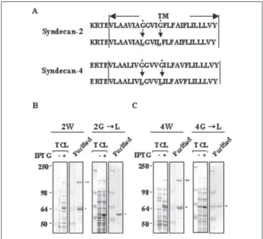
Transmembrane Domains Are Essential for SDS-resistant Dimeriza-tion of Syndecan-2 and Syndecan-4 Core Proteins—Several recombinant syndecan-2 and syndecan-4 constructs were expressed as GST fusion proteins (Fig. 1A) that were purified using glutathione-agarose beads and analyzed by SDS-PAGE following Coomassie blue staining (Fig. 1, B and C). Consistent with the previous reports (14), recombinant full-length syndecan-4 core protein (GST-4W) resolved as SDS-resistant dimers. In addition, five different mutants (4ET, 4eTC, 4TC, 4eT, and 4T) that contained the transmembrane domain showed SDS-resistant dimer formation. On the other hand, neither the cytoplasmic domain (4C) nor the extracellular domain alone (4E) showed SDS-resistant dimerization. Syndecan-2 core proteins showed closely similar results to those of syndecan-4 (Fig. 1C). Therefore, it seems that the transmem-brane domain is important for SDS-resistant dimerization of synde-can-2 and syndecan-4. Similar to syndecan-3 (19), replacement of con-served glycine residues in the transmembrane domain with leucine residues (Gly3 Leu) abolished SDS-resistant dimer formation of both syndecan-2 and syndecan-4 (Fig. 2), supporting the notion that the transmembrane domain is crucial for SDS-resistant dimerization of both syndecan-2 and syndecan-4.
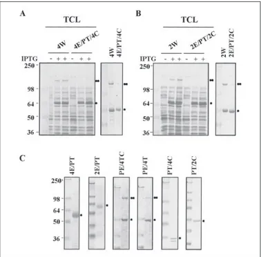
To further investigate the function of the transmembrane domain, each transmembrane domain of syndecan-2 and syndecan-4 was replaced with the transmembrane domain of PDGFR that contains a single transmembrane domain and forms noncovalent dimers in response to ligand binding (9, 10). Consistently, GST-4W and GST-2W showed a strong SDS-resistant dimer formation that was hardly seen in 4E/PT/4C (Fig. 3A) and 2E/PT/2C (Fig. 3B). Furthermore, mutants PE/4T and PE/4TC, which contained the transmembrane domain of syndecan but not that of PDGFR, showed SDS-resistant dimer forma-tion (Fig. 3C). Therefore, these data strongly suggest that the transmem-brane domain is sufficient for inducing SDS-resistant dimerization of both syndecan-2 and syndecan-4 core proteins.
Transmembrane Domains Are Sufficient for Inducing Dimer Forma-tion of Chimeric Proteins in Vivo—Additional syndecan chimeras (4ET/PC and 2ET/PC) were constructed that consisted of the extracel-lular domain and transmembrane domain of each syndecan and the cytoplasmic domain of PDGFR. Either 4ET/PC or 2ET/PC chimera cDNA was cotransfected with Erk1 cDNA into HEK293T cells, and chimera-induced MAPK activation was analyzed. Because it is well known that dimerization of PDGFR cytoplasmic domain is sufficient to activate MAPK activity (11, 12), MAPK activation indicates dimeriza-tion of these chimeras. Western blotting with phospho-Erk anti-body showed that MAPK activation in chimera-transfected cells was increased compared with that in vector-transfected cells (Fig. 4A,
bot-tom section) together with increased tyrosine phosphorylation of several proteins (Fig. 4A, top section). In contrast, transfection of wild type syndecan-4, wild type syndecan-2, or the syndecan-4 chimera with Gly to Leu substitution in the transmembrane domain cDNAs did not affect MAPK activity (Fig. 4B). Interestingly, there was no significant differ-ence in MAPK activation between 4ET/PC and 2ET/PC (Fig. 4B), implying that both transmembrane domains similarly induce dimeriza-tion of chimeras. Because overexpression of the PDGFR cytoplasmic
FIGURE 1. Transmembrane domain is essential for SDS-resistant dimerization of syndecan-4 and syndecan-2. A, schematic representation of GST-syndecan-2 (Syn-2) and GST-syndecan-4 (Syn-4) core proteins. The extracellular domain is represented by the white box (Extracellular), the transmembrane domain by the black box (TM), the cyto-plasmic domain by the shaded box (Cyto), and GST by the circle. Four amino acid residues in membrane flanking region (ERTE, KRTE) and molecular mass estimated from the deduced amino acid sequences are shown. B and C, purification of recombinant GST-syndecan-4 (B) and GST-syndecan-2 (C) core proteins expressed in E. Coli. Purified recom-binant proteins were separated by electrophoresis on 10% SDS-polyacrylamide gels and stained with Coomassie Blue. Molecular mass markers in kilodaltons are shown. Migra-tion posiMigra-tions of SDS-resistant dimer (● ●) and monomer (●) are indicated.
FIGURE 2. Transmembrane mutants fail to form SDS-resistant dimerization of syn-decan-4 and syndecan-2. A, syndecan amino acid sequences of the transmembrane (TM) with four amino acid residues (ERTE, KRTE) in the membrane flanking region are shown. Two of the conserved glycine residues indicated by closed circles were replaced by leucine residues, as indicated by the arrows. B and C, both total cell lysates (TCL) and purified recombinant syndecan or its mutant (Gly3 Leu) were separated by electro-phoresis on 8% SDS-polyacrylamide gels and stained with Coomassie Blue. Migration positions of SDS-resistant dimer (●●) and monomer (●) are indicated. IPTG, isopropyl--D-thiogalactopyranoside.
at Ewha Medical Library on July 17, 2017
http://www.jbc.org/
domain per se might affect chimera-induced MAPK activation, LET/PC was constructed that consisted of the extracellular domain and trans-membrane domain of LAT, which has not been known to form oli-gomers (21, 22), and the cytoplasmic domain of PDGFR (Fig. 4C). Over-expression of LET/PC did not significantly affect MAPK activity,
implying that transmembrane domain induced dimerization is crucial for chimera-induced MAPK activation. Therefore, it is likely that trans-membrane domain is sufficient for inducing the dimer of syndecan-2 and syndecan-4 in vivo.
Transmembrane Domain-induced Dimerization Is Independent of the Extracellular Domain—Because it is known that engagement of syndecan through its extracellular domain induces oligomerization, we investigated the effect of the extracellular domain on chimera-induced dimerization. Vector and chimera (4ET/PC)-transfected cells were plated on either syndecan-2 or syndecan-4 antibody-coated plates to induce clustering of syndecan-2 and syndecan-4 extracellular domains. However, chimera-induced MAPK activities were not affected by engagement of the extracellular domain with the specific antibodies (Fig. 5A). We then made a PDGFR chimera where its transmembrane domain was substituted with that of syndecan-4 (PE/4T/PC) (Fig. 5, B and C). Transfection of this chimera in HEK293T cells showed increased tyrosine phosphorylation of this PDGFR chimera (Fig. 5B, top
section) and MAPK activity (Fig. 5B, bottom section). This chimera-induced MAPK activation was independent on treatment of 50 ng/ml PDGF (Fig. 5C). Thus, it is likely that the transmembrane domain of syndecan-induced dimerization is not affected by the extracellular domain. These results strongly suggest that both syndecan-2 and syn-decan-4 form dimers in vivo mediated by their transmembrane domains alone.
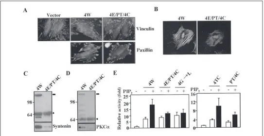
Transmembrane Domain-induced Oligomerization Is Crucial for Syndecan Functions—We next investigated the functionality of trans-membrane domain-induced oligomerization. Because syndecan-4 plays an important role in focal adhesion formation, we analyzed the effect of the altered oligomerization on focal adhesion formation. REF cells transfected with the cDNAs of either 4W or 4E/PT/4C were plated on fibronectin, and the focal adhesions were highlighted by indirect
immu-FIGURE 3. The substitution of transmembrane fails to form SDS-resistant dimeriza-tion of syndecan-4 and syndecan-2. A and B, either total cell lysates (left secdimeriza-tion) or purified recombinant syndecan/PDGFR chimeras (right section) were separated by electrophoresis on 8% SDS-polyacrylamide gels and stained with Coomassie Blue. C, purified proteins were separated by SDS-PAGE and stained with Coomassie Blue. Migration positions of SDS-resistant dimer (●●) and monomer (●) are indicated.
IPTG, isopropyl--D-thiogalactopyranoside.
FIGURE 4. The transmembrane domains of syndecan-2 and syndecan-4 are suffi-cient for chimera-induced MAPK activation. A and B, 1-g cDNA of each chimera, 4ET/PC, 2ET/PC, or 4Gly3 Leu/PC were transiently cotransfected with 1g of Erk1 cDNA into HEK293T cells as described under “Experimental Procedures.” After cells were lysed using radioimmune precipitation assay buffer, whole cell lysates were separated by elec-trophoresis on 10% SDS-polyacrylamide gels, and tyrosine phosphorylation was ana-lyzed using anti-phospho-specific tyrosine antibody (␣-pTyr). MAPK activation was assessed by using phospho-specific antibody (␣-pErk) followed by stripping and reprob-ing with anti-Erk2 antibody. C, cDNAs of either LET/PC or 4ET/PC were transiently trans-fected as indicated. MAPK activation was assessed by using phospho-specific antibody (pErk) followed by stripping and reprobing with anti-Erk2 antibody.
FIGURE 5. The extracellular domain does not affect dimerization tendency of the transmembrane domain. A, either vector or 4ET/PC transfected cells were trypsinized and replated on either bovine serum albumin-coated, syndecan-2 antibody (Syn2 Ab)-coated, or syndecan-4 antibody (Syn4 Ab)-coated plates in the absence of serum. After incubation at 37 °C for 24 h, cells were lysed using radioimmune precipitation assay buffer, and whole cell lysates were separated by electrophoresis on 10% SDS-polyacryl-amide gels. MAPK activation was assessed by using phospho-specific antibody (␣-pErk2) followed by stripping and reprobing with anti-Erk2 antibody (␣-Erk2). B, exponentially growing cells were lysed, and phosphorylation of the PDGFR chimera was analyzed by immunoprecipitation with anti-phosphotyrosine antibody (top section). MAPK activa-tion was assessed as described for panel A. C, exponentially growing cells were serum-starved overnight and then treated with 50 ng/ml PDGF for 15 min. MAPK activation was assessed as described for panel A.
at Ewha Medical Library on July 17, 2017
http://www.jbc.org/
nofluorescence using vinculin or paxillin antibodies. Consistent with previous reports (23, 24), increased focal adhesion formation was observed in 4W-transfected cells as compared with vector-transfected cells. In contrast, expression of an oligomerization mutant 4E/PT/4C slightly decreased focal adhesion formation (Fig. 6A). Exogenously expressed 4W, but not 4E/PT/4C, was retained in the Triton X-100-resistant cytoskeleton (25), implying that the susceptibility of Triton X-100 extraction was significantly increased in the oligomerization mutant (Fig. 6B) and that the interaction with syntenin (26, 27) was evidently decreased in 4E/PT/4C (Fig. 6C). Previous work has shown that, in combination with PIP2, syndecan-4 cytoplasmic domain can interact and strongly activate PKC␣ (14, 16, 18). However, the synde-can-4 oligomerization mutant 4E/PT/4C showed reduced interaction with PKC␣ (Fig. 6D), and 4E/PT/4C, PT/4C, and 4Gly 3 Leu promoted much less in vitro PKC␣ activity (Fig. 6E). The functions of syndecan-2 were also affected by transmembrane domain-induced oligomerization. RIE1 cells transfected with the cDNAs of 2W, but not 2E/PT/2C, showed increased cell migration compared with control cells (Fig. 7A). Similar to 4E/PT/4C, the interaction with syntenin was decreased in 2E/PT/2C (Fig. 7B). Taken together, all these data strongly suggest that transmembrane domain-induced oligomerization is crucial for the functions of both syndecan-2 and syndecan-4 in vivo.
DISCUSSION
Syndecan core proteins are known to have propensity to form non-covalently linked dimers (2, 5, 19). High sequence homology and a pre-vious study with syndecan-3 have suggested that both the transmem-brane domain and the four amino acid residues in the ectodomain flanking region are important for dimerization of syndecan core pro-teins. In this study, we have further investigated the importance of each syndecan domain for functional dimerization. Consistent with previous results (14, 19), recombinant full-length core proteins of syndecan-2 and syndecan-4 (GST-4W and GST-2W) showed a strong SDS-resist-ant dimer formation (Figs. 1–3) that was hardly seen in 4E/PT/4C (Fig. 3A) and 2E/PT/2C (Fig. 3B). Replacement of the conserved glycine res-idues in the transmembrane domain with leucine resres-idues also
abol-ished SDS-resistant dimer formation of recombinant syndecan core proteins (Fig. 2), supporting the notion that the transmembrane domain is crucial for SDS-resistant dimerization of both syndecan-2 and synde-can-4. However, unlike syndecan-3, where mutation of any of the four amino acid residues (ERKE) in the ectodomain immediately external to the transmembrane domain decreased the extent of dimer formation (19), deletion of all four amino acid residues does not decrease SDS-resistant dimer formation of either syndecan-2 or syndecan-4 core pro-teins (Fig. 1). Clearly, the ectodomains do not promote SDS-resistant dimerization of syndecan-2 and syndecan-4 and seem not to be required.
It is clear that syndecans form dimers and the transmembrane domains are crucial for SDS-resistant dimerization of both syndecan-2 and syndecan-4 in vitro. However, there is no direct evidence showing transmembrane domain-induced dimer formation of syndecans in vivo. To define functional dimerization of syndecan core protein in cells, several syndecan-PDGFR chimeras have been used. This receptor has been chosen because its activation is dependent on receptor
dimeriza-FIGURE 7. Functions of syndecan-2 are defective in oligomerization mutant. A, the migration assays of RIE1 cells transfected with cDNAs of either 2W or 2E/PT/2C were performed as described under “Experimental Procedures” using gelatin (1 g/l)-coated Transwell plates. Shown is the relative number of cell migration. B, equal amount of purified GST-2W and GST-2E/PT/2C (top section) were incubated with REF cell lysates for 2 h. Proteins bound were immunoblotted with anti-syntenin antibodies (bottom sec-tion). Migration positions of SDS-resistant dimer (●●) and monomer (●) are indicated. FIGURE 6. Functions of syndecan-4 are defective in oligomerization mutants. A and B, REF cells transfected with the cDNAs of either Myc-tagged 4W or Myc-tagged 4E/PT/4C. Cells were plated on fibronectin-coated plates for 2.5 h and then stained with anti-vinculin (top row) or anti-paxillin antibodies (bottom row) (A). After 24 h plated on coverslips, cells were extracted with 0.1% Triton X-100 in PBS for 10 min and stained with anti-c-Myc antibodies (B). C, equal amount of purified GST-4W and GST-4E/PT/4C (top section) were incubated with REF cell lysates for 2 h. Proteins bound were immunoblotted with anti-syntenin antibodies (bottom section). Migration positions of the SDS-resistant dimer (●●) and monomer (●) are indicated. D, equal amount of purified GST-4W and GST-4E/PT/4C (top section) were mixed with purified PKC␣ for 2 h. Proteins bound were immunoblotted with anti-PKC␣ antibodies (bottom section). Migration positions of SDS-resistant dimer (●) and monomer (●) are indicated. E, PKC assays were performed as described under “Experimental Procedures” with purified 4W or syndecan-4 mutants (0.25 mg/ml) either in the presence or absence of 50M PIP2.Relative activity is indicated by mean⫾ S.E. (n ⫽ 5) compared with
that in the absence of syndecan-4 proteins.
at Ewha Medical Library on July 17, 2017
http://www.jbc.org/
tion and its signaling can be readily analyzed by MAPK activation. Transfection studies with syndecan-PDGFR chimeras clearly show that the transmembrane domains of both syndecan-2 and syndecan-4 are able to induce dimerization of these chimeras, resulting in constitutive MAPK activation (Fig. 4 and 5). Similarly to SDS-resistant dimerization of recombinant core proteins, the extracellular domain is not involved in dimerization of syndecan-PDGFR chimera, as shown by the following three findings. 1) Antibody-induced oligomerization of syndecan extra-cellular domain did not affect chimera-induced MAPK activation (Fig. 5A). 2) Chimera-induced MAPK activation was similarly increased in suspended cells (data not shown). 3) PDGFR chimeras containing the transmembrane domain of syndecan-4 by itself induced MAPK activa-tion independently of PDGF treatment (Fig. 5C). It is believed that growth factor receptors form dimers in a ligand-dependent manner. However, the chimeras containing the transmembrane domain of either sydecan-2 or sydecan-4 likely form constitutive dimers, based on MAPK activation and electrophoretic mobility (Fig. 4 and 5). This result con-firms the hypothesis that dimerization of PDGFR is PDGF-dependent and sufficient for PDGF-mediated MAPK activation. However, it is not the case in syndecan-mediated cell spreading. Because syndecan-4 has a strong tendency for higher order oligomerization (6, 19, 28), both syn-decan-2 and sydecan-4 may form constitutive dimers that, after plating on the extracellular matrix, form higher order oligomers. Because syn-decan-4 can become a high order oligomer both in vivo and in vitro, we could not exclude the possibility that the chimeric proteins described here might also form higher order oligomers. Under our experimental conditions, MAPK activity only reflects dimerization but not higher order oligomerization. Therefore, we cannot discriminate whether the transmembrane domain induces dimerization or higher order oli-gomerization of syndecan core protein in vivo. In addition, although neither the extracellular domain nor the cytoplasmic domain promotes SDS-resistant dimerization of syndecans, it may be possible that both domains regulate higher order oligomerization of syndecan core protein
in vivo.
Syndecan-4 acts as a coreceptor with integrins in focal adhesion for-mation (6, 7), but oligomerization mutants failed to enhance focal adhe-sion formation (Fig. 6A), reside in the Triton X-100-resistant cytoskel-eton (Fig. 6B), interact with PKC␣ (Fig. 6D), or efficiently activate PKC␣
in vitro(Fig. 6E). This implies that transmembrane domain-induced oligomerization may control the biological activity of the syndecan-4 cytoplasmic domain. The syndecan-2 oligomerization mutant was also unable to regulate cell migration (Fig. 7). Therefore, it seems that the transmembrane domain is crucial for inducing functional oligomeriza-tion of the syndecan-2 and sydecan-4 core proteins. This is the first evidence to show the functionality of the transmembrane domain-in-duced oligomerization in vivo.
One of the most important observations in this study is the apparent specificity of the transmembrane domain. The functionality of the transmembrane domain is more significant when contrasted with growth factor receptor-mediated signal transduction. During trans-membrane signaling, extracellular ligands bind to specific receptors on the cell surface, and the binding subsequently alters their oligomeric status to evoke a signal cascade inside the cell (9, 10). A variety of studies have shown that the activation of downstream signaling is dependent on the oligomeric status of the cytoplasmic domain. However, although it plays a major role in controlling oligomeric status of the cytoplasmic domain in the case of syndecans, the transmembrane domain has received relatively little attention. It is clear that oligomerization of the
cytoplasmic domain of receptor is a different issue from that of whole receptor protein, where the transmembrane domains are a key driver of oligomerization. Thus, the transmembrane domain inevitably regulates the activity of the cytoplasmic domain and is crucial for receptor func-tions. How does the transmembrane domain regulate the cytoplasmic domain? Is it only by inducing oligomerization? Transmembrane domains of both PDGFR and syndecan are able to induce oligomeriza-tion of the receptor protein in vivo, although the ligand dependence may be different for the two. The important point is that they both have the potential to do so and that their oligomerization is crucial for activating signaling cascades. Interestingly, however, the unique nature of synde-can function is that the specific activity of syndesynde-can requires the pres-ence of syndecan but not of the PDGFR transmembrane domain. This is demonstrated by the syndecan/PDGFR chimeras where each syndecan transmembrane domain was substituted with that of PDGFR, which showed a clear defect in most of syndecan functions tested (Figs. 6 and 7). This implies that transmembrane domain-induced oligomerization is not just for aggregating the receptors. It may induce a quaternary structure of the cytoplasmic domain, whose overall structure is required for the specific function of each receptor protein. Therefore, syndecan transmembrane domain-induced oligomerization plays a syndecan-specific role in regulating the functions of syndecan that cannot be replaced by other transmembrane oligomerization domains. It remains unclear whether this is a general phenomenon of homo-oligomerization of receptors.
REFERENCES
1. Kim, C., Goldberger, O., Gallo, R., and Bernfield, M. (1994) Mol. Cell. Biol. 5, 797– 805 2. Bernfield, M., Kokenyesi, R., Kato, M., Hinkes, M. T., Spring, J., Gallo, R. L., and Lose,
E. J. (1992) Annu. Rev. Cell Biol. 8, 365–393 3. Carey, D. J. (1997) Biochem. J. 327, 1–16
4. Zimmermann, P., and David, G. (1999) FASEB J. 13, S91–S100 5. Rapraeger, A. C., and Ott, V. L. (1998) Curr. Opin. Cell Biol. 10, 620 – 628 6. Woods, A., and Couchman, J. R. (2001) Curr. Opin. Cell Biol. 13, 578 –583 7. Rapraeger, A. C. (2000) J. Cell Biol. 149, 995–998
8. Simons, M., and Horowitz, A. (2001) Cell. Signal. 13, 855– 862 9. Schlessinger, J. (2000) Cell 103, 211–225
10. Heldin, C. H. (1995) Cell 80, 213–223
11. Schlesinger, T. K., Demali, K. A., Johnson, G. L., and Kazlauskas, A. (1999) Biochem. J. 344,519 –526
12. DeMali, K. A., Balciunaite, E., and Kazlauskas, A. (1999) J. Biol. Chem. 274, 19551–19558
13. Woods, A., and Couchman, J. R. (1994) Mol. Biol. Cell 5, 183–192
14. Oh, E. S., Woods, A., and Couchman, J. R. (1997) J. Biol. Chem. 272, 8133– 8136 15. Oh, E. S., Woods, A., Lim, S. T., Theibert, A., and Couchman, J. R. (1998) J. Biol. Chem.
273,10624 –10629
16. Horowitz, A., and Simons, M. (1998) J. Biol. Chem. 273, 10914 –10918 17. Baciu, P. C., and Goetinck, P. F. (1995) Mol. Biol. Cell 6, 1503–1513
18. Horowitz, A., Murakami, M., Gao, Y., and Simons, M. (1999) Biochemistry 38, 15871–15877
19. Asundi, V. K., and Carey, D. J. (1995) J. Biol. Chem. 270, 26404 –26410 20. Inagaki, M., Watanabe, M., and Hidaka, H. (1985) J. Biol. Chem. 260, 2922–2925 21. Wonerow, P., and Watson, S. P. (2001) Oncogene 20, 6273– 6283
22. Tomlinson, M. G., Lin, J., and Weiss, A. (2000) Immunol. Today 21, 584 –591 23. Longley, R., Woods A., Fleetwood A., Cowling G. J., Gallagher J. T., and Couchman
J. R. (1999) J. Cell Sci. 112, 3421–3431
24. Echtermeyer, F., Baciu, P. C., Saoncella, S., Ge, Y., and Goetinck, P. F. (1999) J. Cell Sci. 112,3433–3441
25. Greene, D. K., Tumova, S., Couchman, J. R., and Woods A. (2003) J. Biol. Chem. 278, 7617–7623
26. Grootjans, J. J., Zimmermann, P., Reekmans, G., Smets, A., Degeest, G., Durr, J., and David, G. (1997) Proc. Natl. Acad. Sci. U. S. A. 94, 13683–13688
27. Grootjans, J. J., Reekmans, G., Ceulemans, H., and David, G. (2000) J. Biol. Chem. 275, 19933–19941
28. Oh, E. S., and Couchman, J. R. (2004) Mol Cells 17, 181–187
at Ewha Medical Library on July 17, 2017
http://www.jbc.org/
doi: 10.1074/jbc.M509238200 originally published online October 27, 2005
2005, 280:42573-42579.
J. Biol. Chem.
10.1074/jbc.M509238200
Access the most updated version of this article at doi:
Alerts:
When a correction for this article is posted
•
When this article is cited
•
to choose from all of JBC's e-mail alerts
Click here
http://www.jbc.org/content/280/52/42573.full.html#ref-list-1
This article cites 25 references, 14 of which can be accessed free at
at Ewha Medical Library on July 17, 2017
http://www.jbc.org/