Amygdalin Contents in Peaches at Different Fruit Development
Stages
Suk-Hee Lee1, Angela Oh2, Seo-Hee Shin2, Ha-Na Kim2, Woo-Won Kang3, and Shin-Kyo Chung2 1Gyeongsangbuk-do Agricultural Research and Extension Services, Daegu 41404, Korea
2School of Food Science and Biotechnology, Kyungpook National University, Daegu 41566, Korea 3Department of Food and Food Service Industry, Kyungpook National University, Gyeongbuk 37224, Korea
Prev. Nutr. Food Sci. 2017;22(3):237-240
https://doi.org/10.3746/pnf.2017.22.3.237
pISSN 2287-1098ㆍeISSN 2287-8602
Research Note
Received 13 March 2017; Accepted 12 July 2017; Published online 30 September 2017 Correspondence to Shin-Kyo Chung, Tel: +82-53-950-5778, E-mail: [email protected]
Copyright © 2017 by The Korean Society of Food Science and Nutrition. All rights Reserved.
This is an Open Access article distributed under the terms of the Creative Commons Attribution Non-Commercial License (http://creativecommons.org/licenses/by-nc/4.0) which permits unrestricted non-commercial use, distribution, and reproduction in any medium, provided the original work is properly cited.
ABSTRACT: Amygdalin contents of the seeds, endocarps, and mesocarps from three peach cultivars (i.e., Stone Peach, Hikawa Hakuho, and Bakhyang) were measured at three stages of fruit development (stone-hardening, fruit enlargement, and ripening). The peach samples were dried and defatted with a Soxhlet apparatus, reflux extracted with methanol, and analyzed using reverse phase high-performance liquid chromatography. During all fruit development stages, the amygdalin contents in the seeds were higher than those in the endocarps and mesocarps. The amygdalin contents of the Stone Peach were comparatively higher than the Hikawa Hakuho and Bakhyang (P<0.05). Further, the amygdalin contents during rip-ening were very low or not detected. Overall, the amygdalin contents of the three peach cultivar samples (seed, endocarp, and mesocarp) increased until the fruit enlargement stage and either remained constant or decreased during ripening. Keywords: amygdalin, fruit development stages, peaches, HPLC, seeds
INTRODUCTION
Due to the abundance of sugar and organic acids, peaches are primarily consumed as fresh produce in Korea. In 2015, the peach production in Korea was 238,000 M/T, and 0.4% was processed (1). Peaches are processed as jam and jelly and fermented to alcoholic drinks (2-4). Unripe peaches contain more nutrients than ripe peaches, such as organic acids, minerals, and polyphenols (5). In Korea, unripe peaches are thinned out and discarded in the orchard mostly from early April to late May. Some studies were carried out on the chemical composition (5-7) and utilization of unripe peaches as functional ma-terials (8,9). Peach seeds were also studied in regard to their use in the prevention of atherosclerosis (10), and were also processed to functional materials (11). Amyg-dalin (D-mandelonitrile-β-D-glucoside-6-β-D-glucoside) is a cyanogenic glycoside plant toxin contained in rela-tively high concentrations in the kernels and seeds of ap-ples, apricots, almonds, cherries, and peaches (12,13), and it is abundant in plum seeds (14). Notably, amygda-lin is hydrolyzed to HCN, benzaldehyde, and D-glucose and can cause acute intoxication and chronic human cen-tral nervous system maladies (15,16). The quantitative profile of amygdalin content during peach development
should be studied in order to utilize unripe peaches as functional materials, and to enhance the quality of proc-essed peach products. In this study, the amygdalin con-tents of three cultivars of peach seeds, endocarps, and mesocarps were analyzed during fruit development (stone-hardening stage, fruit enlargement stage, and rip-ening period).
MATERIALS AND METHODS
Sample materials and reagents
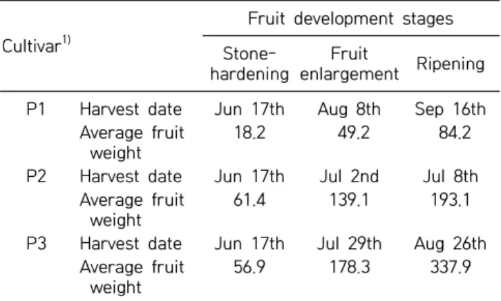
Three cultivars of peaches [Stone Peach (P1), Hikawa Hakuho (P2), and Baekhyang (P3)] were obtained from the Cheongdo Peach Experimental Station of Gyeong-sangbuk-do Agricultural Research Extension Services (Cheongdo, Korea). Table 1 shows the harvest time of each cultivar. Fruits with uniform colors and sizes were selected and divided into mesocarps, endocarps, and seeds. The samples were then air-dried in the shade. Amygdalin standards were purchased from Sigma Co. (St. Louis, MO, USA), and solvents for high-perform-ance liquid chromatography (HPLC) analysis were pur-chased from J.T. Baker (Phillipsburg, NJ, USA). All other chemicals used in this study were obtained from Duksan
238 Lee et al.
Fig. 1. Amygdalin decomposition. Table 1. Harvest date of peaches at different fruit development
stages
Cultivar1)
Fruit development stages
Stone-hardening
Fruit
enlargement Ripening P1 Harvest date Jun 17th Aug 8th Sep 16th
Average fruit weight
18.2 49.2 84.2
P2 Harvest date Jun 17th Jul 2nd Jul 8th Average fruit
weight
61.4 139.1 193.1
P3 Harvest date Jun 17th Jul 29th Aug 26th Average fruit
weight
56.9 178.3 337.9
1)
P1, Stone Peach; P2, Hikawa Hakuho; P3, Baekhyang.
Fig. 2. HPLC chromatograms of amygdalin standard (amygdalin STD) and Stone Peach seed extract (sample; June 17th).
Co. (Ansan, Korea).
Amygdalin extraction and HPLC analysis
The air-dried peach seeds, endocarps, and mesocarps were pulverized, screened with a 60-mesh sieve, and de-fatted with ethyl ether for 3 h using a Soxhlet apparatus. After evaporating the solvent and drying in an oven, the defatted powders were reflux extracted with methanol at 60oC for 6 h, filtered, concentrated with a rotary evapo-rator, filtered, and then injected into an HPLC instru-ment (LC-10A, Shimadzu Co., Kyoto, Japan). The HPLC column was a SupelcosilTM LC-18-S (φ 4.6×250 mm, Supelco, Bellefonte, PA, USA), and the samples were an-alyzed at a UV wavelength of 210 nm with 20% meth-anol (0.7 mL/min) as the mobile phase (17). The amyg-dalin contents were determined with linear regression methods using an amygdalin standard curve.
Statistical analysis
All measured values are reported as mean±standard de-viation, and evaluated by the analysis of variance (ANOVA), followed by Duncan’s multiple range test (P<0.05) using the Statistical Analysis System software
package (version 9.3, SAS Institute Inc., Cary, NC, USA).
RESULTS AND DISCUSSION
Amygdalin analysis by HPLC
As a cyanogenic glycoside, amygdalin is enzymatically hydrolyzed and ultimately transformed into benzalde-hyde and HCN (Fig. 1). The total cyanide content used to be determined using the picrate and acid hydrolysis method for the quantification of cyanogenic glycosides (18). However, since the development of reverse phase HPLC (19), this newer approach has been used to quick-ly determine the amygdalin content in a variety of ma-trices (20). Fig. 2 shows the reverse phase HPLC chro-matograms of the amygdalin standard and a P1 seed. Amygdalin was isolated fairly well in the reverse phase C18 column with a 20% methanol mobile phase. Amyg-dalin contents were obtained from the linear regression curve of the standard amygdalin contents.
Amygdalin contents of peaches during fruit development Amygdalin is mainly contained in the seeds of Rosaceae and is catabolized upon germination as a source of nitro-gen and carbon (21) for later use during flowering and
Amygdalin Contents in Peach Fruits 239
Fig. 3. Amygdalin contents in the seeds of three peach cultivars during different fruit development stages. Means followed by the same letters within the same peaches (a-c) and the same stages (A-C) are not significantly different (P<0.05). P1, Stone Peach; P2, Hikawa Hakuho; P3, Baekhyang.
Fig. 4. Amygdalin contents in the endocarps of three peach cul-tivars during different fruit development stages. Means fol-lowed by the same letters within the same peaches (a-c) and the same stages (A-C) are not significantly different (P<0.05). P1, Stone Peach; P2, Hikawa Hakuho; P3, Baekhyang. 1)Not de-tected.
Fig. 5. Amygdalin contents in the mesocarps of three peach cul-tivars during different fruit development stages. Means fol-lowed by the same letters within the same peaches (a-c) and the same stages (A-C) are not significantly different (P<0.05). P1, Stone Peach; P2, Hikawa Hakuho; P3, Baekhyang. 1)Not de-tected.
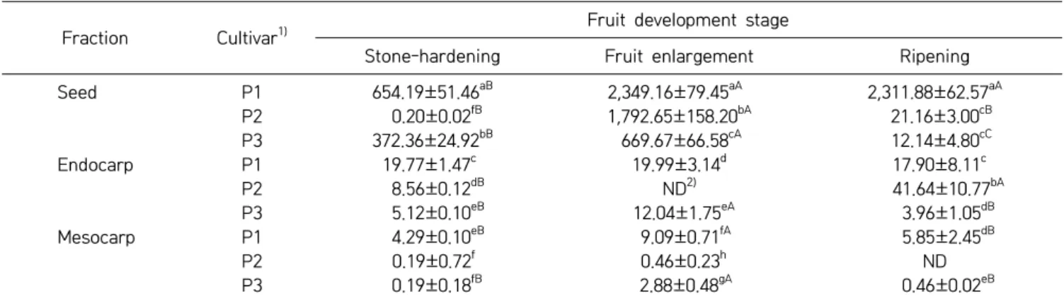
Table 2. Amygdalin contents of three peach cultivars at different fruit development stages (unit: mg/100 g, DW) Fraction Cultivar1)
Fruit development stage
Stone-hardening Fruit enlargement Ripening
Seed P1 654.19±51.46aB 2,349.16±79.45aA 2,311.88±62.57aA P2 0.20±0.02fB 1,792.65±158.20bA 21.16±3.00cB P3 372.36±24.92bB 669.67±66.58cA 12.14±4.80cC Endocarp P1 19.77±1.47c 19.99±3.14d 17.90±8.11c P2 8.56±0.12dB ND2) 41.64±10.77bA P3 5.12±0.10eB 12.04±1.75eA 3.96±1.05dB Mesocarp P1 4.29±0.10eB 9.09±0.71fA 5.85±2.45dB P2 0.19±0.72f 0.46±0.23h ND P3 0.19±0.18fB 2.88±0.48gA 0.46±0.02eB
Means followed by the same letters within the column (a-h) and the row (A-C) are not significantly different (P<0.05).
1)
P1, Stone Peach; P2, Hikawa Hakuho; P3, Baekhyang.
2)
Not detected.
fruit development (22). The amygdalin contents in the seeds, endocarps, and mesocarps from three peach culti-vars during stone-hardening, fruit enlargement, and rip-ening stages are shown in Fig. 3, 4, and 5, respectively. The amygdalin contents in the seeds of the three culti-vars increased until the fruit enlargement stage. During the ripening period, the amygdalin contents of the P1 re-mained constant, but decreased largely for the P2 and P3. In the endocarp of P1, the amygdalin contents showed no significant differences among the fruit development stages. In P3, the amygdalin contents increased slightly to the fruit enlargement stage. But the amygdalin con-tents were not detected in P2. During the ripening peri-od, the amygdalin contents increased in P2, but were not detected in P3. The amygdalin contents of the P1 and P3 mesocarps (flesh) increased until the fruit enlargement stage, and then decreased slightly during ripening. In contrast, no significant differences between the stone- hardening and fruit enlargement stages were observed for the amygdalin contents of the P2 mesocarps, and no amygdalin was detected during the ripening period. The amygdalin contents of the three peach cultivars
depend-ing on their parts at different development stages are shown in Table 2. Overall, the amygdalin contents in the seeds were reasonably higher than those in the endocarp and mesocarp. Relative to P2 and P3, P1 had the
great-240 Lee et al.
est amygdalin content (P<0.05). Additionally, the amyg-dalin contents increased until the fruit enlargement stage and kept constant or decreased slightly during ripening. Likewise, the amygdalin contents of four stone fruit spe-cies (apricot, peach, plum, and bitter apricot trees) also increased during fruit development and remained stable or decreased slightly when they ripened (17,23).
In summary, the amygdalin contents in the seeds, en-docarps, and mesocarps of three peach cultivars (P1, P2, and P3) were determined during fruit development by reverse phase HPLC. For every stage and cultivar, the amygdalin contents in the peach seeds were greater than those in the endocarps and mesocarps (P<0.05). The amygdalin contents of the P1 were moderately greater than those of the other cultivars (P2 and P3). Notably, amygdalin contents were very low or not detected during the ripening periods of the peaches. Further, the amyg-dalin contents of the peaches increased until the fruit enlargement stage and remained constant or decreased slightly during the ripening periods.
AUTHOR DISCLOSURE STATEMENT
The authors declare no conflict of interest.
REFERENCES
1. Ministry of Agriculture, Food and Rural Affairs. 2016. The status of fruits processing in 2015. Sejong, Korea.
2. Yi SH, Ann YG, Choi JS, Lee JS. 1996. Development of peach fermented wine. Korean J Food Nutr 9: 409-412.
3. Jo JW, Kim JK, Kim ID, Kim SD. 2000. Characteristics of peach wine prepared by using different cultivars. Korean J Postharvest Sci Technol 7: 84-88.
4. Chung JH, Mok C, Lim S, Park YS. 2003. Changes of physico-chemical properties during fermentation of peach wine and quality improvement by ulfilteration. J Korean Soc Food Sci Nutr 32: 506-512.
5. Lee JB, Chung HS. 2008. Studies on the components of un-ripe peaches. Korean J Food Preserv 15: 79-83.
6. McCready RM, McComb EA. 1954. Pectic constituents in ripe and unripe fruit. J Food Res 19: 530-535.
7. Kim DM, Kim KH, Choi IJ, Yook HS. 2012. Composition and physicochemical properties of unripe Korean peaches ac-cording to cultivars. J Korean Soc Food Sci Nutr 41: 221-226. 8. Kim HJ. 2007. Isolation and characterization of active whit-ening compound from Prunus persica. MS Thesis. Korea Uni-versity, Seoul, Korea.
9. Kim DM, Kim KH, Kim YS, Koh JH, Lee KH, Yook HS. 2012. A study on the development of cosmetic materials using unripe peaches seed extracts. J Korean Soc Food Sci Nutr 41: 110-115.
10. Yoon IH, Seo BI, Kim SH. 1996. The effect of Persicae Semen on the atherosclerosis in rabbit. J Herbol 11: 79-98.
11. Ryu HM, Jeon DK, Kim SA, Chung HJ. 2013. Antioxidant and quality characteristics of mungbean starch gel added with peach seed powder. Korean J Food Preserv 20: 372-378. 12. Nahrstedt A. 1972. Zur cyanogenese von Prunus avium.
Phyto-chemistry 11: 3121-3126.
13. Jones MB, Fleming ZW, Bailey LF. 1957. Cyanide as a growth inhibiting substance in extracts of peach leaves, bark, and flower buds. Proc Amer Soc Hort Sci 69: 152-157.
14. Bolarinwa IF, Orfila C, Morgan MRA. 2014. Amygdalin con-tent of seeds, kernels and food products commercially-avail-able in the UK. Food Chem 152: 133-139.
15. Concon JM. 1988. Food toxicology: principles and concepts. Marcel Dekker, Inc., New York, NY, USA. p 74.
16. Kwon H, Jo Y. 2007. A study on the decomposition of amyg-dalin using an in vitro assay. J Toxicol Pub Health 23: 47-53. 17. Viorica-Mirela G, Socaciu C, Jianu I, Florica R, Florinela F.
2006. Identification and quantitative evaluation of amygdalin from apricot, plum and peach oils and kernels. Buletin USAMV- CN 62: 246-253.
18. Haque MR, Bradbury JH. 2002. Total cyanide determination of plants and foods using the picrate and acid hydrolysis methods. Food Chem 77: 107-114.
19. Hwang EY, Lee JH, Lee YM, Hong SP. 2002. Reverse-phase HPLC separation of D-amygdalin and neoamygdalin and op-timum conditions for inhibition of racemization of amyg-dalin. Chem Pharm Bull 50: 1373-1375.
20. Kim EJ, Lee HJ, Jang JW, Kim IY, Kim DH, Kim HA, Lee SM, Jang HW, Kim SY, Jang YM, Im DK, Lee SH. 2010. Analytical determination of cyanide in maesil (Prunus mume) extracts. Korean J Food Sci Technol 42: 130-135.
21. Selmar D, Lieberei R, Biehl B. 1988. Mobilization and utiliza-tion of cyanogenic glycosides: the linustatin pathway. Plant Physiol 86: 711-716.
22. Swain E, Poulton JE. 1994. Utilization of amygdalin during seedling development of Prunus serotina. Plant Physiol 106: 437- 445.
23. Zhao Y. 2012. Amygdalin content in four stone fruit species at different developmental stages. ScienceAsia 38: 218-222.