저작자표시-비영리-변경금지 2.0 대한민국 이용자는 아래의 조건을 따르는 경우에 한하여 자유롭게
l 이 저작물을 복제, 배포, 전송, 전시, 공연 및 방송할 수 있습니다. 다음과 같은 조건을 따라야 합니다:
l 귀하는, 이 저작물의 재이용이나 배포의 경우, 이 저작물에 적용된 이용허락조건 을 명확하게 나타내어야 합니다.
l 저작권자로부터 별도의 허가를 받으면 이러한 조건들은 적용되지 않습니다.
저작권법에 따른 이용자의 권리는 위의 내용에 의하여 영향을 받지 않습니다. 이것은 이용허락규약(Legal Code)을 이해하기 쉽게 요약한 것입니다.
Disclaimer
저작자표시. 귀하는 원저작자를 표시하여야 합니다.
비영리. 귀하는 이 저작물을 영리 목적으로 이용할 수 없습니다.
변경금지. 귀하는 이 저작물을 개작, 변형 또는 가공할 수 없습니다.
이학박사 학위논문
시상하부 에너지 대사 조절에서
FAM19A5와 TTF-1의 역할에 관한 연구
Study for the regulation of energy homeostasis:
Role of FAM19A5 and TTF-1 in the hypothalamus
울산대학교 대학원 생명과학과
강다솔
시상하부 에너지 대사 조절에서
FAM19A5와 TTF-1의 역할에 관한 연구
지 도 교 수 이 병 주
이 논문을 이학박사 학위논문으로 제출함
2022 년 8 월
울산대학교 대학원 생명과학과
강다솔
Study for the regulation of energy homeostasis:
Role of FAM19A5 and TTF-1 in the hypothalamus
Supervisor: Byung Ju Lee, Ph.D
A Dissertation
Submitted to
the Graduate School of the University of Ulsan in partial Fulfillment of the Requirements
for the Degree of
Doctor of Philosophy
by Kang, Dasol
Department of Biological science Graduate School
University of Ulsan
August 2022
CONTENTS
Study for the regulation of energy homeostasis:
Role of FAM19A5 and TTF-1 in the hypothalamus
Abstract --- 1
Chapter 1. Brain-specific chemokine FAM19A5 induces hypothalamic inflammation Abstract --- 4
Introduction --- 5
Materials and Methods --- 6
Results --- 10
Figures --- 13
Discussion --- 20
Reference --- 23
Chapter 2. Regulation of AgRP and POMC expression by Sirt1 and TTF-1 interaction according to energy states Abstract --- 27
Introduction --- 28
Materials and Methods --- 30
Results --- 35
Figures --- 38
Discussion --- 44
Reference --- 47
- 1 -
Abstract
Energy imbalance is a serious issue in modern society and is associated with various metabolic disorders such as obesity and diabetes. Hypothalamus, as a center of regulating energy homeostasis, plays an important role in whole-body energy balance and is targeted in various research on metabolic disorders. In the arcuate nucleus (ARC) of the hypothalamus, agouti- related protein (AgRP) neuronal activation strongly promotes the feeding behavior that affects the positive energy balance and activation of pro-opiomelanocortin (POMC) neurons induces the negative energy balance through enhancing energy expenditure. Therefore, balance of positive and negative energy is a crucial role in energy homeostasis.
Here, I reported a role of FAM19A5, as a novel neuro-chemokine, in TNF-α-induced negative energy balance such as sickness responses (chapter 1), and the mechanism regulating positive energy balance through the expression of AgRP and POMC by TTF-1 (chapter 2).
In chapter 1, I demonstrated that FAM19A5 induced negative energy balance by activation of POMC neurons in hypothalamic inflammation. TNF-α-induced hypothalamic inflammation affected FAM19A5 mRNA expression and inhibition of FAM19A5 in hypothalamic inflammation condition reduced sickness response by inhibiting gene expression of inflammatory cytokines. Moreover, FAM19A5 induced POMC neuronal activation that is related to illness-induced anorexia. In summary, FAM19A5 induced negative energy balance in the hypothalamus, and hypothalamic inflammation-induced negative energy balance was restored by inhibition of FAM19A5, as a result, maintaining energy balance.
In chapter 2, I verified that interaction of TTF-1 and Sirt1 controls energy homeostasis by regulating AgRP and POMC gene expression according to energy states. Interaction of Sirt1 with TTF-1 resulted in deacetylation of TTF-1. Furthermore, TTF-1 deacetylation enhanced
- 2 -
AgRP mRNA expression and reduced POMC mRNA expression by promoting nuclear translocation. In summary, AgRP and POMC gene expression was regulated by TTF-1 and Sirt1 interaction, and TTF-1 deacetylation.
Together, these results suggest that FAM19A5 and TTF-1 in the hypothalamus play a pivotal role in energy homeostasis.
- 3 -
Chapter 1
Brain-specific chemokine FAM19A5
induces hypothalamic inflammation
- 4 -
Abstract
The cytokine-like protein FAM19A5 is highly expressed in the brain, but little is known about its functions there. Here, I found that FAM19A5 was expressed in mouse hypothalamic cells expressing proopiomelanocortin (POMC) and neuropeptide Y (NPY)/agouti-related peptide (AgRP), and in the microglia. Tumor necrosis factor-a (TNF-α), which induces inflammatory sickness responses, greatly increased hypothalamic expression of FAM19A5. Knockdown of FAM19A5 expression resulted in decreased TNF-α-induced anorexia, body weight loss, and TNF-α-induced expression of inflammatory factors. In contrast, intracerebroventricular administration of FAM19A5 induced anorexia, body weight loss, and hyperthermia, together with increased expression of inflammatory factors. FAM19A5 injection also induced increases in c-fos activation and POMC mRNA level in hypothalamic POMC neurons. Together, these results suggest that FAM19A5 plays an important role in hypothalamic inflammatory responses.
- 5 -
Introduction
The hypothalamus is a central brain region that controls energy homeostasis and body energy metabolism. Increasing evidence indicates that inflammatory states in the hypothalamus cause dysfunctions in energy metabolism [1]. While chronic metabolic syndromes with leptin and insulin resistance are caused by longlasting metabolic inflammation that affects the hypothalamus, acute inflammation in the hypothalamus leads to a profound negative energy balance typified by sickness responses [2]. Sickness responses are a coordinated state of adaptive behavioral changes with anorexia, fever and hypoactivity to cope with the infection [3-5]. A number of inflammatory signals stimulate local hypothalamic proinflammatory cytokines such as interleukin 1-b (IL-1b), IL-6 and tumor necrosis factor-α (TNF-α), which induce the sickness responses [6,7]. Family with sequence similarity 19 [chemokine (C-C motif)-like] member A5 (FAM19A5) encoded by TAFA genes (TAFA-1 to TAFA-5) is a secretory protein with cytokine-like properties [8] that is highly expressed in the brain and adipose tissues with very low expression in other peripheral organs [8-11]. In adipose tissues, FAM19A5 expression was reduced in rodent models of obesity with high-fat diet-induced obese and leptin receptor-deficient mice [10]. Moreover, FAM19A5 secretion from the adipocytes was decreased by treatment with proinflammatory cytokines such as TNF-α and IL- 6 [10]. Clinical data from human samples also shows that serum FAM19A5 concentration is higher in patients with type 2 diabetes [11]. These peripheral studies indicate that FAM19A5 is involved in the pathophysiology of chronic metabolic inflammation and with inflammatory cytokines. Nonetheless, little is known about its function in the brain. In the current study, I describe how FAM19A5 is involved in hypothalamic inflammatory sickness responses with acute inflammation.
- 6 -
Materials and Methods
Animals
Eight-week-old male C57BL/6 mice were purchased from Hyochang Science (Daegu, South Korea) and used for animal experiments. Transgenic mice expressing enhanced green fluorescent protein in their proopiomelanocortin (POMC) neurons (POMC-EGFP mice,
#008322, Jackson Laboratories, Bar Harbor, ME) and mice expressing humanized Renilla reniformis green fluorescent protein in their neuropeptide Y (NPY) neurons (NPY-hrGFP mice,
#006417, Jackson Laboratories) were used for immunohistochemistry. Animal experiments were conducted in accordance with the regulations of the University of Ulsan Committee for the Care and Use of Laboratory Animals (permission number, BJL-18-010). Mice were housed in a room with a conditioned photoperiod (12 h light/ 12 h dark, lights on from 06:00 to 18:00) and regulated temperature (23-25 ℃) and were allowed ad libitum access to tap water and mouse chow pellets.
Stereotaxic surgery and icv administration of materials
Mice were anesthetized by intraperitoneal injection of tribromoethanol (250 mg/kg, Sigma- Aldrich, ST. Louis, MO) and fixed with a stereotaxic device (Stoelting, Wood Dale, IL). The cannula (26-gauge) was implanted into the right lateral ventricle (0.1 mm lateral, 0.03 mm posterior and 2.4 mm ventral to the bregma) according to the stereotaxic mouse brain atlas [12]
and secured to the skull with dental cement. After a one week recovery period, mice were intracerebroventricularly (icv) injected with sterile saline or recombinant human FAM19A5 (1 mg/2 ml saline, Neuracle Science, Seoul, South Korea) or recombinant human TNF-α (50 ng/2 ml saline, R&D Systems, Minneapolis, MN).
- 7 -
Blockade of FAM19A5 expression with antisense oligodeoxynucleotide
To interfere with FAM19A5 synthesis in the mouse brain, antisense (AS) oligodeoxynucleotide (ODN) (IDT Korea Inc., Gyeonggido, South Korea) was icv delivered into the brain through the right lateral ventricle. The AS ODN (50 -UGU GCA AGT CCA GCC UGA CC-30 ) was designed against the DNA sequence surrounding the translation initiation site based on sequence data (NCBI GenBank™, Accession number BC015306.1). As a control, scrambled (SCR) ODN (50 -GCU GCA TAC GTA CCG AGU AG-30) with identical base composition was used. The ODNs (1 nmol/2 ml) were injected once a day for 2 days. TNF-α or saline was injected one day after ODN injection.
RNA isolation and quantitative real-time polymerase chain reaction (PCR)
Animals were sacrificed 3 h after saline, FAM19A5 or TNF-α injection. The medial basal hypothalamus (MBH) from the brain was dissected out and collected according to the brain atlas under a stereomicroscope. RNA was extracted using Sensi-TriJol reagent (Lugen Sci Inc., Gyeonggi-do, Korea) according to the manufacturer’s protocol. RNA purity and concentration were measured using a spectrophotometer and cDNA was synthesized according to the previously described method [13]. Real-time PCR was performed using the following primer sets: FAM19A5 sense primer, 50 - CGC TGG AAC CTG TGA GAT TG-3’; FAM19A5 antisense primer, 50 - CGA GTG GTG CCT GCT ATC TG-3’; IL-1b sense primer, 50 -AGG GCT GCT TCC AAA CCT TTG AC-3’; IL-1b antisense primer, 50 -ATA CTG CCT GCC TGA AGC TCT TGT-3’; IL-6 sense primer, 50 -GAG ACT TCA CAG AGG ATA CCA C-3’; IL-6 antisense primer, 50 -TCT CAT TTC CAC GAT TTC CCA G-3’; cyclooxygenase-2 (COX-2) sense primer, 50 -TGC TGT ACA AGC AGT GGC AA-3’; COX-2 antisense primer, 50 -AGG GCT TTC AAT TCT GCA GCC A-3’; microsomal prostaglandin E synthase-1 (mPGES-1)
- 8 -
sense primer, 50 -ATC AAG ATG TAC GCG GTG GC-3’; mPGES-1 antisense primer, 50 - GAG GAA ATG TAT CCA GGC GA-3’; POMC sense primer, 50 -GCC TTT CCC CTA GAG TTC AAG-30 ; POMC antisense primer, 50 -ACC GTA ACG CTT GTC CTT G-30 ; b-actin sense primer, 50 -TGG AAT CCT GTG GCA TCC ATG AAA C-3’; b-actin antisense primer, 50 -TAA AAC GCA GCT CAG TAA CAG TCC G-3’. Real time-PCR experiments were performed by BrightGreen 2X qPCR MasterMix-ROX (abm, Vancouver, Canada) using the StepOnePlus Real-Time PCR System (Thermo Scientific, Waltham, MA) for ~40 cycles.
Determination of sickness responses
Mice were anesthetized with tribromoethanol and biotelemetry transmitters (Mini-Mitter, Bend, OR) were implanted into the abdominal cavity. After a week of recovery, temperature and locomotor activity were measured using a data acquisition system (Vital View, Mini-Mitter) at 10-min intervals for 24 h after injection of FAM19A5.
Immunohistochemistry (IHC)
Mice were anesthetized with tribromoethanol, perfused with phosphate buffer (PB, 0.1 M, pH 7.4) and fixed with 4% paraformaldehyde. Brains were dissected out, fixed in 4%
paraformaldehyde for one day, washed with PB and sectioned to 50-mm thickness with a vibratome (VT1000S; Leica Microsystems, Wetzlar, Germany). Sections were incubated for 30 min in 0.2% Triton-X 100 after several washes, washed again with PB and incubated with rabbit anti-FAM19A5 antibody (1:1,000; Neuracle Science), goat anti-ionized calcium binding adaptor molecule 1 (Iba-1) antibody (1:3,000; Abcam, Cambridge, UK), mouse anti-glial fibrillary acidic protein (GFAP) antibody (1:3,000; Sigma-Aldrich) or rabbit anti-c-fos antibody (1:1,000; Millipore, Billerica, MA) on a shaker overnight at room temperature. After
- 9 -
several PB washes, the sections were incubated with anti-rabbit Alexa Fluor 594, anti-goat Alexa Fluor 488 or anti-mouse Alexa Fluor 488 secondary antibodies (1:2,000; Life Technologies, Carlsbad, CA) on a shaker for 2 h at room temperature. POMC neurons and NPY/agouti-related peptide (AgRP) neurons were identified by fluorescence images using POMC-EGFP mice and NPY-hrGFP mice, respectively. The expression of fluorescence was captured with an FV1200 confocal laser scanning microscope (Olympus America, Inc., Center Valley, PA).
Statistical analysis
All data were analyzed using Prism 5.0 software (GraphPad, San Diego, CA). Groups were compared using Student’s t-test.
- 10 -
Results
Hypothalamic FAM19A5 expression is increased by administration of TNF-α
To identify FAM19A5 expression in the hypothalamus, I performed IHC observations of FAM19A5 using transgenic mice expressing GFP in NPY/AgRP and POMC neurons or together with microglial marker Iba-1 and astrocyte marker GFAP (Fig. 1A). Major populations of NPY/AgRP and POMC neurons were immunopositive for FAM19A5 (Fig. 1B). Some Iba- 1 positive microglial cells were also positive for FAM19A5, whereas GFAP positive cells rarely showed FAM19A5 immuno-signals. To assess whether FAM19A5 is involved in hypothalamic inflammation, hypothalamic FAM19A5 expression was observed in mice with icv injection of TNF-α. Administration of TNF-α resulted in a decrease in food intake (Fig. 1C) and body weight (Fig. 1D), and induced increased hypothalamic expression of inflammatory factors such as IL-1b (Fig. 1E) and COX2 (Fig. 1F). These results indicate that TNF-α caused inflammatory sickness responses in mice. In this animal model, hypothalamic FAM19A5 mRNA was also increased by TNF-α (Fig. 1G).
FAM19A5 is involved in TNF-α-induced inflammatory sickness responses in the hypothalamus
Because FAM19A5 expression was increased by icv treatment with TNF-α, FAM19A5 might be involved in the process of TNF-α induced inflammatory sickness responses. To address this possibility, mice received FAM19A5 AS ODN by icv route 24 h prior to icv TNF- α injection. AS ODN effectively decreased endogenous expression of FAM19A5 mRNA in the mouse hypothalamus (Fig. 2A). TNF-α-induced FAM19A5 mRNA level was also inhibited by AS ODN (Fig. 2B). TNF-α-induced anorexia (Fig. 2C) and body weight loss (Fig. 2D) were
- 11 -
partially, but significantly, recovered by AS ODN treatment. Moreover, TNF-α effect on the mRNA level of inflammatory factors was inhibited by the AS ODN-mediated knockdown of FAM19A5 (Fig. 2EeH). These results suggest that FAM19A5 acts as a mediator for the action of TNF-α in hypothalamic inflammation.
FAM19A5 induces sickness responses
To further identify FAM19A5 functioning in inflammatory sickness responses in the hypothalamus, mice were icv-injected with FAM19A5. Administration of FAM19A5 decreased food intake (Fig. 3A) and body weight (Fig. 3B) over 24 h. However, heat denatured FAM19A5 did not cause any significant change in food intake and body weight. Cumulative food intake (Fig. 3C) and body weight (Fig. 3D) were also decreased by FAM19A5. Next, I evaluated body temperature and locomotor activity in mice injected with FAM19A5. Mice given FAM19A5 had increased body temperatures (Fig. 3E), but did not change their locomotor activity (Fig. 3F). To identify whether FAM19A5 is involved in hypothalamic inflammation, I examined its effect on the expression of proinflammatory cytokines and prostaglandin- synthesizing enzymes in the mouse hypothalamus. Icv injection of FAM19A5 strongly increased mRNA expression of proinflammatory cytokines such as IL-1b (Fig. 3G) and IL-6 (Fig. 3H), and also induced increases in mRNA levels of COX2 (Fig. 3I) and mPGES-1 (Fig.
3J), which are involved in prostaglandin synthesis and thus important in the sickness response [14,15]. These results together suggest that FAM19A5 is involved in the generation of sickness responses and in the expression of inflammatory factors.
- 12 -
Administration of FAM19A5 increases c-fos activity of POMC neurons
I assessed whether FAM19A5 exerts an anorectic effect via regulating the hypothalamic POMC neuron that is well-known for its function in inflammation-induced anorexia [3,4,16].
To measure changes in POMC neuronal activity, c-fos was immuno-stained in the hypothalamic POMC neurons of POMC-EGFP mice 30 min after icv treatment with FAM19A5 (Fig. 4A). FAM19A5 stimulated c-fos activity of POMC neurons (Fig. 4B). FAM19A5 also increased hypothalamic POMC mRNA level (Fig. 4C). However, FAM19A5 induced no changes in c-fos activity of the NPY/AgRP neurons or in AgRP mRNA level (data not shown).
These results suggest that FAM19A5-induced anorexia is mediated through hypothalamic POMC neurons.
- 13 -
Figures
Figure. 1 Hypothalamic FAM19A5 expression is enhanced by TNF-α treatment.
(A, B) To identify FAM19A5 expression in the hypothalamus, IHC was performed with FAM19A5 antibody on hypothalamic sections derived from transgenic mice expressing GFP in the NPY/AgRP (NPY-hrGFP) and POMC (POMC-EGFP) neurons. Microglia and astrocytes were identified with antibodies against their marker proteins such as Iba-1 and GFAP.
Representative IHC images (A) and counted results (B) revealed expression of FAM19A5 in
- 14 -
NPY/AgRP and POMC neurons and microglia. (C-G) Effect of icv TNF-α administration on hypothalamic inflammatory responses and FAM19A5 expression. Food intake (C) and body weight (D) were measured for 24 h after icv-injection of TNF-α (n= 6). (E-G) RNA samples were extracted from the hypothalamus of mice 3 h after icv-injection of TNF-α and were analyzed with real-time PCR amplification to determine mRNA species encoding IL-1b (E), COX-2 (F) and FAM19A5 (G). Data are represented as mean ± SEM (n = 3). *, p < 0.05; **, p < 0.01; ***, p < 0.001 vs control mice injected with 0.9% saline solution. AU = arbitrary units.
- 15 -
Figure. 2 Effect of FAM19A5 synthesis blockade on TNF-α-induced hypothalamic inflammation.
To identify involvement of FAM19A5 in TNF-α-induced sickness responses, mice were icv- injected with antisense (AS) FAM19A5 oligodeoxynucleotide (ODN) or scrambled (SCR) ODN 3 h prior to icv-administration of TNF-α. (A) Effect of AS ODN on hypothalamic FAM19A5 mRNA level. (B) AS ODN effect on TNF-α-induced hypothalamic FAM19A5 mRNA expression. (C, D) Food intake (C) and body weight (D) were measured for 1 day after TNF-α injection. (E-H) Changes in mRNA expression of inflammatory factors were determined with real-time PCR analysis 3 h after administration of TNF-α: IL-1b (E), IL-6 (F),
- 16 -
COX-2 (G) and mPGES-1 (H). All data are represented as mean ± SEM (n= 3). *, p < 0.05; **, p < 0.01; ***, p < 0.001. AU = arbitrary units.
- 17 -
Figure. 3 FAM19A5 effect on hypothalamic inflammatory responses.
(A-D) Effect of FAM19A5 on food intake and body weight. Food intake (A) and body weight (B) were measured in mice icv-injected with FAM19A5 or heat-denatured (boiled) FAM19A5 (saline, n= 3 mice; FAM19A5, n= 4; boiled FAM19A5, n= 4). Cumulative changes in food intake (C) and body weight (D) for 24 h were measured after icv-administration with FAM19A5 (saline, n= 3 mice; FAM19A5, n= 4). (E, F) Effect of FAM19A5 on body temperature and locomotor activity. Body temperature (E) and locomotor activity (F) were
- 18 -
determined for 24 h after icv-injection of FAM19A5 (n= 4). Shaded area represents the night period. (G-J) FAM19A5-induced changes in hypothalamic mRNA levels of proinflammatory cytokines and prostaglandin-synthesizing enzymes. RNA samples were extracted from the hypothalamus of mice 3 h after icv-injection of FAM19A5 and analyzed with real-time PCR amplification to determine mRNA species for IL-1b (G), IL-6 (H), COX-2 (I) and mPGES-1 (J). Data are represented as mean ± SEM (n= 3). *, p < 0.05; **, p < 0.01; ***, p < 0.001. N.S
= not significant. AU = arbitrary units.
- 19 -
Figure. 4. Effect of FAM19A5 on POMC neurons.
(A, B) To identify the effects of FAM19A5 on POMC neurons, c-fos immunoreactivity was measured by IHC with c-fos antibody in the POMC-EGFP mouse hypothalamus 30 min after icv-injection of FAM19A5. Representative images of c-fos IHC (A) and calculated data (B) revealed that c-fos positive POMC cells were increased by FAM19A5 (n = 4 sections/each of 3 mice). Scale bar= 100 mm. (C) POMC mRNA levels were determined using mouse hypothalamic extracts 3 h after icv-administration of FAM19A5. Data are represented as mean
± SEM (n= 3). *, p < 0.05; ***, p < 0.001. AU = arbitrary units.
- 20 -
Discussion
Since FAM19A5 expression was first reported in the brain [8], a limited number of studies have reported possible brain functions of FAM19A5 in pathophysiological and psychiatric conditions; genetic analyses show involvement of FAM19A5 in human Alzheimer’s disease [17,18], a chromosomal mutation that disrupts the FAM19A5 gene is correlated with low body weight and neuropsychiatric problems such as aggression and autistic symptoms [19] and a mutation with multiple copies of FAM19A5 is associated with glioma in some patients [20]. A recent study described FAM19A5 expression in neurons, microglia, and astrocytes using X-gal staining in FAM19A5-LacZ knock-in mice, finding that X-gal expression increased in cortical neurons following traumatic brain injury [21].
In this study, I identified functions for FAM19A5 in hypothalamic inflammation. The powerful inflammatory signal of TNF-α induced an increase in FAM19A5 expression in the hypothalamus, while AS ODN-mediated knockdown of FAM19A5 mitigated TNF-α-induced anorexia and blocked a TNF-α-induced increase in inflammatory factors. Icv administration of FAM19A5 caused inflammatory sickness responses characterized by reduced body weight and food intake together with increased body temperature. FAM19A5 treatment caused a dramatic increase in the expression of inflammatory factors such as proinflammatory cytokines and prostaglandin-synthesizing enzymes. Therefore, FAM19A5 is likely to be involved in inflammatory responses in the hypothalamus.
FAM19A5 expression was high in POMC and NPY/AgRP neurons and moderate in microglia, but not in astrocytes. Administration of FAM19A5 induced anorexia and increased POMC neural activity and POMC mRNA expression but did not affect neural activity and AgRP mRNA level in the NPY/AgRP neurons. It has previously been reported that the POMC
- 21 -
neuron is an essential unit in controlling the negative energy balance of disease-related metabolic syndromes such as anorexia [3,4,13,22]. Therefore, it is plausible that FAM19A5 released from the cells as stated above affected POMC neurons via autocrine and paracrine routes, as suggested by previous research [10] on possible functions of perivascular adipose tissue-derived FAM19A5. However, it has so far been unclear how FAM19A5 elicits its anorectic effect on target cells such as POMC neurons. In a previous report, FAM19A5 was found to regulate cardiometabolic disease through sphingosine-1-phosphate receptor 2 in adipocytes [10]. However, sphingosine-1-phosphate receptor 2 has very low expression, and little is known about its function in the hypothalamus [23]. Therefore, FAM19A5 may act through different mechanisms in the hypothalamus.
In this study, FAM19A5 caused an increase in body temperature but did not affect locomotor activity. Because body temperature and locomotor activity can be influenced by many different factors, it is hard to clarify how these different responses might be caused by FAM19A5. In the hypothalamus, POMC neurons are also involved in the neuronal process of these physiological changes, resulting in energy expenditure [3,13,24]. Different subsets of the POMC neuronal population are involved in the regulation of feeding behavior and control of energy expenditures [25,26]. Therefore, it is possible that FAM19A5 may differently affect different subsets of POMC neurons and other cellular units involved in inflammatory sickness responses in the hypothalamus.
In the present study, FAM19A5 also stimulated inflammatory factors such as proinflammatory cytokines and prostaglandin synthesizing enzymes, which are important in inflammatory sickness responses [3,13,27]. TNF-α-induced increases in inflammatory factor mRNA levels were decreased by AS ODN-mediated FAM19A5 knockdown. These results indicate that FAM19A5 is also important in the local production of inflammatory factors in the
- 22 -
hypothalamus, though mechanisms of FAM19A5 action on these factors are largely unknown.
Therefore, I cannot exclude the possibility that FAM19A5 may indirectly affect important units of hypothalamic sickness responses such as POMC neurons via regulation of inflammatory factors in other hypothalamic cells. Further studies are certainly required to elucidate the detailed mechanisms underlying FAM19A5 functions in hypothalamic inflammatory responses.
- 23 -
References
1. M. Valdearcos, A.W. Xu, S.K. Koliwad, Hypothalamic inflammation in the control of metabolic function, Annu. Rev. Physiol. 77 (2015) 131e160.
2. D. Cai, T. Liu, Hypothalamic inflammation: a double-edged sword to nutritional diseases, Ann. N. Y. Acad. Sci. 1243 (2011) E1eE39.
3. B.S. Park, S.H. Jin, J.J. Park, et al., Visfatin induces sickness responses in the brain, PLoS One 6 (2011), e15981.
4. P.G. Jang, C. Namkoong, G.M. Kang, et al., NF-kappaB activation in hypothalamic pro- opiomelanocortin neurons is essential in illness-and leptin induced anorexia, J. Biol.
Chem. 285 (2010) 9706e9715.
5. T.M. Reyes, P.E. Sawchenko, Involvement of the arcuate nucleus of the hypothalamus in interleukin-1-induced anorexia, J. Neurosci. 22 (2002) 5091e5099.
6. J.P. Konsman, P. Parnet, R. Dantzer, Cytokine-induced sickness behaviour: mechanisms and implications, Trends Neurosci. 25 (2002) 154e159.
7. D.C. Poon, Y.S. Ho, K. Chiu, et al., Sickness: from the focus on cytokines, prostaglandins, and complement factors to the perspectives of neurons, Neurosci.
Biobehav. Rev. 57 (2015) 30e45.
8. Y. Tom Tang, P. Emtage, W.D. Funk, et al., TAFA: a novel secreted family with conserved cysteine residues and restricted expression in the brain, Genomics 83 (2004) 727e734.
9. S.J. Paulsen, M.T. Christensen, N. Vrang, et al., The putative neuropeptide TAFA5 is expressed in the hypothalamic paraventricular nucleus and is regulated by dehydration, Brain Res. 1199 (2008) 1e9.
- 24 -
10. Y. Wang, D. Chen, Y. Zhang, et al., Novel adipokine, FAM19A5, inhibits neointima formation after injury through sphingosine-1-phosphate receptor 2, Circulation 138 (2018) 48e63.
11. Y.B. Lee, H.J. Hwang, J.A. Kim, et al., Association of serum FAM19A5 with metabolic and vascular risk factors in human subjects with or without type 2 diabetes, Diabetes Vasc. Dis. Res. 16 (2019) 530e538.
12. G. Paxinos, K.B.J. Franklin, The Mouse Brain in Stereotaxic Coordinates, 2 eds, Academic Press, San Diego, 2001.
13. S. Jin, J.G. Kim, J.W. Park, et al., Hypothalamic TLR2 triggers sickness behavior via a microglia-neuronal axis, Sci. Rep. 6 (2016) 29424.
14. C.B. Saper, A.A. Romanovsky, T.E. Scammell, Neural circuitry engaged by prostaglandins during the sickness syndrome, Nat. Neurosci. 15 (2012) 1088e1095.
15. E. Pecchi, M. Dallaporta, A. Jean, et al., Prostaglandins and sickness behavior: old story, new insights, Physiol. Behav. 97 (2009) 279e292.
16. X. Shi, X. Wang, Q. Li, et al., Nuclear factor kappaB (NF-kappaB) suppresses food intake and energy expenditure in mice by directly activating the Pomc promoter, Diabetologia 56 (2013) 925e936.
17. C. Herold, B.V. Hooli, K. Mullin, et al., Family-based association analyses of imputed genotypes reveal genome-wide significant association of Alzheimer’s disease with OSBPL6, PTPRG, and PDCL3, Mol. Psychiatry 21 (2016) 1608e1612.
18. J. Mez, J. Chung, G. Jun, et al., Two novel loci, COBL and SLC10A2, for Alzheimer’s disease in African Americans, Alzheimers Dement 13 (2017) 119e129.
19. A.A. Kashevarova, E.O. Belyaeva, A.M. Nikonov, et al., Compound phenotype in a girl with r(22), concomitant microdeletion 22q13.32-q13.33 and mosaic monosomy 22,
- 25 -
Mol. Cytogenet. 11 (2018) 26.
20. T. Díaz de Ståhl, C. Hartmann, C. de Bustos, et al., Chromosome 22 tiling-path array- CGH analysis identifies germ-line- and tumor-specific aberrations in patients with glioblastoma multiforme, Genes Chromosomes Cancer 44 (2005) 161e169.
21. A. Shahapal, E.B. Cho, H.J. Yong, et al., FAM19A5 expression during embryogenesis and in the adult traumatic brain of FAM19A5-LacZ knock-in mice, Front. Neurosci. 13 (2019) 917.
22. B.E. Wisse, R.S. Frayo, M.W. Schwartz, et al., Reversal of cancer anorexia by blockade of central melanocortin receptors in rats, Endocrinology 142 (2001) 3292e3301.
23. L. Fagerberg, B.M. Hallstrom, P. Oksvold, et al., Analysis of the human tissue- € specific expression by genome-wide integration of transcriptomics and antibody-based proteomics, Mol. Cell. Proteom. 13 (2014) 397e406.
24. R. Banno, D. Zimmer, B.C. De Jonghe, et al., PTP1B and SHP2 in POMC neurons reciprocally regulate energy balance in mice, J. Clin. Investig. 120 (2010) 720e734.
25. J.W. Sohn, Y. Xu, J.E. Jones, et al., Serotonin 2C receptor activates a distinct population of arcuate pro-opiomelanocortin neuron via TRPC channels, Neuron 11 (2011) 488e497.
26. J.W. Sohn, Network of hypothalamic neurons that control appetite, BMB Rep 48 (2015) 229e233.
27. H.R. Kim, D.H. Kim, K.K. Kim, et al., Tonicity-responsive enhancer binding protein (TonEBP) regulates TNF-alpha-induced hypothalamic inflammation, FEBS Lett. 593 (2019) 2762e2770.
- 26 -
Chapter 2
Regulation of AgRP and POMC expression
by Sirt1 and TTF-1 interaction according to energy states
- 27 -
Abstract
Agouti-related peptide (AgRP) and proopiomelanocortin (POMC) neurons are expressed in the hypothalamus and act according to the energy state of the whole body. Previous studies reported that these neurons were regulated by thyroid transcription factor-1 (TTF-1). However, mechanisms of how TTF-1 responds to the change in the body’s energy states are not fully understood. Here, I reported that NAD+-dependent deacetylase Sirtuin 1 (Sirt1) is a key regulator for the energy-dependent TTF-1 activation for the regulation of AgRP and POMC gene expression. I verified that Sirt1 and TTF-1 genes were highly expressed during energy starvation using qPCR and western blot. Moreover, I demonstrated that TTF-1 regulated AgRP and POMC mRNA expression and promoter activity in response to activation of Sirt1. In addition, TTF-1 interacted with Sirt1 depending on energy states and was deacetylated by energy starvation and by Sirt1 activator. Intriguingly, nuclear translocation of TTF-1 was induced by energy starvation and Sirt1 activator while energy starvation-induced nuclear translocation of TTF-1 was attenuated by Sirt1 inhibitor. Notably, I observed that transactivity of TTF-1 on AgRP and POMC expression was diminished by acetyl-lysine point mutation of TTF-1. Together, these observations suggest that energy starvation induces the interaction between TTF-1 and Sirt1, which regulates the expression of AgRP and POMC genes.
- 28 -
Introduction
Thyroid transcription factor-1 (TTF-1) is a member of the NKx2 transcription factor family, which is expressed in the thyroid, lung, and hypothalamus [1,2]. Specifically, TTF-1 was detected in some neuronal cells of the hypothalamus such as neurons expressing gonadotropin- releasing hormone (GnRH), POMC, and AgRP [3-5]. Notably, the expression of TTF-1 was energy-dependent in the hypothalamus and highly expressed in a state of insufficient energy such as fasting [4]. Furthermore, energy-dependent TTF-1 plays an important role in controlling energy homeostasis through the regulation of AgRP and POMC genes [6].
Therefore, TTF-1 synthesis blockade by antisense oligodeoxynucleotide resulted in a decrease in food intake and body weight. However, the nutrient-responsive mechanism that regulates TTF-1 is not clearly understood.
The energy-dependent metabolites such as ATP/ADP, and oxidized nicotinamide adenine dinucleotide (NAD+)/reduced NAD (NADH) are in a dynamic state in the hypothalamus, and energy-sensing mechanisms including cellular signaling pathway of transcription factors of the forkhead box, class O (FOXO), protein kinase B (AKT), and mammalian target of rapamycin (mTOR) are closely correlated with the change in cellular level of metabolites [7]. However, effect of energy metabolites on TTF-1 is not fully understood. According to recent literature, TTF-1 was verified as a novel interaction partner of Sirtuin 1 (Sirt1) through proteomic screening, and activated Sirt1 deacetylated TTF-1, which resulted in stimulation of orexin 2 receptor transcription [8,9]. Sirt1 is a nutrient-sensitive NAD+-dependent class Ⅲ deacetylase and plays a role in energy balance [10,11]. Especially, Sirt1 is induced by diet restriction in the hypothalamus, and a decrease in Sirt1 expression or activity resulted in decreased food intake and body weight gain in the lean rodents, whereas pharmacological activation of Sirt1 resulted
- 29 -
in increased food intake and body weight gain [12-16]. In particular, Sirt1 is essential for the metabolic functions of AgRP and POMC neurons [11,17]. Thus, the inhibition of Sirt1 specifically decreased energy intake and body weight by regulation of AgRP and POMC neurons [18-20]. However, metabolic roles of Sirt1 action on the TTF-1 deacetylation remain largely unknown.
Here, I identified the mechanism of energy state-responsive TTF-1. Moreover, energy starvation-induced TTF-1 regulates AgRP and POMC expression through interaction with Sirt1. Therefore, I suggest that Sirt1 and TTF-1 interaction that resulted in TTF-1 acetylation is involved in the regulation of AgRP and POMC according to energy status.
- 30 -
Materials and Methods
Animals
Animals were fed a standard diet (STD, Feedlab, Gyeonggi-Do, Korea) ad libitum and given free access to tap water. All animals were maintained in temperature- and humidity-controlled rooms with a 12 h/12 h light-dark cycle, with the lights on from 7:00 a.m. to 7:00 p.m. and regulated temperature (23-25 oC). To analyze the Sirt1 and TTF-1 expression in the AgRP neurons by energy states, AgRP-Cre mice (Stock No. 012899, Jackson Laboratory, Bar Harbor, ME, USA) were crossbred with Rosa26LSL-Cas9-EGFP mice (Stock No.026175, Jackson Laboratory). These mice (AgRP-Cre; Cas9-EGFP) specifically expressed enhanced green fluorescence protein (EGFP) in AgRP neurons. All animals and procedures used were in accordance with the guidelines and approval of the Institutional Animal Care and Use Committee at the University of Ulsan (permission number, BJL-20-030).
Immunohistochemistry (IHC)
To identify the Sirt1 and TTF-1 expression by energy starvation in the AgRP neurons, mice were anesthetized with tribromoethanol, perfused with phosphate buffer (PB, 0.1 M, pH 7.4), and fixed with 4% paraformaldehyde. Brains were dissected out, fixed in 4 % paraformaldehyde for one day, washed with PB, and sectioned to 50 μm thickness with a vibratome (VT1000P; Leica Microsystems, Wetzlar, Germany). Brain sections were incubated in 10 mM citrate buffer, pH 6.0, for 30 min at 80 oC and allowed to cool to room temperature for 30 min washed with PB, and incubated with 0.3 % hydrogen peroxide (H2O2) in 0.1 M phosphate buffer (PB, pH 7.4) for 30 min to block endogenous peroxidase activity. Brain sections were incubated for 30 min in 0.3 % Triton-X 100 after several washed with 0.1 M PB
- 31 -
and incubated with rabbit anti-Sirt1 (#07-131, Sigma-Aldrich, ST. Louis, MO, USA; 1:1000 dilution), mouse anti-TTF-1 (#MA5-13961, Invitrogen, Gaithersburg, MD, USA; 1:1000 dilution), and goat anti-NPY (#sc-14728, Santacruz, Dallas, TX, USA; 1:1000) on a shaker overnight at 4 oC. After several 0.1 M PB washes, the brain sections were incubated with anti- rabbit Alexa Fluor 647 (Thermo Scientific, Waltham, MA; 1:2000 dilution), anti-mouse Alexa Fluor 594 (Thermo Scientific; 1:2000 dilution), anti-goat Alexa Fluor 488 (Thermo Scientific;
1:2000 dilution) secondary antibodies on a shaker for 2 h at room temperature. The expression of fluorescence was captured with an FV1200 confocal laser scanning microscope (Olympus America, Inc., Center Valley, PA, USA).
Cell culture and transfection
To verify the energy-sensing mechanism of TTF-1 by energy states in hypothalamic neurons, mHypoA 2/28 cells (CELLutions Biosystems Inc., Burlington, ON, Canada) that were originally generated from the hypothalamus and contained a broad library of hypothalamic neuronal phenotypes were used [21,22]. mHypoA 2/28 cells were maintained in high-glucose Dulbecco’s Modified Eagle Medium (Welgene, Gyeongsangbuk-do, South Korea) supplemented with 10% fetal bovine serum (Welgene) and 100 U/ml penicillin-streptomycin (Welgene) in a humidified atmosphere with 5 % CO2 at 37 °C. To determine effects of Sirt1 on TTF-1 expression and acetylation, mHypoA 2/28 cells were treated with resveratrol (RSV, 100 uM, Enzo Life Sciences, Inc., Plymouth Meeting, PA, USA) for 24 h or EX-527 (EX, 100 uM, Sellekchem, Houston TX, USA). To investigate the effect of TTF-1, mHypoA 2/28 cells had grown to 70 % confluence, they were transiently transfected with expression vectors using jetPRIME® reagent (Polyplus, New York, NY, USA): expression vector pcDNA4-V5/HIS containing the TTF-1 coding region (300 ng) or control pcDNA4-V5/HIS (Invitrogen).
- 32 -
NAD+ and NADH measurements
To determine the effect of energy states change, mHypoA 2/28 cells were treated with 5 mM glucose without serum for 24 h, cells were scraped and mice were fasted for 24 h of fed ad libitum before being sacrificed. The medial basal hypothalamus (MBH) from the brain was dissected and collected according to the brain atlas under a stereomicroscope. NAD+ and NADH levels were quantified by using the EnzyFluo™ NAD+: NADH assay kit (BioAssay Systems, Hayward, CA, USA) according to the manufacturer’s protocol.
RNA isolation and quantitative real-time polymerase chain reaction (qPCR)
RNA was extracted using RNAiso reagent (Takara Bio Inc., Shiga, Japan) according to the manufacturer’s protocol. RNA purity and concentration were measured using a Take3 Micro- Volume Plate (Agilent Technologies, Inc., Santa Clara, CA, USA) and cDNA was synthesized by using MMLV Reverse Transcriptase (Beams biotechnology, Gyeonggi-do, South Korea) according to the manufacturer’s protocol. Real-time PCR was performed using the following primer sets: Sirt1 sense primer, 5’- GAC AGA ACG TCA CAC GCC AG -3’; Sirt1 antisense primer, 5’- TTG TTC GAG GAT CGG TGC CAA -3’; TTF-1 sense primer, 5’- AAC AGA AGT ACC TGT CGG CG -3’; TTF-1 antisense primer, 5’- ACC AGA TCT TGA CCT GCG TG -3’; AgRP sense primer, 5’- TGT GTA AGG CTG CAC GAG TC -3’; AgRP antisense primer, 5’- GGC AGT AGC AAA AGG CAT TG-3’; POMC sense primer, 5’- GAG TTC AAG AGG GAG CTG GA – 3’; POMC antisense primer, 5’- GGT CAT GAA GCC ACC GTA AC - 3’; ꞵ-actin sense primer, 5’- GGG GTG TTG AAG GTC TCA AA -3’; ꞵ-actin antisense primer, 5’- GAT CTG GCA CCA CAC CTT CT -3’. Real time-PCR experiments were performed by BlasTaq 2X qPCR MasterMix (Applied Biological Materials Inc., Vancouver, Canada) using the StepOnePlus Real-Time PCR System (Thermo Scientific) for ~40 cycles.
- 33 -
Immunoprecipitation (IP)
To analyze the TTF-1 and Sirt1 interaction, and TTF-1 acetylation by energy state change, cells were harvested and lysed with RIPA buffer (Lugen, Gyeonggi-do, South Korea). 500 µg of protein extract were immunoprecipitated with anti-TTF-1 (#MA5-13961, Invitrogen) antibodies for overnight at 4 oC followed by incubation with protein G-agarose (Lugen) 48 h at 4 °C. Immunoprecipitates were washed 5 times each with 650 µl RIPA buffer (Lugen) with protease inhibitor (Roche, Basel, Switzerland) using a rotator, after which the pellets were resuspended with an SDS-PAGE sample loading buffer.
Western blotting analysis
mHypoA 2/28 cells and MBH were harvested and lysed with RIPA buffer (Lugen). Protein concentration was measured by the Bradford dye-binding assay (Bio-Rad, CA, USA), and then, 30 µg of protein from each sample were separated by SDS-PAGE and transferred onto the nitrocellulose membranes (GE Healthcare Life science, Madison, WI, USA) by electrophoretic transfer. The membrane was blocked with 5 % bovine serum albumin in TBS-tween buffer, and then incubated with mouse anti-TTF-1 (Invitrogen; 1:1000 dilution), rabbit anti-Sirt1 (Sigma- Aldrich; 1:1000 dilution) and rabbit anti-acetyl-lysine (#9441, Cell Signaling Technology, Danvers, MA, USA). The membrane was incubated with HRP-conjugated mouse secondary antibody (#sc-516102 Santa Cruz; 1:4000 dilution), HRP-conjugated rabbit secondary antibody (#sc-2357 Santacruz; 1:4000 dilution), and the immunoreactive signals were detected by Chemiluminescent detection reagent (Thermo Scientific). Protein density was normalized using an anti-ꞵ-actin antibody (#A5441, Sigma-Aldrich; 1:5000 dilution).
- 34 -
Promoter Assays
To determine whether Sirt1 and TTF-1 regulate AgRP and POMC transcription, mHypoA cells were co-transfected with an AgRP promoter-luciferase reporter vector (300 ng) or a POMC promoter-luciferase reporter vector (500 ng) and a TTF-1 expression vector (300 ng) using jetPRIME® (Polyplus). Transfection efficiency was normalized by co-transfecting a Renilla reporter plasmid (pRL-SV40 vector; Promega, Madison, WI, USA) at 100 ng/well.
Transfected cells were harvested 24 h after transfection, and luciferase activity was measured using a luciferase reporter assay system (Promega) according to the manufacturer’s protocols.
Statistical Analysis
Statistical analyses were performed in GraphPad Prism 9 software (GraphPad Software, San Diego, CA, USA). All data are expressed as the mean ± SEM. Data were analyzed with a one- way or two-way analysis of variance followed by Tukey’s multiple comparison test for unequal replications. A Student’s t-test was used to compare the two groups.
- 35 -
Results
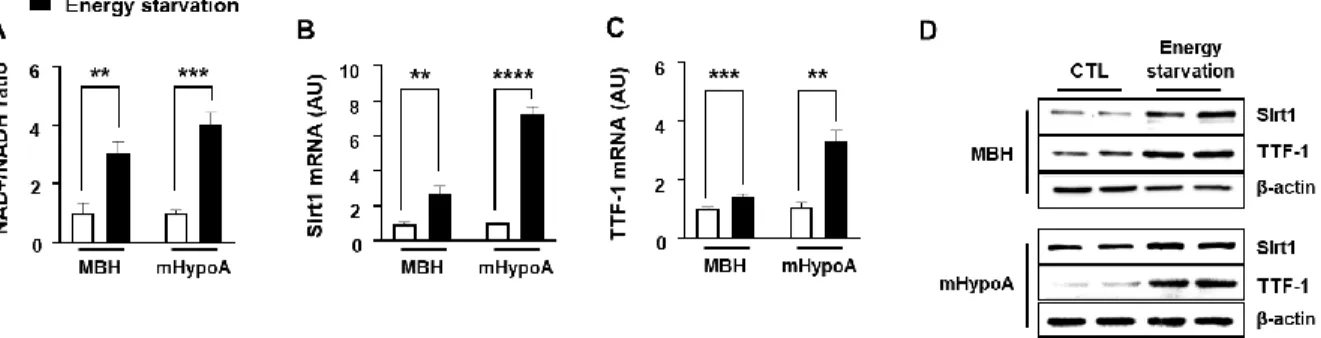
Expression of Sirt1 and TTF-1 genes according to energy states in the hypothalamus According to recent literature, Sirt1 and TTF-1 play pivotal roles in longevity and energy homeostasis and were expressed in the hypothalamus [4,13,23]. Furthermore, Sirt1-mediated TTF-1 deacetylation is important in longevity through regulation of orexin 2 receptor expression in the dorsomedial hypothalamic nucleus (DMH), and lateral hypothalamic area (LHA) [9]. Sirt1 activity is dependent upon NAD+ and NAD+/NADH ratio is changed by energy states. Therefore, to identify whether Sirt1 and TTF-1 are involved in the hypothalamic nutrient-responsive regulation, I first measured NAD+ and NADH in the medial hypothalamus (MBH) and hypothalamic neuronal cell line (mHypoA). The NAD+/NADH ratio was increased in mouse MBH by 24 h fasting and in mHypoA cells by low-glucose and low-serum (Energy starvation) (Fig. 1A). Energy starvation increased Sirt1 and TTF-1 mRNA, and protein expression (Fig 1B-D). These results indicate that Sirt1 and TTF-1 responded to changes in energy states in the mouse hypothalamus.
Effect of Sirt1 activator and inhibitor on TTF-1, AgRP, and POMC expression
To maintain energy homeostasis, brain regulates various aspects of the metabolism, such as food intake and energy expenditure through AgRP and POMC neurons [24,25]. Previous reports showed that AgRP and POMC were regulated by Sirt1 and TTF-1, but the relationship between them is not fully understood [6,15,19].To investigate their relationship, I measured the changed expression of Sirt1 and TTF-1 by treatment of RSV that is a well-known Sirt1 activator.
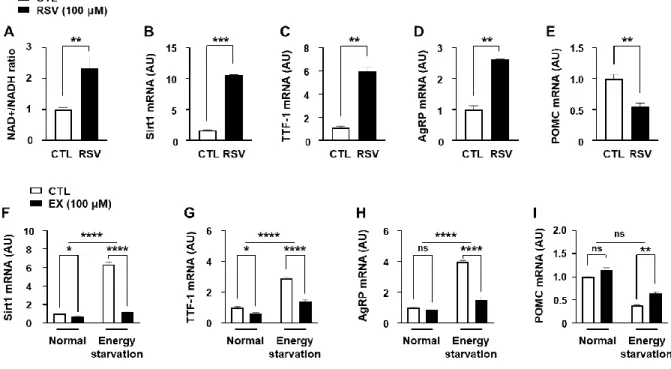
RSV increased Sirt1 (Fig 2B) by increasing the NAD+/NADH ratio (Fig 2A). I observed that
- 36 -
RSV also increased TTF-1 (Fig. 2C) and AgRP (Fig. 2D), and decreased POMC (Fig 2E) mRNA expression. Thus, I assessed whether TTF-1, AgRP, and POMC were affected by the Sirt1 inhibitor in the energy starvation. The increased Sirt1 (Fig. 2F), TTF-1 (Fig. 2G), and AgRP (Fig. 2H) and decreased POMC (Fig 2I) mRNA expression according to energy starvation were attenuated by EX. These results suggest that TTF-1, AgRP, and POMC were regulated by energy state-sensitive Sirt1.
Regulation of AgRP and POMC expression by Sirt1 and TTF-1
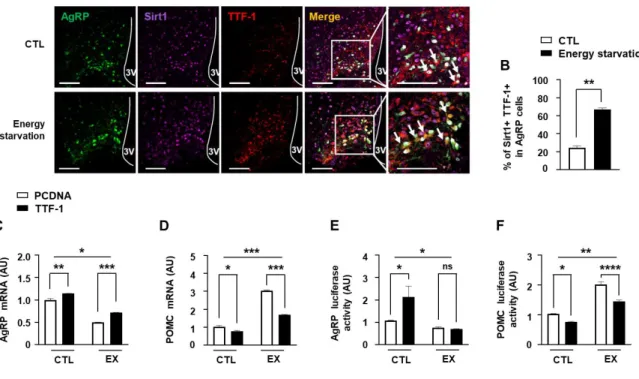
Inhibition of Sirt1 by EX administration into the third ventricle diminished AgRP expression [12], Thus, I observed that EX decreased TTF-1 gene expression and hypothesized that TTF-1 mediates Sirt1 effect on the regulation of AgRP. I performed IHC observations of Sirt1 and TTF-1 expression in AgRP neurons and revealed that fasting elevated Sirt1 and TTF-1 expression (Fig. 3A-B) in the AgRP neurons. To assess role of TTF-1 and Sirt1 in the AgRP expression, the mHypoA cells were transfected with TTF-1 expression vector, and then cells were treated with EX. Inhibition of AgRP mRNA expression (Fig. 3C) and AgRP promoter activity (Fig. 3E) by EX was ameliorated by TTF-1 overexpression. On the other way, EX- induced POMC mRNA (Fig 3D) and POMC promoter activity (Fig 3F) were attenuated by TTF-1 overexpression. These results suggest that TTF-1 plays an important role in the Sirt1- induced regulation of AgRP and POMC expression.
Effect of Sirt1 activator and inhibitor on TTF-1 acetylation
Sirt1 was reported to modulate activity of several transcription factors by changing their
- 37 -
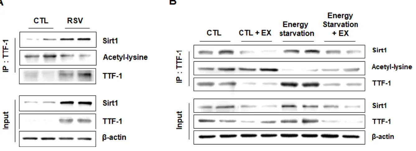
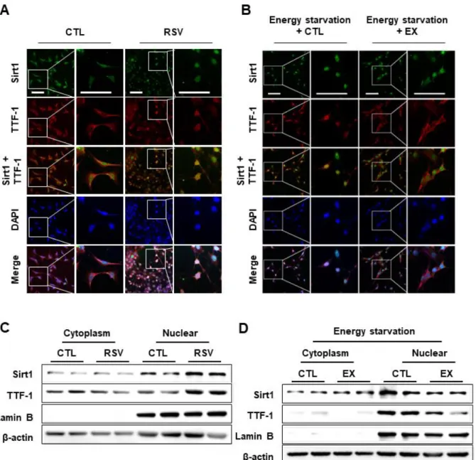
acetylation states [23,26]. Therefore, to verify whether Sirt1 regulates TTF-1 acetylation according to energy states, I determined Sirt1 and TTF-1 interaction and TTF-1 acetylation using immunoprecipitation. Sirt1 and TTF-1 protein expression, and interaction between Sirt1 and TTF-1 were increased by RSV (Fig. 4A) and energy starvation (Fig. 4B). Energy starvation-induced Sirt1 and TTF-1 protein expression, and interaction between Sirt1 and TTF- 1 were attenuated by the Sirt1 inhibitor (Fig. 4B). In addition, TTF-1 acetylation was reduced by RSV (Fig. 4A), and the energy starvation-induced decrease in TTF-1 acetylation was restored by EX (Fig. 4B). These results suggest that TTF-1 acetylation was affected by energy states and was regulated by Sirt1.
Effect of Sirt1 on nuclear translocation of TTF-1
Previous reports showed that change in TTF-1 acetylation affects DNA-binding affinity and transcriptional activity of TTF-1 that require its nuclear translocation [27,28]. To identify effect of TTF-1 deacetylation on its nuclear translocation, I performed ICC using mHypoA cells treated with RSV or EX. Deacetylation of TTF-1 by RSV induced nuclear translocation of TTF-1 (Fig 5A, C). On the other hand, energy starvation-induced TTF-1 nuclear translocation was attenuated by EX (Fig 5B, D). These results suggest that the nuclear translocation of TTF- 1 was regulated by energy starvation-induced Sirt1 induced deacetylation in energy-starved condition.
- 38 -
Figures
Figure 1. Change of Sirt1 and TTF-1 expression by energy state.
Mice were fasted for 24 h (Energy starvation) compared to ad libitum access to food (Normal).
mHypoA cells were treated with 25 mM glucose + 10 % serum medium (Normal) or 5 mM glucose + 0 % serum medium (Energy starvation) for 24 h. (A) Intracellular NAD+ and NADH levels were measured using a microplate reader for mouse MBH tissues and mHypoA cells exposed to either normal conditions or energy starvation (n= 4-5/each group). NAD+/NADH ratio was calculated based on the concentration of NAD+ and NADH. (B-C) Sirt1 (B) and TTF- 1 (C) mRNA was analyzed by real-time qPCR and normalized to ꞵ-actin mRNA (n= 4~6/each group). (D) Protein was extracted from MBH tissues and mHypoA cells exposed to either normal conditions or energy starvation (n= 3/each group) and Sirt1 and TTF-1 protein expression was measured by western blot analysis. β-actin intensity was used as a loading control (n= 3/each group). **, p<0.01, ***P<0.001 ****P<0.0001. AU= arbitrary units.
- 39 -
Figure 2. Effect of Sirt1 on the TTF-1 expression and acetylation.
(A-E) mHypoA cells were treated with RSV (100 μM) for 24 h. (A) NAD+/NADH ratio was measured for mHypopA cells (n= 4/each group). (B-E) Sirt1 (B), TTF-1 (C), AgRP (D), and POMC (E) mRNA levels were measured by qPCR using mHypoA cells that were treated with RSV 24 h (A-D) (n= 4/each group). (F-I) To identify involvement of Sirt1 in the energy starvation-induced TTF-1 expression, levels of Sirt1 (F), TTF-1 (G), AgRP (H), and POMC (I) mRNA expression were measured using cells treated with 5 mM glucose + 0 % serum medium (energy starvation) for 24 h before EX treatment for 3 h. mRNA was analyzed by real-time qPCR and normalized to ꞵ-actin mRNA (n= 3/each group). *, p<0.05, **, p<0.01, ***P<0.001
****P<0.0001. ns= non-significant. AU= arbitrary units.
- 40 -
Figure 3. Regulation of AgRP and POMC expression by Sirt1 and TTF-1.
(A-B) To identify the effect of energy starvation on the Sirt1 and TTF-1 expression in AgRP neurons, Sirt1 and TTF-1 immunoreactivity were measured by IHC with Sirt1 and TTF-1 antibodies in AgRP GFP mouse hypothalamus after 24 h fasting. Immunohistochemical analyses were performed to determine changes in AgRP neurons in the hypothalamic ARC caused by 24 h fasting. Representative images (A) and calculated data (B) indicate that Sirt1 and TTF-1 immunoreactivities in AgRP neurons were increased by fasting. Scale bar = 100 μm. (C-F) To demonstrate the role of TTF-1 in AgRP and POMC regulation by Sirt1, I performed qPCR (C, D) and promoter assay (E, F). AgRP (C) and POMC (D) mRNA levels and AgRP promoter activity (E), and POMC promoter activity (F) were measured using mHpoA cells that were transfected with TTF-1 expression vector or control vector before treatment with EX or vehicle (n= 3-4/each group). *, p<0.05, **, p<0.01, ***P<0.001
****P<0.0001. ns= non-significant. AU= arbitrary units.
- 41 -
Figure 4. Effect of Sirt1 activation on interaction of TTF-1 with Sirt1 and TTF-1 acetylation.
TTF-1, Sirt1, and acetyl-lysine were measured by western blot analysis using mHypoA cell extracts before (input) or after immunoprecipitated with TTF-1 antibody (IP:TTF-1). (A) mHypoA cells were treated with RSV or vehicle (CTL). (B) mHypoA cells were treated with 5 mM glucose + 0 % serum medium (energy starvation) for 24 h before EX treatment for 3 h.
β-actin intensity was used as a loading control.
- 42 -
Figure 5. Effect of Sirt1 on nuclear translocation of TTF-1.
To identify effect of TTF-1 deacetylation on the TTF-1 function, I determined a change in nuclear translocation according to TTF-1 acetylation. (A, B) ICC was performed with Sirt1 and TTF-1 antibodies on mHypoA cells that were treated with RSV (A) for 24 h or EX with 5 mM glucose + 0 % serum medium (energy starvation) for 3 h (B), respectively. (C, D) Western blotting was performed on cytoplasmic and nuclear TTF-1 by RSV (B) or EX with a 5 mM
- 43 -
glucose + 0 % serum medium for 3 h (D). β-actin and nuclear lamin B were internal controls for loading control and nuclear fractions, respectively. Scale bar = 100 μm.
- 44 -
Discussion
Several studies reported that TTF-1 function includes energy homeostasis and inflammation in the hypothalamus [4,6,29,30]. Although role of TTF-1 in the regulation of hypothalamic control of energy homeostasis has been studied for more than 10 years, the mechanisms that regulate TTF-1 in response to energy states are still not fully understood. In this study, I identified energy-responsive mechanism of TTF-1 via interaction with Sirt1 and for the regulation of AgRP and POMC expression. I observed that Sirt1-positive TTF-1 expression was increased by fasting in the AgRP neurons, and energy starvation-induced TTF-1 expression was attenuated by Sirt1 inhibitor. Furthermore, TTF-1 deacetylation and nuclear translocation were regulated by interaction with Sirt1. These findings indicate that TTF-1 and Sirt1 interaction plays an important role in the energy states-sensing, and thus, as a result, regulates AgRP and POMC expression.
These results showed that TTF-1 is affected by increasing NAD+/NADH in energy starvation, in particular, NAD+-dependent Sirt1 expression. NAD is a vital cofactor involved in brain bioenergetics for metabolism and ATP production and exists in an oxidized (NAD+) or reduced (NADH) form, with NAD+/NADH (the redox ratio) is an important determinant of metabolic homeostasis [31]. Specifically, NAD+ concentrations increase in response to conditions associated with low nutrient availability such as calorie restriction, fasting, glucose or serum starvation, and exercise [32,33]. Therefore, the increase in the NAD+/NADH ratio means that the energy-starvation state is induced.
Previous studies reported that hypothalamic Sirt1 interacts with TTF-1 and Sirt1-induced TTF-1 deacetylation plays an important role in longevity and aging in DMH and LHA [9].
However, its function in regulation of energy homeostasis is unknown. To elucidate Sirt1 and
- 45 -
TTF-1 relationship and potential roles in energy homeostasis in the hypothalamus, I used RSV and EX. RSV is well-known to induce an increase in the activity of the NAD+ synthetase nicotinamide mononucleotide adenylyltransferase (NMNAT1) and consequently increases Sirt1 through an increase in the NAD+/NADH ratio. Therefore, RSV has been used as an activator of Sirt1 [34]. On the contrary, EX inhibits Sirt1 by closing the NAD+ binding site of Sirt1, and has been widely used as a selective Sirt1 inhibitor [35,36]. Intriguingly, I observed that EX also inhibited Sirt1 gene expression. Several studies have reported that EX inhibits Sirt1 expression as well as Sirt1 activity. However, detailed mechanisms how inhibits the Sirt1 gene expression have yet been unknown [37-41].
According to recent literatures, AgRP expression was increased and POMC expression was decreased by Sirt1 activation through diverse Sirt1 activators [12,20]. I observed that TTF-1 induced AgRP expression and reduced POMC expression and these effects were attenuated by Sirt1 inhibition, suggesting that Sirt1 is essential in the regulation of AgRP and POMC expression by TTF-1. Thus, unmasking the underlying mechanism for Sirt1-dependent regulation of TTF-1 is required to better understand how TTF-1 regulates energy homeostasis by sensing energy states.
Acetylation of transcription factors is important for transactivity via affecting DNA binding affinity and nuclear translocation [42,43]. Additionally, regulation of acetylation is important in the control of key physiological processes such as metabolism, circadian rhythm, and cell cycle along with gene regulation in various organisms [44]. In line with these notions, in this study, TTF-1 dependent change in AgRP and POMC expression might be due to that Sirt1 activation accelerates TTF-1 nuclear translocation through TTF-1 deacetylation. Therefore, Sirt1 inhibition suppressed TTF-1 nuclear translocation and reversed effect of TTF-1 deacetylation on the AgRP and POMC expression in the energy-starved condition. Particularly,
- 46 -
it is currently accepted that protein acetylation was regulated by energy states [45,46]. In line with this notion, the present results indicate energy starvation induced TTF-1 deacetylation.
Given the importance of TTF-1 in energy control, decreased TTF-1 acetylation might affect expression of diverse genes for energy homeostasis. Nonetheless, little is known about the function of TTF-1 acetylation in the brain. Previous literatures reported that the TTF-1 homeodomain has six conserved acetylation sites on lysine residues and several phosphorylated sites on serine residues [47]. Thus, TTF-1 has diverse co-factors that regulate TTF-1 post-translational modifications including acetylation and phosphorylation [48].
Especially, TTF-1 acetylation was regulated by cyclic adenosine monophosphate (cAMP), activator of the thyroid and retinoic acid receptor (ACTR), steroid receptor coactivator (SRC), CREB-binding protein (CBP), and Sirt1 [9,27,49]. However, I could not exclude possibility that other post-translational modifications of TTF-1 are affected by Sirt1, and that TTF-1 responds to the energy state by interacting with the other factors. Therefore, further studies are required to identify other co-factors that affect acetylation and post-translational modification of TTF-1 such as phosphorylation for the energy-responsive action of TTF-1.
In summary, the current study demonstrates that regulation of TTF-1 acetylation by Sirt1 plays an important role in hypothalamic nutrient-sensitive response of TTF-1, resulting in the regulation of AgRP and POMC. The results provide a new insight into an energy sensing- responsive mechanism caused by TTF-1 and Sirt1 in the hypothalamus.
- 47 -
References
1. Guazzi, S., et al., Thyroid nuclear factor 1 (TTF-1) contains a homeodomain and displays a novel DNA binding specificity. EMBO J, 1990. 9(11): p. 3631-9.
2. Guan, L., et al., Thyroid Transcription Factor-1: Structure, Expression, Function and Its Relationship with Disease. Biomed Res Int, 2021. 2021: p. 9957209.
3. Lee, B.J., et al., TTF-1, a homeodomain gene required for diencephalic morphogenesis, is postnatally expressed in the neuroendocrine brain in a developmentally regulated and cell-specific fashion. Mol Cell Neurosci, 2001. 17(1): p. 107-26.
4. Kim, J.G., et al., TTF-1, a homeodomain-containing transcription factor, regulates feeding behavior in the rat hypothalamus. Biochem Biophys Res Commun, 2006.
349(3): p. 969-75.
5. Matagne, V., et al., Thyroid transcription factor 1, a homeodomain containing transcription factor, contributes to regulating periodic oscillations in GnRH gene expression. J Neuroendocrinol, 2012. 24(6): p. 916-29.
6. Kim, J.G., et al., Thyroid transcription factor-1 regulates feeding behavior via melanocortin pathway in the hypothalamus. Diabetes, 2011. 60(3): p. 710-9.
7. Efeyan, A., W.C. Comb, and D.M. Sabatini, Nutrient-sensing mechanisms and pathways. Nature, 2015. 517(7534): p. 302-10.
8. Armour, S.M., et al., A high-confidence interaction map identifies SIRT1 as a mediator of acetylation of USP22 and the SAGA coactivator complex. Mol Cell Biol, 2013. 33(8):
p. 1487-502.
9. Satoh, A., et al., Sirt1 extends life span and delays aging in mice through the regulation of Nk2 homeobox 1 in the DMH and LH. Cell Metab, 2013. 18(3): p. 416-30.
- 48 -
10. Canto, C., et al., AMPK regulates energy expenditure by modulating NAD+
metabolism and SIRT1 activity. Nature, 2009. 458(7241): p. 1056-60.
11. Dietrich, M.O., et al., Agrp neurons mediate Sirt1's action on the melanocortin system and energy balance: roles for Sirt1 in neuronal firing and synaptic plasticity. J Neurosci, 2010. 30(35): p. 11815-25.
12. Cakir, I., et al., Hypothalamic Sirt1 regulates food intake in a rodent model system.
PLoS One, 2009. 4(12): p. e8322.
13. Satoh, A., et al., SIRT1 promotes the central adaptive response to diet restriction through activation of the dorsomedial and lateral nuclei of the hypothalamus. J Neurosci, 2010. 30(30): p. 10220-32.
14. Sasaki, T., et al., Induction of hypothalamic Sirt1 leads to cessation of feeding via agouti-related peptide. Endocrinology, 2010. 151(6): p. 2556-66.
15. Cyr, N.E., et al., Central Sirt1 regulates body weight and energy expenditure along with the POMC-derived peptide alpha-MSH and the processing enzyme CPE production in diet-induced obese male rats. Endocrinology, 2015. 156(3): p. 961-74.
16. Katsyuba, E., et al., NAD(+) homeostasis in health and disease. Nat Metab, 2020. 2(1):
p. 9-31.
17. Ramadori, G., et al., SIRT1 deacetylase in POMC neurons is required for homeostatic defenses against diet-induced obesity. Cell Metab, 2010. 12(1): p. 78-87.
18. Sasaki, T., et al., Hypothalamic SIRT1 prevents age-associated weight gain by improving leptin sensitivity in mice. Diabetologia, 2014. 57(4): p. 819-31.
19. Toorie, A.M. and E.A. Nillni, Minireview: Central Sirt1 regulates energy balance via the melanocortin system and alternate pathways. Mol Endocrinol, 2014. 28(9): p. 1423- 34.
- 49 -
20. Quinones, M., et al., Hypothalamic Actions of SIRT1 and SIRT6 on Energy Balance.
Int J Mol Sci, 2021. 22(3).
21. Belsham, D.D., et al., Ciliary neurotrophic factor recruitment of glucagon-like peptide- 1 mediates neurogenesis, allowing immortalization of adult murine hypothalamic neurons. FASEB J, 2009. 23(12): p. 4256-65.
22. Kim, H.R., et al., Tonicity-responsive enhancer binding protein (TonEBP) regulates TNF-alpha-induced hypothalamic inflammation. FEBS Lett, 2019. 593(19): p. 2762- 2770.
23. Xu, J., et al., Brain SIRT1 Mediates Metabolic Homeostasis and Neuroprotection. Front Endocrinol (Lausanne), 2018. 9: p. 702.
24. Quarta, C., X. Fioramonti, and D. Cota, POMC Neurons Dysfunction in Diet-induced Metabolic Disease: Hallmark or Mechanism of Disease? Neuroscience, 2020. 447: p.
3-14.
25. Quarta, C., et al., POMC neuronal heterogeneity in energy balance and beyond: an integrated view. Nat Metab, 2021. 3(3): p. 299-308.
26. Elibol, B. and U. Kilic, High Levels of SIRT1 Expression as a Protective Mechanism Against Disease-Related Conditions. Front Endocrinol (Lausanne), 2018. 9: p. 614.
27. Yi, M., et al., Role of CBP/p300 and SRC-1 in transcriptional regulation of the pulmonary surfactant protein-A (SP-A) gene by thyroid transcription factor-1 (TTF-1).
J Biol Chem, 2002. 277(4): p. 2997-3005.
28. Park, K.S., et al., TAZ interacts with TTF-1 and regulates expression of surfactant protein-C. J Biol Chem, 2004. 279(17): p. 17384-90.
29. Yun, C.H., et al., TTF-1 action on the transcriptional regulation of cyclooxygenase-2 gene in the rat brain. PLoS One, 2011. 6(12): p. e28959.
30. Jeong, B., et al., Role of thyroid transcription factor-1 in transcriptional regulation of
- 50 -
heme oxygenase-1. Biochem Biophys Res Commun, 2018. 496(1): p. 147-152.
31. Canto, C., K.J. Menzies, and J. Auwerx, NAD(+) Metabolism and the Control of Energy Homeostasis: A Balancing Act between Mitochondria and the Nucleus. Cell Metab, 2015. 22(1): p. 31-53.
32. Anderson, K.A., et al., Metabolic control by sirtuins and other enzymes that sense NAD(+), NADH, or their ratio. Biochim Biophys Acta Bioenerg, 2017. 1858(12): p.
991-998.
33. Xie, N., et al., NAD(+) metabolism: pathophysiologic mechanisms and therapeutic potential. Signal Transduct Target Ther, 2020. 5(1): p. 227.
34. Nanjan, M.J. and J. Betz, Resveratrol for the Management of Diabetes and its Downstream Pathologies. Eur Endocrinol, 2014. 10(1): p. 31-35.
35. Vachharajani, V.T., et al., SIRT1 inhibition during the hypoinflammatory phenotype of sepsis enhances immunity and improves outcome. J Leukoc Biol, 2014. 96(5): p. 785- 96.
36. Hall, A.M., et al., The Role of Sirt1 in Epileptogenesis. eNeuro, 2017. 4(1).
37. Kemelo, M.K., et al., D-galactosamine/lipopolysaccharide-induced hepatotoxicity downregulates sirtuin 1 in rat liver: role of sirtuin 1 modulation in hepatoprotection.
Physiol Res, 2014. 63(5): p. 615-23.
38. Kim, H.W., S.A. Kim, and S.G. Ahn, Sirtuin inhibitors, EX527 and AGK2, suppress cell migration by inhibiting HSF1 protein stability. Oncol Rep, 2016. 35(1): p. 235-42.
39. Yang, X., et al., SIRT1 inhibition promotes atherosclerosis through impaired autophagy.
Oncotarget, 2017. 8(31): p. 51447-51461.
40. Kang, X., et al., Sirtuin-1 (SIRT1) stimulates growth-plate chondrogenesis by attenuating the PERK-eIF-2alpha-CHOP pathway in the unfolded protein response. J
- 51 -
Biol Chem, 2018. 293(22): p. 8614-8625.
41. Fan, W., et al., Tetrahydroxystilbene glucoside alleviates angiotensin II induced HUVEC senescence via SIRT1. Can J Physiol Pharmacol, 2021. 99(4): p. 389-394.
42. Filtz, T.M., W.K. Vogel, and M. Leid, Regulation of transcription factor activity by interconn