157
Received:June 15, 2016, Revised:August 30, 2016, Accepted:September 20, 2016
Corresponding to:Yoon-Kyoung Sung, Department of Rheumatology, Hanyang University Hospital for Rheumatic Diseases, 222 Wangsimni-ro, Seongdong-gu, Seoul 04763, Korea. E-mail:[email protected]
Copyright ⓒ 2017 by The Korean College of Rheumatology. All rights reserved.
This is a Open Access article, which permits unrestricted non-commerical use, distribution, and reproduction in any medium, provided the original work is properly cited.
Case Report
pISSN: 2093-940X, eISSN: 2233-4718
Journal of Rheumatic Diseases Vol. 24, No. 3, June, 2017 https://doi.org/10.4078/jrd.2017.24.3.157
A Case of Refractory Oral Aphthous Ulcer Successfully Treated with Adalimumab
Jinyeong Kim1, Myung-jin Song1, Hye-Jin Jeong2, Soo-Kyung Cho1,3, Yoon-Kyoung Sung1,3
1Department of Internal Medicine, Hanyang University College of Medicine, Seoul, 2Department of Rheumatology, Keimyung University Dongsan Medical Center, Daegu, 3Department of Rheumatology, Hanyang University Hospital for Rheumatic Diseases, Seoul, Korea
Oral aphthous ulcer is a common lesion characterized by loss of dermal tissue of various shapes in many diseases. It is not seri- ous, but may affect quality of life through pain and discomfort. In many cases, it heals spontaneously without treatment or with topical agents such as antiseptics, analgesics and corticosteroid in a few days. However, rarely, there are a few cases of aphthous ulcer that remain refractory despite appropriate treatment. Tumor necrosis factor-α inhibitors have recently been used in treat- ment of severe mucosal ulcer in Behçet’s disease. Herein, we report a patient suffering from refractory oral aphthous ulcers that were resistant to topical and systemic agents, but were successfully treated with adalimumab. (J Rheum Dis 2017;24:157-160) Key Words. Oral ulcer, Aphthous stomatitis, Tumor necrosis factor-alpha inhibitor, Adalimumab
INTRODUCTION
Oral ulcer is a defect of surface tissue characterized by various depth, yellow-gray color fibrous membrane and relatively well-marked margin. It is associated with vari- ous conditions such as aphthous, trauma, drug, infection, malignancy and rheumatic disease including Behçet’s disease. Among these, aphthous ulcer is the most com- mon type which occurs in 20% of the general population at any time during their lifetime. Over than 80% of aph- thous ulcers belong to minor type which tends to be smaller than 10 mm in diameter, self-limited and dis- appear not leaving a scar. Approximately 15% of aph- thous ulcers are a major type which is larger than 10 mm in diameter, lasts from 2 weeks to several months and heals with scar [1]. In severe case of recurrent aphthous ulcers, particularly the major type, it is difficult to treat with topical agents alone. Therefore, systemic therapy with corticosteoid or immunesuppressive agent is neces- sary in recurrent aphthous ulcer [2]. Recently, it has been shown that tumor necrosis factor (TNF)-α inhibitors are used effectively for management of refractory and chroni-
cally recurrent oral ulcers [3]. As anti-TNF-α therapy is largely effective for Behçet’s disease characterized with multiple mucosal ulcers, we consider the use of TNF-α inhibitors for the patient without underlying disease, who is suffering from persistent oral ulcers. This case shows the successful management of refractory oral aph- thous ulcers with adalimumab which is monoclonal anti- body to TNF-α.
CASE REPORT
A 62-year-old man, complaining persistent deep oral ul- cers lasting for more than 2 years, was referred to our hospital. At the first time, oral ulcers were spontaneously developed on right border and tip of his tongue without any traumatic events. He visited dental clinic and was treated with local corticosteroid, anesthetics and antiseptics.
Since there was no improvement in his symptoms, sev- eral times of local steroid injection and systemic cortico- steroid was used besides topical agents. And additional supportive care was provided such as non-steroidal an- ti-inflammatory drug and analgesics for severe pain, pilo-
Jinyeong Kim et al.
158 J Rheum Dis Vol. 24, No. 3, June, 2017
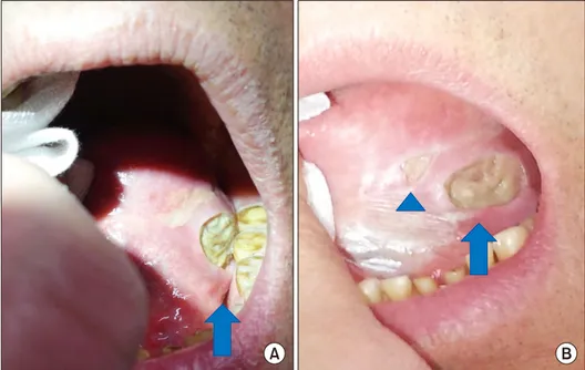
Figure 1. A 20×10 mm sized oral ulcer (arrows) with deep clean-cut margin on his left bor- der of tongue (A), there was no any improvement but small ex- tra-ulcer (arrow head) devel- oped during systemic steroid therapy (B).
carpine for dry mouth and antibiotic agent and anti-fun- gal agent for preventing secondary infection. Also, he was treated with surgical procedure that is excision with su- ture of mucous membrane three times, but it was failed due to wound dehiscence. Although various therapies were tried, oral ulcers were not improved and new lesions were developed at another site. Then he visited our hospi- tal for challenging new treatments after 2 years from onset.
He had diabetes mellitus for over than 10 years with in- sulin therapy and it was not controlled strictly (hemoglobin A1c 7.7). He had not been diagnosed with hypertension, tuberculosis infection, chronic liver diseases, malignancy, sexually transmitted diseases and rheumatic diseases.
There are no family history of severe oral-ulcer diseases and any rheumatic diseases.
A 20×10 mm sized ulcer was located on left lower bor- der of his tongue which had ovoid shape and clean-cut margin with soft tissue defect and whitish plaques were also accompanied around ulcer when he first visited. He had his weight loss about 5 kg for 2 years because of poor oral intake.
Initial laboratory findings were as follow: white blood cell count was 7,100/mm3 (absolute neutrophil count was 3,670/mm3); hemoglobin was 13.9 g/dL; platelet count was 248,000/mm3; erythrocyte sedimentation rate was 2 mm/hr; C-reactive protein was <0.8 mg/dL; serum creatinine was 0.72 mg/dL (0.5∼1.2 mg/dL); serum total protein was 6.8 g/dL (6.4∼8.5 g/dL); serum albumin was 4.2 g/dL (3.2∼5.5 g/dL); serum lactate dehydrogenase
was 214 IU/L (60∼200 U/L).
Several systemic diseases which can cause characteristic oral ulcers should be ruled out. Clinical manifestations suggesting Behçet’s disease including genital ulcers, eye lesions such as uveitis or retinal vasculitis, cutaneous le- sions like erythema nodosum were not detected. Human leukocyte antigen-B51 was negative. Any cutaneous symptoms such as malar or discoid rash, arthritis and evi- dence of neurologic or hematologic diseases were not existed. Autoantibodies including anti-nuclear antibody, anti-SS-A/Ro antibody, anti-SS-B/La antibody and rheu- matoid factor were all negative. He did not have systemic signs like febrile sensation or chilling. Human im- munodeficiency virus and venereal disease research labo- ratory were negative. Simple chest X-ray appeared normal without old tuberculosis (TB) or other infectious sequelae. Sputum acid fast bacillus staining, purified pro- tein derivative skin test and Interferon-gamma release as- says of Mycobacterium tuberculosis were negative.
Magnetic resonance imaging of his neck did not show the pathologic lymphadenopathy or malignant lesions. The Biopsy result of ulcer was benign which is ulcerative change with granulation tissue (chronic oral ulcer) rather than malignancy.
In our clinic, a medical treatment started with oral pre- dnisolone up to 10 mg, colchicine 0.6 mg twice a day and folic acid. After two months later, oral ulcer did not show any improvement and he felt still painful (Figure 1). We tried to apply azathioprine, but it was also not very helpful.
Then, we discussed with patient to use TNF-α inhibitor
Refractory Oral Aphthous Ulcer Treated with Adalimumab
www.jrd.or.kr 159
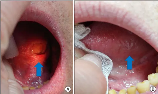
Figure 2. Healing ulcer (arrows) at 2 months (A), and 7 months (B) from administration of adali- mumab.
and started subcutaneous injection of adalimumab 40 mg every 2 weeks. The dose of oral corticosteroid got tapered steadily out after 5th injection and completely stopped af- ter 11th injection. Interval of administration of adalimu- mab also extended from every 2 to 4 weeks. After two months later starting treatment with adalimumab, the pa- tient had an improvement of pain. And oral ulcer was com- pletely healed within seven months of treatment (Figure 2). He has remained in remission state and followed up in the outpatient department maintaining adalimumab 40 mg every 4 weeks without systemic corticosteroid.
DISCUSSION
Oral ulcers are classified into two groups which are acute and chronic according to their duration. Acute ul- cers develop within short time and last less than 2 weeks.
On the other hand, chronic ulcers slowly develop and progress consistently. Common causes of oral ulcer are trauma including mechanical and chemical, drug-in- duced, autoimmune disorders, infectious diseases such as tuberculosis, mycosis, other bacterial and parasite, and malignancy [4]. Our patient had 2-years lasting oral ulcer which was not easily cured with appropriate therapy. We tried to find his underlying disease which can induce in- tractable and persisting ulcer. We should have focused on aphthous ulcer itself because we could not find specific systemic disease to him for evaluation.
Generally, treatment of oral aphthous ulcer starts with topical agents such as local anesthetics, antiseptic/anti-in- flammatory agents, sucralfate and local corticosteroid. If
painful ulcers prolong due to failure of topical medi- cations, a systemic therapy is indicated which includes colchicine, corticosteroid, pentoxifylline, dapsone, an- ti-metabolites, cyclosporine and alkylating agents. He was treated with from topical agents to systemic cortico- steroid and colchicine. All these medical treatment and even surgical repair were failed. Moreover, we could not increase the usage of corticosteroid because of his dia- betes mellitus. Because it is generally accepted that high- er occurrence of oral mucosal lesions was significantly as- sociated with poor metabolic control of diabetes mellitus [5]. Azathioprine also was added to decrease dose of cor- ticosteroid, but it did not affect at all. So, It was proven that his oral aphthous ulcer was refractory to conven- tional treatments.
TNF-α is an immune-modulating cytokine as important mediator in various aspects of immunity. The un- regulated TNF-α expression develops immune diseases.
One study reported that oral ulcer is characterized by high expression of TNF-α. This major inflammatory mediator may contribute to the activation and recruitment of leu- kocytes that are found in oral ulcers [6]. There are cur- rently 5 TNF-α inhibitors approved in rheumatic dis- eases which are divided two strategies: soluble receptors (etanercept) and targeting monoclonal antibodies (infli- ximab, adalimumab, golimumab, certolizumab). A recent report has shown that the effectiveness of adalimumab therapy in autoimmune disease had better outcomes than that of infliximab and etanercept from a molecular view [6]. The more effective antigen-antibody interface in ada- limumab provided information at a molecular structural
Jinyeong Kim et al.
160 J Rheum Dis Vol. 24, No. 3, June, 2017
level for further understanding the clinical advantages of adalimumab therapy compared with infliximab [7].
Based on such studies, we chose adalimumab among TNF-α inhibitors in spite of the cost from its off-label use.
Like all biologics, anti TNF-α agents should be used with caution because of their side effects. The common side effects are injection site reaction and upper respira- tory infections. They do not necessitate the termination of injection but need supportive supervision by specialists.
More serious side effects include tuberculosis reac- tivation, deep fungal injections and infection from other atypical pathogens. Rarely, worsening or initiation of con- gestive heart failure, medically significant cytopenia, de- velopment of lymphoma can be possibly notable [8].
Patient was observed closely by medical expert during first administration of adalimumab. To prevent possible infection, we screened his latent tuberculosis infection and vaccinated him from pneumococcus infection. Also serial laboratory monitoring for liver enzymes was done.
Besides medical problems, high cost from off-label use might be bother patient absolutely.
One study shows the characteristics of patient-group of recurrent aphthous ulcer [9]. It causes subjective pain in 90% of patients, as well as emotional stress. It can induce difficulty to eat and swallow in 80% of them, causing mal- nutrition and poor quality of their lives. Considering his occupation―professor, it might become his handicap causing a pronunciation problem. Although it is not being life-threatening diseases, these ulcers must be treated aggressively. With intractable patient with oral ulcer, TNF-α inhibitors are brought up and those are able to prevent unnecessary use of systemic corticosteroid.
There has been two similar cases that intractable oral ul- cers without underlying diseases such as Behçet’s or Crohn’s disease were treated with adalimumab [10,11]. In all cases, ulceration seemed to be resistant to conventional therapies, prior to use adalimumab. The difference of our case from those reports is the route of administration of adalimumab-subcutaneously injected in reference cases.
This study is the first to report the remission of intract- able aphthous oral ulcer without underlying disease by administrating adalimumab in Korea. There has been a limitation to assess an effect of adalimumab in oral aph- thous ulcer from a lack of cases or studies. Therefore, fur- ther studies based on large number of patients for longer duration in order to confirm long-term safety and effec- tiveness in oral aphthous ulcers are necessary.
SUMMARY
Here we report the case of successful treatment of a 62-year old man with intractable oral aphthous ulcer with adalimumab. This suggests that application of an- ti-TNF-α therapy can be effective for the management of recalcitrant oral ulcer.
CONFLICT OF INTEREST
No potential conflict of interest relevant to this article was reported.
REFERENCES
1. Field EA, Allan RB. Review article: oral ulceration-- aetiopa- thogenesis, clinical diagnosis and management in the gas- trointestinal clinic. Aliment Pharmacol Ther 2003;18:949- 62.
2. Beguerie JR, Sabas M. Recurrent aphthous stomatitis: an update on etiopathogenia and treatment. J Dermatol Nurses Assoc 2015;7:8-12.
3. Altenburg A, Abdel-Naser MB, Seeber H, Abdallah M, Zouboulis CC. Practical aspects of management of re- current aphthous stomatitis. J Eur Acad Dermatol Venereol 2007;21:1019-26.
4. Muñoz-Corcuera M, Esparza-Gómez G, González-Moles MA, Bascones-Martínez A. Oral ulcers: clinical aspects. A tool for dermatologists. Part II. Chronic ulcers. Clin Exp Dermatol 2009;34:456-61.
5. Al-Maweri SA, Ismail NM, Ismail AR, Al-Ghashm A.
Prevalence of oral mucosal lesions in patients with type 2 diabetes attending hospital universiti sains malaysia.
Malays J Med Sci 2013;20:39-46.
6. Natah SS, Häyrinen-Immonen R, Hietanen J, Malmström M, Konttinen YT. Immunolocalization of tumor necrosis factor-alpha expressing cells in recurrent aphthous ulcer le- sions (RAU). J Oral Pathol Med 2000;29:19-25.
7. Hu S, Liang S, Guo H, Zhang D, Li H, Wang X, et al.
Comparison of the inhibition mechanisms of adalimumab and infliximab in treating tumor necrosis factor α-associ- ated diseases from a molecular view. J Biol Chem 2013;
288:27059-67.
8. Scheinfeld N. Adalimumab: a review of side effects. Expert Opin Drug Saf 2005;4:637-41.
9. Safadi RA. Prevalence of recurrent aphthous ulceration in Jordanian dental patients. BMC Oral Health 2009;9:31.
10. Vujevich J, Zirwas M. Treatment of severe, recalcitrant, ma- jor aphthous stomatitis with adalimumab. Cutis 2005;76:
129-32.
11. Sánchez-Cano D, Callejas-Rubio JL, Ruiz-Villaverde R, Ortego-Centeno N. Recalcitrant, recurrent aphthous sto- matitis successfully treated with adalimumab. J Eur Acad Dermatol Venereol 2009;23:206.