Inhibitory Effects of Allicin on TNF-α-induced ICAM-1 Expression is Associated with Catalase
Nam-Sung Kang1, Suhkneung Pyo1 and Eun-Hwa Sohn2*
1Division of Immunopharmacology, College of Pharmacy, Sungkyunkwan University, Suwon, Korea
2Department of Herbal Medicine Resource, Kangwon National University, Samcheok, 245-711, Republic of Korea
Abstract - Allicin, a garlic componente, is believed to provide protection against various diseases including inflammation.
Since interactions of the cell adhesion molecules are known to play important roles in mediating inflammation, inhibiting adhesion protein upregulation is a possible therapeutic target. In this study, we demonstrate that TNF-α- and catalase-induced expression of ICAM-1 on human lung epithelial cells (A549) in a dose-dependent manner and catalase expression and activity were also increased in TNF-α-treated cells. Treatment of the TNF-α-treated cells with catalase inhibitor 3-amino-1,2,4-triazole resulted in a significant decreased the level of ICAM-1. These data suggest that induction of ICAM-1 expression by TNF-α is associated with catalase. In addition, allicin was found to inhibit the TNF-α induced expression of ICAM-1 on the A549 cells. This compound also inhibited the production of catalase induced by TNF-α, which suggests that the inhibition of ICAM-1 expression by allicin may be due to the modulated production of catalase.
Key words - allicin, TNF-α, ICAM-1, catalase
*Corresponding author. E-mail : [email protected]
Introduction
Garlic has been used as a general food and for therapeutic purposes in Oriental for a long time. Previous investigations have shown that garlic plays an important pharmacological role as an anti-microbial (Cellini et al., 1996), anti-thrombotic (Bordia et al., 1996), anti-hypertensive (Mcmahon and Vargas, 1993; Foushee et al., 1982), anti-hyperglycemic (Chang and Johnson, 1980), and anti-hyperlipemic (Yeh and Yeh, 1994;
Eilat et al., 1995) agent. It has also been suggested that allium derivatives from garlic regulate nuclear factors involved in the immune and inflammatory functions, as well as proli- feration (Pinto and Rivlin, 2001). In addition, it has been recently demonstrated that garlic extracts reduce the migration of leukocyte through the endothelial cell monolayer (Hobauer et al., 2000).
An injury to or a dysfunction of the endothelium is believed to be one of the first events in the development of the inflammation, and the inflammatory mechanisms are an integral part of this process (Berliner et al., 1995). An important event in this inflammatory response is the locali-
zation of leukocytes at the sites of the inflammatory lesions through a multistep process (Muller, 2003). Adhesion mole- cules on the endothelial and nonvascular cells play a major role in the inflammatory reaction. Intercellular adhesion molecule-1 (ICAM-1/CD54), which is one of several cell surface molecules, mediates the cell-to-cell interaction and plays an important role in facilitating the immune response at the inflammation sites (Van de Stolpe and Van der Saag, 1996). Since reactive oxygen including superoxide, hydrogen peroxide and catalase are involved in acute, and chronic inflammation, there is a need to define the role of these oxidants and free radicals in the pathophysiology of inflamma- tory diseases. However, despite many studies, the precise roles of these species in the expression adhesion molecules induced by TNF-α are not clearly understood. Therefore, the aim of this study was to clarify the mechanism underlying regulation of TNF-α-induced ICAM-1 expression and the effects of allicin in human epithelial cells.
Materials and Methods
Reagents
Allicin extract was prepared according to the methods of
Prasad et al (Prasad et al, 1995). Briefly, allicin, diallyl disulfide-oxide, was extracted from a garlicin tablet which contained 2500 μg of allicin per tablet (Madaus Murdock, Inc., 10 Mountain Spring Parkway, Springville, Utah, 84663 USA). The uncoated tablet was uncoated, crushed (0.75 g) and homogenized in test tube containing 5.0 ml of distilled water. The homogenate was then centrifuged for 10 min at 13,000 x g, and the supernatant recentrifuged for 5 min at 13,000 x g and the concentration adjusted to 500 μg/㎖ of allicin. The TNF-α and anti-ICAM-1 (BBA3) antibodies were purchased from R & D Systems, USA. Anti-mouse IgG-HRP and p-nitrophenyl phosphate were purchased from Sigma Chemical Co. Fetal bovine serum was purchased from Gibco, USA.
Cells and Cell culture
The human lung epithelial cell line A549 was purchased from the American. Tissue Culture Collection (ATCC, Manassas, VA) and cultured in DMEM supplemented with 10% fetal bovine serum (FBS), penicillin (100 IU/ml), and streptomycin (100 μg/ml) (GIBCO BRL, Life Technologies, Grand Island, NY) in a humidified atmosphere containing 5% CO2 in an incubator at 37°C.
Determination of Cell Surface Expression of ICAM-1 by ELISA
The cell surface expression of the adhesion molecules on the endothelial monolayers was quantified using ELISA using a modification of the methods described previously (Son et al., 2001). The A549 cells were seeded at a concentration of 2x10⁴ cells/well in 96-well, flat bottom, gelatincoated plates (Nalgen Nunc International, IL). The cells were incubated with the control media, and TNF-α for 20 h in the presence or absence of the various doses of the allicin in order to measure ICAM-1 expression. The cells were washed with phosphate buffer saline pH 7.4 (PBS) and fixed with 10% glutaral- dehyde for 30 min at 4°C. Bovine serum albumin (1.0% in PBS) was added to the cells in order to reduce the non- specific binding. The cells were incubated with anti-ICAM-1 monoclonal antibodies or the isotype matched control anti- bodies (0.25 mg/mL, diluted in blocking buffer) overnight at 4°C, washed with PBS followed by incubation with the
alkaline phosphataseconjugated goat anti-mouse secondary antibodies (1 g/mL, diluted in PBS). The cells were then washed with PBS and exposed to the peroxidase substrate (p-nitrophenyl phosphate 1 mg/mL in 0.1 M glycin buffer, pH 10.4 containing 1 mM MgCl2, and 1 mM ZnCl2). The absorbance was determined at 405 nm using a Molecular device microplate reader (Menlo Park, CA). The absorbance values of the isotype matched control antibodies were taken as blank and were subtracted from the experimental values.
Measurement of Catalase Activity
The catalase activity was determined spectrophotometrically by monitoring the decomposition of H2O2 (Aebi, 1984). The homogenate (50–00 μg proteins) was added to a cuvette containing potassium phosphate (50 mM, pH 7) with 25 mM H2O2. The total volume of the reaction mixture was 1 ml. The disappearance of H2O2 was monitored at a wavelength of 240 nm for 1 min at 25°C. The CAT activity was expressed as micromoles of H2O2 consumed per minute per milligram of cellular protein with a molar extinction coefficient of 43.6 M-1cm-1. Cellular protein content was quantified using a Bio-Rad protein assay kit (Bio-Rad Lab, Hercules, CA) with bovine serum albumin (BSA) as the standard.
Measurement of mRNA Levels by Reverse-Transcription Polymerase Chain Reaction (RT-PCR)
The total RNA was extracted using a Trizol reagent kit (Invitrogen, Carlsbed, CA), according to the manufacturer’
recommendations. The yield and purity of the RNA were confirmed by measuring the ratio of the absorbances at 260 and 280 nm. Polymerase chain reaction (PCR) was performed using ICAM-1-or catalase-specific primers to identify their respective specific cDNA. The following ICAM-1-specific primers were synthesized: sense primer, 5′-CTGCTGGGAA TTTTCTGGCCAC-3′ antisense primer, 5′-CTATGGCAAC GACTCCTTCTCG-3′. The following catalase-specific primers were synthesized: sense primer, 5′-ACCTGGCATTGAGGC CAGTC-3′ antisense primer, 5′-GGATGTGGCTCCCGTA GTCA-3′. Each primer pair was chosen to span the introns of their respective rat genes. Using these primers, it was ex- pected that the DNA fragments of 584, 442, and 313 bp would be amplified from the ICAM-1, catalase, and β-actin
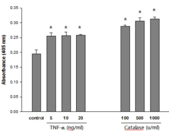
Fig. 1. Effects of TNF-α and Catalase on ICAM-1 expression in A549 cells. (A) The cells were treated with various dose of TNF-α or catalase and cultured for 24 hr. The cell-surface ICAM-1 expression level was measured by ELISA using the anti-ICAM-1 antibody as described in Materials and Methods.
The results are expressed as the mean ± S.E.M. of three independent experiments performed in quintuplicate. *Signifi- cantly different from the control cells.
TNF-α (ng/ml) Cellular Catalase Activity (M/min/mg protein)
0.000 0.005 0.010 0.015 0.020 0.025 0.030 0.035
control 5 10
∗
∗
Fig. 2. Induction of cellular catalase by TNF-α in A549 cells.
The cells were treated with TNF-α and then cultured for 24 hr.
The cellular catalase activity was measured as described in Materials and Methods. The data represents the mean ± S.E.M of three independent experiments performed in quintuplicate.
* Significantly different from the control cells.
RNA, respectively. Samples were stored at -20°C, after amplification
Western Blot Analysis
Western blot analysis was performed. After treatment, the cells were washed twice in PBS and suspended in a lysis buffer (50 mM Tris, pH 8, 150 mM NaCl, 0.1% sodium dodecyl sulfate, 0.5% sodium deoxycholate, 1% NP40, 100 μg/ml phenylsulfonyl fluoride, 2 μg/ml aprotinin, 1 μg/ml pepstatin, and 10 μg/ml leupeptin), and placed on ice for 30 min. The supernatant was collected after centrifugation at 15,000 × g for 20 min at 4°C. The protein concentration was determined using the Bio-Rad protein assay (Bio-Rad Lab) with BSA as the standard. The whole lysates (20 μg) were resolved on a 7.5% SDS–olyacrylamide gel, transferred to an immobilon polyvinylidene difuride membrane (Amersham, Arlington Heights, IL), and probed with the appropriate antibodies. The blots were developed using an enhanced chemiluminescence (ECL) kit (Amersham). In all immuno- blotting experiments, the blots were reprobed with an anti-β -actin antibody to control for the protein loading.
Statistical Analysis
Results are presented as means + S.E.M. Statistical difference between groups was determined by one-way analysis of variance (ANOVA) and significant values are indicated with an asterisk (*p<0.05).
Results and Discussion
Induction of ICAM-1 Expression in A549 Cells by TNF-α and Catalase
ELISA was used to examine the effects of TNF-α and catalase on ICAM-1 expression in the A549 cells. All the determinations were made on the same 96-well plate. ELISA showed the ICAM-1 was expressed at low levels on the nonstimulated endothelial cells and was significantly induced by TNF-α. and catalase in dose dependant manners (Fig. 1).
Enhancement of Catalase activity by TNF-α
Although TNF-α was previously shown to induce several cellular antioxidative enzymes in other experimental models (Mo et al., 2003), the inducibility of cellular antioxidants in lung epithelial cells by TNF-α has not been reported in the literature. As shown in Figure 2, treatment of the A549 cells with TNF-α resulted in the significant induction of cellular
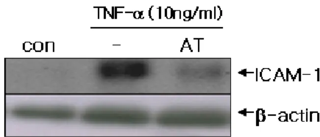
Fig. 3. Inhibition effect of 3-amino-1,2,4-triazole (AT) on the expression of ICAM-1 induced by TNF-α. A549 cells were preincubated for 1hr with 3-amino-1,2,4-triazole (AT; 50 mM), a catalase activity inhibitor. and then incubated with TNF-α for 24 hrs. The expression of ICAM-1 and β-actin were analyzed by Western blotting.
Fig. 4. Effect of allicin on TNF-α-induced ICAM-1 expression by A549 cells. The cells were cultured for 24 h in the medium or in the medium supplemented with TNF-α (10 ng/mL) in the presence or absence of the allicin. The total RNA was prepared and used for ICAM-1 or beta-actin RT-PCR. Similar findings were observed in three separate experiments.
Fig. 5. Effect of allicin on TNF-α-induced catalase expression by A549 cells. The cells were cultured for 24 h in the medium or in the medium supplemented with TNF-α (10 ng/mL) in the presence or absence of the allicin. The total RNA was prepared and used for catalase or beta-actin RT-PCR. Similar findings were observed in three separate experiments.
catalase activity in a dose-dependent manner.
Inhibitory effects of AT, catalase inhibitor, on ICAM-1 expression by TNF-α
In order to determine if the TNF-α-induced increase in the expression and activity of catalase affects ICAM-1 expression in cells, the A549 cells were pretreated with 3-amino-1,2,4- triazole (AT), catalase inhibitor, for 1 h. The cells were treated with TNF-α and then incubated for 24 h. This was followed by measuring the ICAM-1 protein levels. AT pretreatment partially but effectively suppressed the ICAM-1 expression induced by TNF-α in the A549 cells (Fig. 3).
Allicin inhibits TNF-α-induced ICAM-1 and Catalase expression on epithelial cells
To examine the effect of allicin, A549 were incubated without or with various concentrations (0.01, 0.1, 1 μg/ml) of allicin for 24hrs in the presence of TNF-α. These concentra- tions were based on the concentrations of allicin determined in previous studies (Kang et al, 2001). As detected by RT- PCR, mRNA level of ICAM-1 and catalase were significantly induced by TNF-α, and allicin reduced TNF-α-induced ICAM-1 and catalase expression in a dose dependent manner (Fig. 4).
Allicin inhibits TNF-α-induced catalase production Since catalase is known to be an important modulator of inflammatory response to various stimuli and ICAM-1 ex- pression (Son et al, 2006), we determined the effect of allicin
on catalase production in epithelial cells. As shown in Fig. 2, treatment of A549 cells with TNF-α resulted in increased catalase activity. Moreover, this increased catalase production was inhibited by allicin in a dose dependent manners (Fig. 5).
Discussion
Garlic extract has been found to promote healing of inflammation in the colon (Khan and Ali, 1999). However, although garlic extract has been found to have such anti- inflammatory properties, very little is known with regard to the effect of allicin, a major component of garlic, on the induction of cell adhesion molecules by TNF-α. In the present study, allicin was found to block the TNF-α-induced expression of the leukocyte adhesion molecules, ICAM-1. Therefore, allicin exhibit anti-inflammatory effects on the expression of the adhesion proteins induced by TNF-α. These compounds also inhibited catalase induction in the TNF-α-induced A549 human epithelial cells.
TNF-α, which participates in the inflammatory response, induces the expression of the adhesion molecules when added to the endothelial cells in the culture (Sherman et al., 1991;
Bevilacqua et al., 1989). Catalase is an antioxidant enzyme that protects the cell from oxidative damage by removing H2O2 (Halliwell and Gutteridge, 1999). It was reported that catalase induces ICAM-1 expression in human endothelial cells (Aoki et al., 1997). Based on these findings, these results suggest involvement of catalase in γIR-induced ICAM-1 expression. However, the mechanistic relationship between the TNF-α-induced ICAM-1 expression and catalase activation is unclear. Interestingly, the results presented in this article show that treating the cells with TNF-α enhanced catalase activity and expression. It was also found that TNF-α-induced ICAM-1 expression were reduced by AT, which is a catalase inhibitor. In additon, we detected that the treatment of allicin resulted in a significant decrease ICAM-1 expression and catalase induction. Therefore, it appears that allicin inhibited the catalase induction induced by TNF-α and thereby ICAM-1 expression.
It is possible that the balance between the pro- and antioxidative mechanisms of the cell may determine the expression level of ICAM-1 induced by TNF-α. However, the specific downstream elements that are activated by the enhancement of catalase as well as its functional role for the cell remain to be identified. It is expected that results in this study will provide new insights into the mechanisms by which allicin mediates its effect on the human lung epithelial cell line A549, which might be used to identify potential targets for the development of novel drugs for treating various inflammatory disorders.
Literature Cited
Aebi, H. 1984. Catalase in vitro. Methods Enzymol. 105:
121-126.
Aoki, T., Suzuki, Y., Nishio, K., Suzuki, K., Miyata, A., Oyamada, Y., Mori, M., Fujita, H. and Yamaguchi, K. 1997.
Effect of antioxidants on hyperoxia-induced ICAM-1 ex- pression in human endothelial cells. Adv. Exp. Med. Biol.
411: 503-511.
Berliner, J.A., Navab, M., Fogelman, A.M., Frank, J.S., Demer, L.L., Edwards, P.A., Watson, A.D. and Lusis, A.J. 1995.
Atherosclerosis: basic mechanisms. oxidation, inflammation, and genetics. Circulation, 91(9): 2488-2496.
Bevilacqua, M.P., Pober, J.S., Wheeler, M.E., Cotran, R.S. and Gimbrone, M.A. 1985. Jr. Interleukin 1 acts on cultured human vascular endothelium to increase the adhesion of polymorphonuclear leukocytes, monocytes and related cell lines. J. Clin. Invest., 76: 2003-2009.
Bordia, T., Mohammed, N. and Thomson, M. 1996. An evalua- tion of garlic and onion as antithrombotic agents. Prota- glandins Leukot Essent Fatty Acids 54: 183-186.
Cellini, L., Di Campli, E. and Masulli, M. 1996. Inhibition of Helicobacter pylori by garlic extracts (Allium sativum).
FEMS Immunol Med Microbiol 13: 273-277.
Chang, M.L.W. and Johnson, M.A. 1980. Effect of garlic on carbohydrate metabolism and lipid synthesis in rats. J Nutr110: 931-936.
Eilat, S., Oestraicher, Y. and Rabinkov, A. 1995. Alteration of lipid profile in hyperlipidemic rabbits by allicin, an active constituent of garlic. Coronay Artery Dis 6: 985-990.
Foushee, D.B., Ruffin, J. and Banerjee, U. 1982. Garlic as a natural agent for the treatment of hypertension: A preliminary report Cytobios 34: 145-152.
Halliwell, B. and Gutteridge, J.M.C. 1999. Free radicals in biology and medicine. New York: Oxford University Press.
Hobauer, R., Frass, M., Gmeiner, B., Kaye, A.D. and Frost, E.A. 2000. Garlic extract (allium sativum) reduces migration of neutrophils through endothelial cell monolayers. Middle East J Anesthesiol 15: 649-658.
Kang, N.S., Moon, E.Y., Cho, C.G. and Pyo, S. 2001. Immuno- modulating effect of garlic component, allicin on murine peritoneal macrophages. Nutr Res 21: 617-626.
Khan, I. and Ali, M. 1999. Altered expression of the Na+/H+
exchanger isoform-3 in experimental colitis: effect of garlic.
Mol Cell Biochem 200: 77-84.
Mcmahon, F.G. and Vargas, R. 1993. Can garlic lower blood pressure? A pilot study. Pharmacotherapy 13: 406-407.
Mo, S.J., Son, E.W. and Rhee, D.K. 2003. Modulation of TNF-alpha-induced ICAM-1 expression, NO and H2O2
production by alginate, allicin and ascorbic acid in human endothelial cells. Pyo S. Arch Pharm Res. 26(3): 244-251.
Muller, W.A. 2003. Leukocyte-endothelial-cell interactions in leukocyte transmigration and the inflammatory response.
Trends Immunol. 24: 327-334.
Pinto, J.T. and Rivlin, R.S. 2001. Antiproliferative effects of allium derivatives from garlic. J Nutr 131: 1058S-1060S.
Prasad, K., Laxdal, V.A. and Yu, M. 1995. Antioxidant activity of allicin, an active principle in garlic. Mol Cell Biochem 148: 183-189.
Sherman, M.L., Datta, R., Hallahan, D.E., Weicheslbaum, R.R.
and Kufe, D.W. 1991. Regulation of tumor necrosis factor gene expression by ionizing radiation in human myeloid leukemia cells and peripheral blood monocytes. J. Clin.
Invest., 81: 506-510.
Son, E.W., Cho, C.K., Rhee, D.K. and Pyo, S. 2001. Inhibition of gamma-irradiation induced adhesion molecules and NO production by alginate in human endothelial cells. Arch.
Pharm. Res., 24: 466-471.
Son, E.W., Rhee, D.K. and Pyo, S. 2006. Gamma-irradiation- induced intercellular adhesion molecule-1 (ICAM-1) expre- ssion is associated with catalase: activation of Ap-1 and JNK. J Toxicol Environ Health A. 69(24): 2137-2155.
Van de Stolpe, A. and Van der Saag, P.T. 1996. Intercellular adhesion molecule-1. J. Mol. Med. 74: 13-33.
Yeh, Y.Y. and Yeh, S,M, 1994. Garlic reduces plasma lipids by inhibiting hepatic cholesterol and triacylglycerol synthesis.
Lipids 29: 189-193.