THP-1 Cell과 HUVEC을 이용한 Co-Culture Model System에서 최종당화산물에 의한 Cytokines와 RAGE 발현
이광원1․이현순2†
1
고려대학교 식품공학부
2
고려대학교 식품영양학과 및 보건과학연구소
Co-Culture Model Using THP-1 Cell and HUVEC on AGEs-Induced Expression of Cytokines and RAGE
Kwang-Won Lee1 and Hyun-Sun Lee2†
1Division of Food Bioscience & Technology, College of Life Science and Biotechnology and
2Dept. Food and Nutrition and Institute of Health Science, Korea University, Seoul 136-703, Korea
Abstract
Although monoculture methods have been remarkably useful due to their simplicity, they have serious limi- tation because of the different types of cells communication with each other in many physiological situations.
We demonstrated levels of markers of endothelial dysfunction such as tumor necrosis factor-α (TNF-α) and interleukin-1β (IL-1β) as well as stimulation of receptor of advanced glycation endproducts (AGEs) on mono- and co-culture system such as only monocyte (THP-1) cultivation system, only endothelial cell (HUVEC) culti- vation system, and co-cultivation system of THP-1 and HUVEC. The mRNA levels of TNF-α and IL-1β on HUVEC increased by the co-culture with monocyte after 4 hr at 100 μg/mL glyceraldehyde-AGE. The secreted protein contents into medium of TNF-α and IL-1β increased after 8 hr approximately 2~2.5 times compared to mono-cultivation. In contrast, the mRNA level of receptor of AGE (RAGE) was relatively insensitive on the co-culture system. The mediators by which monocytes activate endothelial cell have not been fully elucidated.
In this study we confirmed production of soluble cytokines such as TNF-α and IL-1β by monocytes. Use of monocyte conditioned medium, which contains both cytokines, can activate endothelial cell.
Key words: advanced glycation endproducts (AGEs), endothelial dysfunction, co-culture, endothelial cell, monocyte
†
Corresponding author. E-mail: [email protected]
†
Phone: 82-2-940-2858, Fax: 82-2-940-2850
서 론
글라이케이션 반응, 혹은 non-enzymatic glycation 반응 이란 환원당의 carbonyl기가 단백질의 유리아미노기인 ly- sine이나 arginine과 반응하여 초기 Shiff base를 형성하고, 계속적으로 축합, 재전위, 산화, 분열, 고리화 등의 일련의 복잡한 반응을 통하여 갈색의 비가역적 최종당화산물(ad- vanced end-products, AGEs)을 만드는 반응이다(1). 이 반 응은 모든 단백질의 ε-amino group이나
N-terminal group 에서 일어나며 또한 아미노산을 함유하고 있는 콜라젠, DNA나 low-density lipoprotein(LDL)과 같은 혈청 단백질 과 쉽게 공유결합 하여 체내에서도 AGEs 화합물을 만든다 고 알려져 있다(2). 생체내의 glycation 반응은 몇 주 정도에 걸쳐 일어날 정도로 천천히 일어나지만 일단 반응이 시작되 면 비가역적 반응으로 AGEs 화합물이 체내에 축적되게 되 면서 여러 가지 질병을 일으킨다(3). 특히 만성적인 고혈당
인 당뇨의 경우 AGEs가 급속도로 생성되며, 당뇨합병증이 진행되는 환자의 신장, 망막, 경화된 동맥에서 AGEs의 축적 이 관찰되고 있다(4-6). AGE에 의해 동맥경화는 다음과 같 은 기전에 의해 일어난다고 알려져 있다. 먼저 혈액 내에 존재하는 LDL의 인지질 성분에서 glycation 반응에 의해 LDL의 산화변성이 일어나며(7), 이렇게 변성된 LDL은 혈관 내피세포에 존재하는 receptor of AGEs(RAGE)를 통해 내 막(intima)으로 흡수되어 거품세포(foam cell)의 형성을 촉 진하며(8,9), 콜레스테롤에스터(cholesterol ester)의 축적 (10) 등 혈관조직의 정상적인 기능을 저하시켜 최종적으로 는 동맥경화와 같은 질환을 일으키는 것으로 보고되고 있다 (11-13).
최근 식품소재로부터 AGEs의 생성을 억제하거나 AGEs
에 의한 혈관 내피세포의 기능상실에 대한 연구가 활발히
진행되고 있다. 녹차에서 추출한 epigallocatechin gallate
(EGCG)와 같은 flavonol은 AGEs의 생성 및 RAGE signal
을 억제하여 AGEs의 체내 생성 및 축적을 감소시켜(14), AGEs에 의한 다양한 질병을 억제하는 것으로 알려져 있다 (15). 또한 숙성된 마늘 추출물은 AGEs 생성을 억제하여 당뇨합병증을 억제한다고 알려져 있으며(16), 이외에도 가 자나무(
Terminalia chebula) 열매 추출물(17), anthocyanin cyanidin-3-
O-glucoside(18)는 혈관내피세포의 기능상실 을 억제한다고 알려져 있다.
그러나 기존의 연구는 대부분 AGEs에 의한 단구세포의 변화 혹은 혈관내피세포의 변화를 단독 system에서 연구되 고 있으나 몇몇 연구자들의 보고에 의하면 AGEs에 의해 혈액 내 단구세포는 IL-1, TNF와 같은 cytokine을 생성하며 이렇게 형성된 cytokine은 혈관내피세포의 기능상실을 가속 화시킬 것으로 예측하고 있다(19,20). 따라서 실제의 경우를 반영하는 모델인 단구세포와 혈관내피세포의 co-culture system이 중요하다. 따라서 AGEs 화합물에 의한 혈관내피 세포의 기능상실의 연구를 위해서는 co-culture system이 확립되어야 하며, 이 system을 이용하여 AGEs에 의한 기전 연구 및 이를 억제할 수 있는 식품소재에 대한 연구가 진행 되어야 할 것이다.
세포배양을 이용한 연구는 기능성 소재나 기전 연구에 많 이 이용되는 실험 방법이지만 한 가지 세포만을 이용하는 실험방법은 cell communication이 배재된 실험 방법이다. 특 히 혈관성 질환의 연구의 경우 혈관내피세포와 단구세포의 상호작용이 매우 중요하다. 따라서 본 연구는 혈관내피세포 의 기능상실에 매우 중요한 cytokine인 IL-1β과 TNF-α의 발현 정도와 AGEs 화합물인 조직으로 들어가는 receptor인 RAGE 발현 정도를 단구세포/혈관내피세포의 단독과 co- culture system을 비교하였다. 또한 AGEs에 의한 혈관성질 환을 억제할 수 있는 효능물질 검색을 위한 최적의 time point를 전체적으로 조사하고자 하였다.
재료 및 방법
재료
본 실험에 사용한 DL-glyceraldehyde, 2-deoxy-ribose, glycoaldehyde, glycoxal, diethylene triamine pentaacetic acid(DTPA), bovine serum albumin(BSA, low-endotoxin, fatty acid free)는 Sigma Chemical Co.(St. Louis, MO, USA)에서 구입하였으며, 혈관내피세포인 human umbilical vein endothelial cell(HUVEC), endothelial growth medium 2(EGM-2)는 Clonetics(San Diego, CA, USA), 단구세포인 THP-1(human acute monocytic leukemia cell line)은 American Type Culture Collection(Rockville, MD, USA)에 서 구입하여 사용하였다. 이외의 세포 배양을 위한 배지 및 기타재료는 GIBCO-BRL(Gaithersburg, MD, USA)에서 구 입하여 사용하였다. HUVEC은 EGM-2 배지에서 배양하였 으며 80∼90%의 confluence에서 실험하고, passage가 10번
을 넘기지 않은 세포만 사용하였다. THP-1은 Dulbecco's modified Eagle's medium(DMEM)에 10% fetal bovine serum(FBS), 100 U/mL penicillin 및 100 μg/mL strepto- mycin을 혼합한 배지를 사용하였다.
Advanced glycation endproduct(AGE)의 제조
최종당화산물 즉, AGEs의 제조는 Takeuchi 등(21)이 보 고한 방법에 따라 제조하였다. 그 과정을 간략히 기술하면, 10 mg/mL BSA, 5 mM DTPA과 각각의 당류(DL-glyc- eraldehyde, 2-deoxy-ribose, glycoaldehyde 혹은 glycoxal) 를 농도를 달리하여 0.2 M phosphate buffer(pH 7.4)에 혼합 한 후 0.2 μm를 이용하여 멸균한 다음 37
oC에서 7일간 당화 시켰다. 당화의 정도는 형광분석기(VICTOR
TM, Perkin Elmer, Rockville, MD, USA)를 이용하여 형광도(Ex 370 nm, Em 440 nm)를 측정하였다. Glycer-AGE의 제조는 0.1 M glyceraldehyde와 상기의 방법과 동일한 조건으로 진행 하였으며 당화과정이 종료된 후 잔존하는 glyceraldehyde를 제거하기 위하여 투석(MWCO: 1000 KDa) 후 단백질 양을 측정한 다음 사용하였다. Glycer-AGE의 처리 농도는 많은 선행 연구자들이 처리하고 있는 100 μg/mL로 처리하였다 (17,22,23).
Co-culture system
HUVEC은 EGM-2 배지를 이용하여 1×10
6cell/60 mm dish에 seeding한 다음 24시간을 배양하였다. 그 후 THP-1 (1×10
6) 단구세포를 HUVEC과 1:1 비율로 넣고(24), 4시간 후 100 μg/mL glycer-AGE를(17) 첨가한 후 24시간 동안 배양하였다. 각 time point 마다 각각의 세포와 배지를 회수 하여 측정 전까지 -70
oC에서 보관하였다. 대조군으로 100 μ g/mL BSA를 동일한 조건에서 24시간 처리하였다.
Reverse-transcription polymerase
Total RNA 분리는 Trizol-base protocol을 이용하여
chloroform/isopropanol 법(25)으로 추출하여 cDNA를 합성
하였다. 실험에 사용한 primer sequence는 TNF-α의 for-
ward는 GGCAGTCAGATCATCTTCTCGAA, reverse는
GAAGGCCTAAGGTCCACTTGT를 사용하였으며(26), IL-
1β의 forward는 ATGGCAGAAGTACCTGAGCTC, re-
verse는 TTCCTTGAGGCCCAAGGCCAC를 사용하였으
며(27), RAGE의 forward는 AGCCCTCTCCTCAAATC-
CACT, reverse는 ACTACTCTCGCCTGCCTCAG를 사용
하였다(28). 내부 표준 유전자로 사용한 GAPDH의 forward
primer는 AGGTCGGAGTCAACGGATTTG, reverse는
ACAGTCTTCTGGGTGGCAGTG이었다. PCR은 94
oC에
서 3분(1 cycle), 94
oC에서 1분, TNF-α는 63
oC, IL-1β는
67
oC, RAGE 62
oC에서 1분 그리고 72
oC에서 1분씩(25
cycles), 72
oC에서 5분(1 cycle)간 실시하였다. PCR 산물은
1.2% agarose gel에 100 V에서 30분간 전기영동 후 자외선
광으로 유전자 발현 정도를 알아보았다.
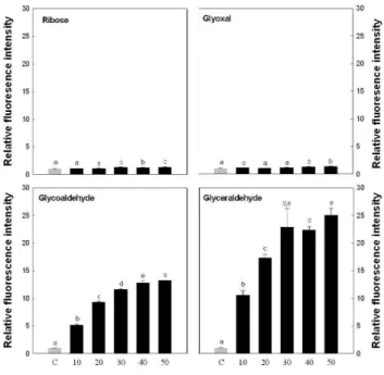
Fig. 1. Formation of various carbonyl compound-derived AGEs. AGEs-modified albumin was prepared by incubating BSA (10 mg/mL) with ribose, glyoxal, glycoaldehyde and glycer- aldehyde. Formative intensity of each AGEs was estimated using a fluorescence spectrophometry with 370 nm excitation and 440 nm emission. Values are mean±SD and means with different let- ters (a-e) on bars are significantly different at p<0.05 by Dun- can's multiple range tests.
Cytokine 및 RAGE의 측정
60 mm dish에서 glycer-AGE를 100 μg/mL로 처리한 다 음, 24시간까지 처리하였으며 처리 후 배지를 회수하여 cytokine을 ELISA kit(Pierce endogen, Rockford, IL, USA) 를 사용하여 측정하였다.
통계 분석
실험 결과는 SPSS 10.0(SPSS Inc., Chicago, IL, USA)을 이용하여 통계 처리하였으며 data는 평균(mean)과 표준편 차(standard deviation, SD)로 표시하였다. 군간의 유의성은 ANOVA test 후 구체적인 사후 검증은 p<0.05 수준에서 Duncan's multiple range test로 실시하였다.
결과 및 고찰
최종 당화산물의 당화물 제조
글라이케이션 반응은 체내의 단백질과 당(포도당)이 반응 해서 먼저 Schiff base를 형성하고 이 과정이 더 진행되면 Amadori product가 된다. 이것이 몇몇 과정을 거치면서 AGEs가 형성되는데 이것이 우리가 일반적으로 말하는
“classic”한 Hodge pathway이다(29). 그렇지만 최근에는 이 pathway 외에도 강력한 전구체인 dicarbonyl compound를 통해서도 체내에서 AGEs가 만들어지는 것으로 보고되었 다. 이 과정은 포도당이 체내에서 자동산화 되면서 dicar- bonyl이 생성되는 ball pathway, Schiff base에서 dicarbonyl 이 생성되는 Namiki pathway, Amadori product에서 dicarbonyl이 만들어져서 AGE로 가는 다른 경로도 존재하 며 이러한 dicarbonyl compound에 의해 AGEs가 더욱 빠르 게 형성된다고 알려져 있다(30).
따라서 co-culture system의 연구에 앞서 AGEs 화합물 을 제조하기 위한 당화물을 선정하였다. 4종의 당화물을 이 용하여 AGEs 생성 속도를 측정한 결과(Fig. 1), ribose와 glycoxal은 7일간의 반응에서도 AGE의 형성이 급격히 증가 하지 않았으나 glycoaldehyde와 glyceraldehyde는 급격히 AGE 화합물이 형성되는 것을 확인하였으며 특히 glycer- aldehyde가 가장 강력하게 AGE 화합물을 형성하였다.
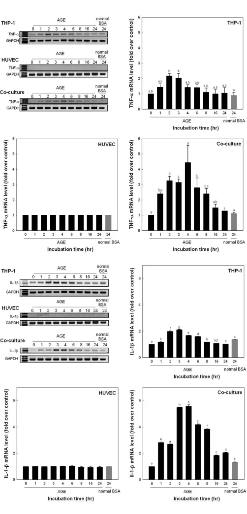
각 배양 조건에 따른 TNF-α와 IL-1β의 발현
Glyceraldehyde를 이용하여 제조된 AGEs 화합물(glycer- AGEs)을 THP-1, HUVEC 및 두 세포가 동시에 배양된 system에 100 μg/mL의 농도로 처리 후 24시간까지 처리시 간을 달리하고 각 세포를 회수하여 TNF-α와 IL-1β 발현 pattern을 mRNA 수준에서 조사하였다. Fig. 2의 결과를 보 면, 단구세포인 THP-1의 경우 100 μg/mL의 glycer-AGE와 배양시킬 때 배양 2~3시간에서 배양전보다 유의적 차이를 보이면서 증가하였으며 대조구에 비해 2.2배 증가되는 것을 알 수 있었다. 혈관내피세포인 HUVEC의 경우에는 24시간 배양하는 동안 발현정도의 유의적인 차이가 없었다. 그러나
두 세포를 동시에 배양한 system에서 혈관내피세포인 HUVEC의 경우에는 배양 1시간부터 유의적 차이를 보이면 서 증가하였으며 배양 4시간에서 대조구보다 4.4배 정도까 지 TNF-α의 발현이 증가된 후 다시 감소되는 것을 확인할 수 있었다.
IL-1β의 발현 또한 Fig. 3에서와 같이 THP-1의 경우 배 양 1시간부터 유의적 차이를 보이면서 증가하여 배양 3시간 에서 대조구에 비해 2.1배까지 증가되는 것을 알 수 있었다.
혈관내피세포인 HUVEC의 경우에는 24시간 배양하는 동안 발현정도의 유의적인 차이가 없었다. 그러나 두 세포를 동시 에 배양한 system에서 혈관내피세포인 HUVEC의 경우에 는 배양 1시간째부터 증가하여 배양 3~4시간에서 대조구보 다 5.5배 정도까지 IL-1β의 발현이 증가되는 것을 확인할 수 있었다.
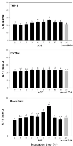
TNF-α와 IL-1β의 배지 내에서의 농도를 측정해본 결과
THP-1만 배양한 경우 대조구의 배지 내 TNF-α 함량이 배
양 6시간에 98.2 pg/mL로 대조구 53.8 pg/mL보다 증가하였
으며 HUVEC의 경우 배양 8시간에 93.3 pg/mL로 증가하였
다. 그러나 co-culture의 경우 배양 4시간부터 증가하여 배
양 8시간에 199.2 pg/mL로 증가하였다 다시 감소하는 것을
확인할 수 있었다(Fig. 4). 배지 내 IL-1β 함량은 HUVEC의
경우 24시간 동안 큰 변화를 보이지 않았으나 co-culture의
경우 배양 6시간부터 증가하여 배양 8시간에 39.5 pg/mL로
증가하였다 배양 24시간에는 배양전과 같은 수준으로 감소
Fig. 2. mRNA expression of TNF-α.
Each system was incubated with 100 μg/
mL glyceraldehyde-AGEs. Values are mean±SD and means with different let- ters (a-d) on bars are significantly dif- ferent at p<0.05 by Duncan's multiple range tests.
Fig. 3. mRNA expression of IL-1β.
Each system was incubated with 100 μg/
mL glyceraldehyde-AGEs. Values are
mean±SD and means with different let-
ters (a-h) on bars are significantly dif-
ferent at p<0.05 by Duncan's multiple
range tests.
Incubation time (hr)
Fig. 4. TNF-α content in medium. Each system was incubated with 100 μg/mL glyceraldehyde-AGEs. Values are mean±SD and means with different letters (a-h) on bars are significantly different at p<0.05 by Duncan's multiple range tests.
Incubation time (hr)
Fig. 5. IL-1β content in medium. Each system was incubated with 100 μg/mL glyceraldehyde-AGEs. Values are mean±SD and means with different letters (a-f) on bars are significantly different at p<0.05 by Duncan's multiple range tests.
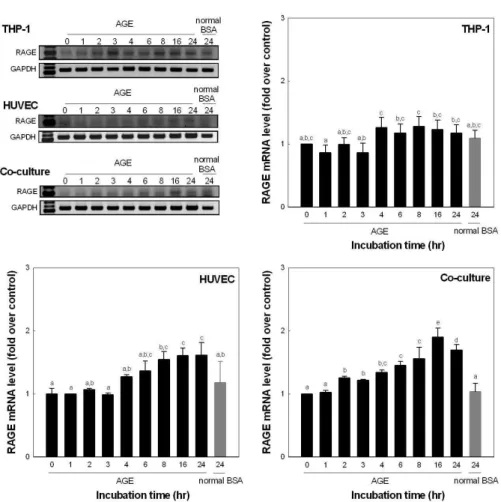
하는 것을 알 수 있었다(Fig. 5). RAGE는 TNF-α와 IL-1β 의 발현과 다르게 혈관내피세포인 HUVEC은 단독 및 co- culture에서 배양 16시간에 대조구에 비해 각각 1.6배, 1.9배 증가하였다(Fig. 6).
글라이케이션 반응에 의한 단백질의 가교결합(cross- linking)은 세포의 정상적인 기능의 저하를 일으켜 결과적으 로는 당뇨 합병증(4-6), 동맥경화(7,8)와 같은 혈관성 질환 이외에도 알츠하이머(31), 요독증(uremia)(32) 등 많은 성인 병의 원인으로 알려져 있다. 특히 당뇨 합병증이나 동맥경화 증은 혈관성 질환으로 AGEs에 의해 혈관내피세포에서 생 성되는 혈관 활성 조절 물질의 이상에 따른 것으로 설명된다 (4-8). AGEs에 의한 동맥경화증이나 당뇨 합병증과 같은 혈관성 질환을 예방하거나 진행을 억제시킬 수 있는 유효성 분의 효능연구에 있어 혈관내피세포의 기능 상실을 측정하 는 방법은 매우 중요하다.
혈관내피세포의 기능상실에 혈구세포와의 상호작용이 매 우 중요한 것이 이미 선행 연구자들에 의해 보고되어 있다.
혈구세포에 의해 활성화된 TNF-α와 IL-1β와 같은 cy-
tokine류는 혈관내피세포에 영향을 주어 E-selectin과 P-
selectin과 같은 혈관내피세포의 adhesion receptors나 vas-
cular cell adhesion molecule-1(VCAM-1), IL-8과 같은
chemokine을 생산하게 한다(24,33). Tsouknos 등(24)의 연
구 결과에 의하면 혈구세포와 혈관내피세포의 1:1 배양이
혈관내피세포에 100 U/mL의 TNF-α를 처리한 것과 유사한
수준으로 E-selectin과 VCAM-1이 발현되는 것을 확인하
였다. 또한 배양 4시간과 24시간을 비교해본 결과 배양 24시
간에 더 증가하였다고 보고하였다(이때의 측정방법은 im-
munofluorescence임). 그러나 본 연구자들이 검색해 본 결
과 혈관내피세포와 혈구세포의 co-culture system은 보고
된 논문을 찾을 수 있었으나 이 두 세포주와 AGEs와의 검색
Fig. 6. mRNA expression of RAGE.
Each system were incubated was 100 μg/
mL glyceraldehyde-AGEs. Values are mean±SD and means with different let- ters (a-e) on bars are significantly dif- ferent at p<0.05 by Duncan's multiple range tests.
조건은 명확하게 검색할 수 없었다.
본 연구 결과 AGEs에 의한 혈관내피세포의 기능상실의 연구에 있어서 혈관내피세포를 단독으로 연구하는 것보다 단구세포와 같이 co-culture할 때 TNF-α와 IL-1β 같은 cytokine의 발현이 더욱 증가하는 것을 확인할 수 있었다.
TNF-α와 IL-1β의 m-RNA 수준에서는 배양 4시간에서 가 장 높은 증가량을 보였고, 방출된 단백질의 수준에서는(배지 내에 함유된 단백질 양) 배양 8시간에 가장 많은 증가량을 보였다. 특히 단구세포인 THP-1에 glycer-AGE를 처리한 경우 처리 2∼3시간 후에 TNF-α와 IL-1β가 증가하였다.
그러나 HUVEC의 경우 유의적인 증가가 확인되지 않았으 나 co-culture할 때에는 대조군에 비해 5.5배까지 증가하는 것을 확인할 수 있었다. 이것은 AGEs에 의한 혈관내피세포 의 cytokine의 발현은 단구세포와의 cell communication이 중요한 역할을 하는 것으로 추정할 수 있었다. 그러나 RAGE 의 경우 TNF-α와 IL-1β의 결과와는 다르게 co-culture나 단독배양이나 큰 차이가 없음을 알 수 있었다. 그러나 배양 시간이 길어질수록 co-culture system에서 혈관내피세포에 서 AGEs의 receptor의 발현이 증가하는 것으로 보아 증가된 cytokine의 영향은 있는 것으로 추정할 수 있었다.
본 연구결과 최종당화산물에 의해 혈관내피세포의 기능 상실의 연구에 있어 식품소재나 천연소재로부터 효능물질 의 유효성 검색 시 본 연구결과에서 얻은 co-culture 조건이
유용하게 사용될 것이며 특히 mRNA 수준에서는 처리 후 4시간에, protein 수준에서는 8시간에 효능을 측정하면 유효 성 있는 연구 성과를 얻을 있을 것으로 기대된다.
요 약
Glyceraldehyde를 이용하여 제조된 AGEs 화합물(glycer-
AGEs)을 단구세포인 THP-1, 혈관내피세포인 HUVEC 및
이 두 세포가 동시에 배양된 system에서 100 μg/mL로 처리
후 24시간까지 처리시간을 달리하여 처리하였다. 배양시간
동안 각 세포와 배양액을 회수하여 TNF-α와 IL-1β의 발현
을 mRNA 수준에서 조사하였다. 그 결과, THP-1의 경우
배양 2시간에서 TNF-α나 IL-1β의 mRNA 발현이 대조구
에 비해 증가되었으나 혈관내피세포인 HUVEC의 경우에는
24시간 배양하는 동안 유의적인 차이가 없었다. 그러나 두
세포를 동시에 배양한 system에서 혈관내피세포인 HUVEC
의 경우에는 배양 4시간에서 대조구보다 TNF-α의 발현은
4.4배, IL-1β의 경우 5.5배 정도 증가되는 것을 확인할 수
있었다. TNF-α와 IL-1β의 배지 내에서의 농도를 측정해
본 결과, THP-1만 배양한 경우 대조구의 배지 내 TNF-α
함량이 배양 6시간에 98.2 pg/mL로 대조구 53.8 pg/mL보다
증가하였으며 HUVEC의 경우 배양 8시간에 93.3 pg/mL로
증가하였다. 그러나 co-culture의 경우 배양 4시간부터 증가
하여 배양 8시간에 199.2 pg/mL로 증가하였다. RAGE는 TNF-α와 IL-1β의 발현 pattern과 다르게 단독 및 co- culture에서 배양 16시간에 대조구에 비해 각각 1.6배, 24시 간에 1.9배 증가하였다. 따라서 본 연구 결과 최종당화산물 에 의해 혈관내피세포의 기능상실의 연구에 있어 co- culture 조건이 유용하며, 특히 mRNA 수준에서는 4시간에, protein 수준에서는 8시간에 효능을 측정하면 유효성 있는 연구 성과를 얻을 있을 것으로 예측할 수 있었다.
감사의 글
이 논문은 2008년도 정부재원(교육인적자원부 학술연구 조성사업비)으로 한국학술진흥재단의 지원을 받아 연구되 었으며(KRF-2008-532-F00019) 이에 감사드립니다.
문 헌