Received on May 18, 2012. Revised on June 4, 2012. Accepted on June 7, 2012.
CC This is an open access article distributed under the terms of the Creative Commons Attribution Non-Commercial License (http://creativecommons.org/licenses/by-nc/3.0) which permits unrestricted non-commercial use, distribu- tion, and reproduction in any medium, provided the original work is properly cited.
*Corresponding Author. Tel: 82-2-710-9560; Fax: 82-2-2077-7322; E-Mail: [email protected] Keywords: Silica nanoparticles, Dendritic cells, Apoptosis, Inflammatory response
Induction of Functional Changes of Dendritic Cells by Silica Nanoparticles
Kyeongah Kang and Jong-Seok Lim*
Department of Biological Science and the Research Center for Women’s Disease, Sookmyung Women’s University, Seoul 140-742, Korea
Silica is one of the most abundant compounds found in nature. Immoderate exposure to crystalline silica has been linked to pulmonary disease and crystalline silica has been classified as a Group I carcinogen. Ultrafine (diameter <100 nm) silica particles may have different toxicological proper- ties compared to larger particles. We evaluated the effect of ultrafine silica nanoparticles on mouse bone marrow-derived dendritic cells (BMDC) and murine dendritic cell line, DC2.4.
The exposure of dendritic cells (DCs) to ultrafine silica nano- particles showed a decrease in cell viability and an induction of cell death in size- and concentration-dependent manners.
In addition, in order to examine the phenotypic changes of DCs following co-culture with silica nanoparticles, we added each sized-silica nanoparticle along with GM-CSF and IL-4 during and after DC differentiation. Expression of CD11c, a typical DC marker, and multiple surface molecules such as CD54, CD80, CD86, MHC class II, was changed by silica nanoparticles in a size-dependent manner. We also found that silica nanoparticles affect inflammatory response in DCs in vitro and in vivo. Finally, we found that p38 and NF-κB activation may be critical for the inflammatory re- sponse by silica nanoparticles. Our data demonstrate that ul- trafine silica nanoparticles have cytotoxic effects on den- dritic cells and immune modulation effects in vitro and in vivo.
[Immune Network 2012;12(3):104-112]
INTRODUCTION
Nanotechnology is one of the exponentially developing tech- nologies of the 21st century. Nanoparticles sized between 1
and 100 nm are already in use for the most part including cosmetics, food industry, medicine, electronics, etc (1).
However, because nanoparticles have large surface areas, high chemical reactivity, internal pore volumes, and en- hanced cell penetrability, it may induce much more toxic ef- fects (2-5).
Silica (SiO2) is one of the most abundant compounds found in nature as crystalline or amorphous silica (6). Silica has been considered an ideal nanoparticle for biomedical applica- tions such as gene therapy, drug delivery, and biomedical imaging (7). However, it is becoming apparent that silica has cytotoxic and genotoxic effects. In human lymphoblastoid cells, ultrafine crystalline silica inhibited cell viability and growth, and also induced apoptosis and DNA strand breaks of cells (8). Ultrafine silica induced oxidative stress and pro-inflammatory responses in macrophages, mice and rats (6,9,10). Also, pulmonary inflammation, emphysema, alveolar hyperinflation, and apoptosis of alveolar and granulomatous cells have been found in animals exposed to silica (11,12).
According to several toxicity results, silica has been classified as a Group 1 carcinogen by the International Agency for Research on Cancer (IARC) in 1997 (6).
Dendritic cells (DCs) are potent antigen-presenting cells, which reside in most tissues including blood and lymphoid organ (13-15). They function as sentinels of immune system and initiators of innate and adaptive immune responses.
Mature DCs that have recognized antigen in peripheral tissue migrate to secondary lymphoid tissues and present the anti- gen to naïve T cells. In consequence, T cell responses are initiated (16,17). In mice, CD11c has been acknowledged as
a typical DC marker. When DCs mature, they express high levels of MHC molecules, CD54, CD80, CD86, etc (18).
Through expression of co-stimulatory molecules and cytokine secretion such as IL-12, IL-10, and IL-23, DCs induce activa- tion of naïve T cells (15).
Vallhov et al. have reported size-dependent effects of mes- oporous silica nano- (270 nm) and microparticles (2.5μm) on human dendritic cells (5). The viability of monocyte-de- rived DC was inhibited by mesoporous silica. Also, the meso- porous silica induced immune regulatory signals through an alteration of co-stimulatory molecule expression and pro- duction of IL-12p70. Of the plentiful nanoparticles available, those sized 2∼50 nm are of special interest in biotech- nologies. Therefore, in the present study, we evaluated whether ultrafine (20∼50 nm) silica induces a cytotoxic effect and inflammation in mouse dendritic cells. Ultrafine silica de- creased the viability of DCs and increased the amount of cell deaths. Moreover, we have found that it has a differential ef- fect on surface molecules on DCs after exposing them to sili- ca in different frequency and concentration. In addition to the effect on DC differentiation, it induced TNF-α production in dendritic cells and led to inflammatory responses in vitro and in vivo, suggesting that ultrafine silica nanoparticles have a cytotoxic effect on dendritic cells and an immune modulation effect.
MATERIALS AND METHODS Mice and cell lines
C57BL/6 mice were purchased from Samtako (Osan, Republic of Korea). Mice were maintained in specific pathogen-free conditions and used at 5∼7 weeks. The experiments employ- ing the mice were performed in accordance with institutional guidelines. The DC2.4 cell line, which was established as a murine dendritic cell line, was kindly provided by Dr. K.
Rock of Harvard Medical School (19).
Preparation of silica nanoparticles and cell culture All silica nanoparticles were purchased from Sigma-Aldrich (St. Louis, MO). Transmission electron microscopy (TEM) im- age of the same nanoparticles has already been reported else- where (6). Silica nanoparticles were suspended in distilled water and were autoclaved to inactivate any contaminating endotoxin. Dendritic cells and DC2.4 cells were cultured in RPMI 1640 (Gibco/Invitrogen, Carlsbad, CA) supplemented with 10% heat-inactivated FBS (HyClone, Logan, UT). Growth
factors used in the primary culture of DCs were recombinant mouse GM-CSF and IL-4 (R&D Systems, Minneapolis, MN).
FITC- conjugated antibodies to CD11c, CD54, CD80, and PE-conjugated antibodies to CD86, and MHC class II were purchased from BD Pharmingen (Palo Alto, CA).
Dendritic cell preparation from bone marrow To obtain bone marrow-derived DCs, we used a method by Inaba et al. (20). Briefly, bone marrow cells isolated from fe- murs of C57BL/6 mice were harvested and incubated for 30 min at 4oC with an antibody cocktail containing seven mono- clonal antibodies, designated RA3-3A1/6.1, J11d.2, J1J.10, GK1.5, M5/114.15.2, F4/80, and 3.168. The cells were wash- ed with culture medium and treated with rabbit complement (Low-ToxR-M, Cedarlane, Ontario, Canada) according to the manufacturer’s instruction. Viable cells were then isolated by a density gradient centrifugation on Histopaque 1077 (Sigma-Aldrich) and washed twice with culture medium de- void of serum. The lymphocyte-depleted bone marrow cells were distributed in 24-well plates at 5-10×105 cells/ml. The cells were incubated in RPMI 1640 medium, supplemented with 10% heat-inactivated FBS (Hyclone). The media were supplemented with mouse GM-CSF (10 ng/ml) and IL-4 (10 ng/ml). Every 2 days, culture media were replaced. On days 6 or 7, the nonadherent cells were harvested by a gentle swirling and used in subsequent experiments.
MTT assay
DC 2.4 cells were seeded at a density of 1×10⁴ cells/ml in 96-well plates and incubated with RPMI-1640 medium con- taining 10% FBS in the presence of various concentrations and sizes of silica nanoparticles. After 24 h incubation, 0.5 mg/ml of MTT solution was added to each well. After in- cubation for 3∼4 h at 37oC, the MTT solution was removed.
The incorporated formazan crystals in viable cells were solu- bilized with 100μl of dimethyl sulfoxide. The absorbance was determined using VICTOR3TM (PerkinElmer, Waltham, MA).
Trypan blue exclusion assay
To analyze the growth in serum-containing medium, the cells were plated in a 100 mm dish in RPMI 1640 medium contain- ing 10% FBS in the presence of various concentrations and sizes of silica nanoparticles. Cells were harvested after 24 and 48 h and then stained with 0.4% trypan blue and counted using a hematocytometer.
Analysis of cell death by annexin V and 7-AAD Stain- ing
Cells cultured on culture dishes with silica nanoparticles for 24 h were harvested and stained with annexin V (BD Biosciences, Bedford, MA) and 7-AAD (BD Biosciences). The cell death was analyzed by flow cytometry using FACSCantoTMII (BD Biosciences).
Flow cytometry
DCs produced by in vitro culturing were subjected to flow cytometric analysis using a FACSCantoTMII. Cells were al- lowed to react with appropriate antibodies against CD11c, CD54, CD80, CD86, and MHC class II at 4oC for 30 min and analyzed for their antigen expression.
RT-PCR
RNA was isolated with Trizol (Gibco/Invitrogen). The first strand cDNA was synthesized from 1μg of total RNA using M-MLV reverse transcriptase (Promega, Madison, WI). β -actin was used as a loading control. PCR products were elec- trophoresed on a 1% agarose gel and visualized by ethidium bromide staining.
Matrigel plug assay
Silica nanoparticles, cell supernatants, or silica nanoparti- cle-treated DC2.4 cells were mixed with Growth Factor Reduced MatrigelTM Matrix (BD Biosciences). The Matrigels (700μl each) were injected subcutaneously into the abdomi- nal region of C57BL/6. After 9∼11 days, mice were euthan- ized and matrigel plugs were removed. To evaluate hemoglo- bin level in matrigel plug, the Drabkin's reagent kit (Sigma) was used. The plugs were homogenized in 0.5 ml of distilled water and it was dissolved at 4oC. After then, homogenates were centrifuged and solution separated from homogenates was incubated with 0.5 ml of Drabkin s solution for 15 min at room temperature. Absorbance values were measured at 540 nm.
H&E staining
Silica nanoparticle-treated DC2.4-matrigel plugs were fixed with 10% formaldehyde overnight at 4oC. Samples were dehy- drated, paraffin-embedded, sectioned, and stained with hema- toxylin and eosin (H&E).
Western blot analysis
DC2.4 cells were exposed to silica nanoparticles (40μg/ml)
for 6 h. Cells were washed with DPBS and lysed in protein extraction solution (iNtRON Biotechnology, Seongnam, Re- public of Korea). Proteins were separated on a 12% SDS-pol- yacrylamide gel and blotted onto a PVDF membrane, which was then blocked by incubating with TBST (Tris-buffered sal- ine and 0.05% Tween-20) containing 5% skim milk.
Membranes were incubated with specific antibodies and washed with TBST. All antibodies for MAPK were purchased from Cell Signaling Technology Inc. (Beverly, MA) and anti- bodies for IκB-α and actin were purchased from Santa Cruz Biotechnology, Inc (Santa Cruz, CA). Antigen-antibody com- plexes were visualized after incubating the membrane with diluted secondary antibody coupled to horseradish perox- idase and detected by enhanced chemiluminescence.
Statistical analysis
One-way ANOVA was used for statistical analysis. It was rep- resented as mean±SD. p<0.05 was considered significant.
RESULTS
Cytotoxic effects of silica nanoparticles
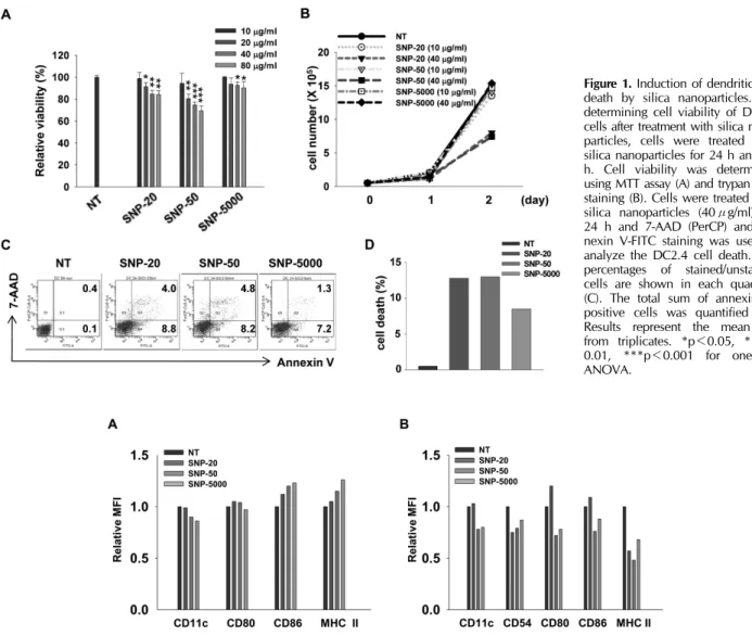
We evaluated cytotoxic effects of silica nanoparticles on mur- ine dendritic cell line, DC2.4. The names and the sizes of silica nanoparticles used are as follows: SNP-20, 10∼20 nm;
SNP-50, 50 nm; SNP-5000, 1∼5μm. We performed MTT as- say and trypan blue exclusion assay to estimate cell pro- liferation and viability. DC2.4 cells were cultured with silicas of different concentrations and sizes for 24 or 48 h. Ultrafine silica nanoparticles significantly decreased cell viability, de- pending on size and concentration (Fig. 1A). Because silica nanoparticles can interfere with MTT assay (21,22), trypan blue exclusion assay was also performed. Similar to the pre- ceding MTT assay, as the size and the concentration of ultra- fine silicas increased, the proliferation of DC2.4 cells was re- duced (Fig. 1B). However, 1∼5μm sized silica particles had little toxic effect compared to ultrafine silica nanoparticles (Fig. 1A and B).
The cell death induced by ultrafine silicas was evaluated by measuring the binding of 7-amino-actinomycin D (7-AAD) and annexin V. As shown in Fig. 1C and D, DC2.4 cells showed an increase in cell death after being exposed to silica for 24 h. From these data, it is obvious that ultrafine silica nanoparticles have cytotoxic effect on dendritic cells.
Figure 1. Induction of dendritic cell death by silica nanoparticles. For determining cell viability of DC2.4 cells after treatment with silica nano- particles, cells were treated with silica nanoparticles for 24 h and 48 h. Cell viability was determined using MTT assay (A) and trypan blue staining (B). Cells were treated with silica nanoparticles (40μg/ml) for 24 h and 7-AAD (PerCP) and an- nexin V-FITC staining was used to analyze the DC2.4 cell death. The percentages of stained/unstained cells are shown in each quadrant (C). The total sum of annexin V- positive cells was quantified (D).
Results represent the mean±SD from triplicates. *p<0.05, **p<
0.01, ***p<0.001 for one-way ANOVA.
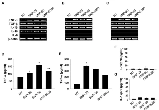
Figure 2. Silica nanoparticles affect the expression of surface antigens on dendritic cells. Bone marrow-derived DCs were generated by culturing bone marrow cells with 10 ng/ml of GM-CSF and 10 ng/ml of IL-4 for 6 days. Thereafter, cells were co-cultured with silica nanoparticles (40μg/ml) for 24 h (A). On the other hand, bone marrow-derived cells were cultured with 10 ng/ml of GM-CSF, 10 ng/ml of IL-4, and silica nanoparticles (10μg/ml) for 7 days (B). Cells were allowed to react with appropriate antibodies at 4oC for 30 min for the detection of CD11c, CD54, CD80, CD86 and MHC class II. Cells were analyzed using a FACSCantoTMII. The data are represented as relative Mean Fluorescence Intensity (MFI).
Effect of silica nanoparticles on DC differentiation In order to examine the phenotypic changes of bone mar- row-derived DCs (BMDC) following exposure to silicas, we added silicas along with GM-CSF and IL-4 during DC differ- entiation or added for 24 h after the complete DC differen- tiation. In case of exposure to silicas after DC differentiation, immature DCs were co-cultured with 40μg/ml of silica for 24 h. In case of exposure to silicas during DC differentiation, cells were co-cultured with 10μg/ml of silica every 2 days, repeatedly. In DCs exposed to silicas after differentiation, ex- pression of CD11c was inhibited by silicas which have a size
of 10∼20 nm and above (Fig. 2A). The expression of CD86 and MHC class II slightly increased after exposure to all rang- es of particle size. There was no meaningful effect on the expression of CD80. In DCs exposed to silicas during differ- entiation, not only the expression of CD11c, but also the ex- pression of all co-stimulatory molecules and MHC class II were reduced by silicas (Fig. 2B). Thus, DCs with different differ- entiation status exposed to silicas induced dissimilar changes of surface molecule expression. However, our findings showed that the effects of silica particles on the expression of DC sur- face molecules did not occur in nanoparticle-specific manner.
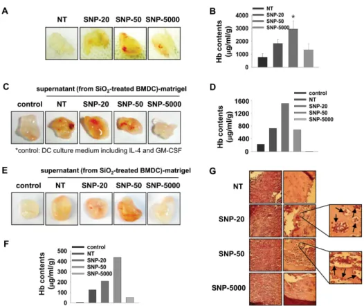
Figure 3. Silica nanoparticles induce TNF-α production in dendritic cells. DC2.4 cells (A) and bone marrow-derived dendritic cells (B, D, and F) were exposed to silica nanoparticles (40μg/ml) for 24 h.
Then, bone marrow-derived cells were cultured with 10 ng/ml of GM-CSF, 10 ng/ml of IL-4, and 10 μg/ml of silica nanoparticles for 7 days (C, E, and G). Cells were harvested for RNA preparation.
Transcriptional levels of cytokines were detected using RT-PCR (A∼C).
The supernatants of BMDCs were collected for 24 h. The levels of TNF-α (D and E) and IL-12p70 (F and G) in culture supernatants were determined by ELISA. Data repre- sent the mean±SD of duplicates. *p
<0.05, **p<0.01 for one-way ANOVA.
Effect of silica nanoparticles on cytokine production in DC
We further investigated whether silica nanoparticles are able to affect inflammatory cytokine production at transcriptional and translational levels in the dendritic cell line DC2.4 and bone marrow-derived DCs. Just like the preceding experi- ments, the cells were exposed to silicas together with GM-CSF and IL-4 during DC differentiation or exposed to silicas only for 24 h after the complete DC differentiation. The super- natants of dendritic cells were obtained from supernatants during the last 24 h. DC2.4 cells were co-cultured with 40 μg/ml of silica for 24 h for RT-PCR and ELISA such as in the case of exposure after differentiation. As shown in Fig.
3A∼C, TNF-α expression in DC2.4 and BMDC was in- creased by all silica nanoparticles. However, they showed no effects on the mRNA expression of other cytokines including IL-1β or TGF-β. Secretion of TNF-α was enhanced by the exposure to silica (Fig. 3D and E), and production of IL-12p70 was below detectable levels by exposure to silica (Fig. 3F and G). Moreover, repeated exposure to silica in low dose after media change induced higher TNF-α production in DC than one exposure to silica in high dose (Fig. 3E).
From these results, we have demonstrated that silica nano- particles are able to increase the production of pro-in- flammatory cytokine such as TNF-α in dendritic cells, and
as a result, they may induce inflammatory responses.
Effect of silica nanoparticles on vessel formation and immune cell infiltration in matrigel plugs transplant- ed into mice in vivo
We also investigated whether silica nanoparticles induce in- flammatory response in vivo. To observe vessel formation and immune cell infiltration by nanoparticle-induced in- flammation, we used a growth factor-reduced matrigel. It is in liquid state at low temperature but becomes a gel at room temperature to form a genuine reconstituted basement mem- brane (23). The major components of this material are lam- inin, entactin, TGF-β, etc. With these characteristics, it has been used to support in vivo angiogenesis and recruitment of lymphocytes (23-26). First of all, we evaluated the effect of silica nanoparticles by itself on angiogenesis and lympho- cyte infiltration in matrigel plugs transplanted into mice. As shown in Fig. 4A, silica nanoparticles induced vessel for- mation into the matrigel. In order to examine the recruitment of lymphocytes into matrigel, gel-formed matrigel was ex- changed to liquid at 4oC with Dulbecco s Phosphate Buffered Saline (DPBS) and subcutaneously injected into the abdomi- nal region of mice. We could find some cells infiltrated into the matrigel, but cell population was not detectable because of its rare number. Lymphocyte infiltration was indirectly ana-
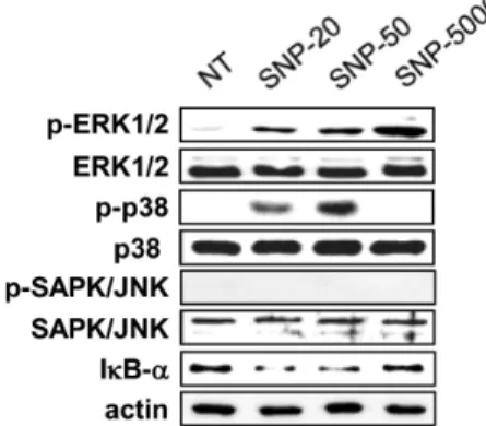
Figure 4. Silica nanoparticles elicit inflammatory responses in vivo. C57BL/6 mice were injected subcutaneously with 700μl liquid matrigel containing silica nanoparticles (10 mg/kg). After 10 days, gels were excised (A) and hemoglobin in matrigel plugs was determined using Drabkin’s reagent (B). BMDCs were exposed to silica nanoparticles (40μg/ml) for 24 h (C and D). In addition, bone marrow-derived cells were cultured with 10 ng/ml of GM-CSF, 10 ng/ml of IL-4, and 10μg/ml of silica nanoparticles for 7 days (E and F). The culture supernatants were collected for last 24 h and concentrated with centricon centrifugal filter devices. A mixture of 500μl liquid matrigel and 200μl concentrated supernatant was injected to C57BL/6 mice subcutaneously. After 9∼10 days, gels were excised (C and E) and hemoglobin in matrigel plugs was determined (D and F). DC 2.4 cells were treated with silica nanoparticles (40μg/ml) for 24 h. DC2.4 cells treated with silica nanoparticles were isolated by a density gradient centrifugation on ficoll to remove contamination of silica nanoparticles. C57BL/6 mice were injected subcutaneously with 700μl liquid matrigel containing 5×10⁵ DC2.4 cells. After 10 days, gels were excised and matrigel plugs were stained with hematoxylin &
eosin (H&E) (G). Results represent the mean±SD from duplicates. *p<0.05 for one-way ANOVA.
lyzed via the measurement of hemoglobin (Hb) concentration in matrigel. As a result, Hb concentration in matrigel was in- creased by a treatment with ultrafine silica particles, and silica with 1∼5μm size also induced an increased Hb concen- tration in matrigel compared to the non-treated controls.
However, Hb concentration was the highest in the matrigel treated with silica particles, SNP-20 and SNP-50.
Next, we examined the effect of supernatant of silica nano- particle-treated BMDC on vessel formation and Hb concen- tration in matrigel plugs when they are transplanted into mice in vivo. Supernatants were collected from BMDC exposed to silica nanoparticles which were added after differentiation or during differentiation. Those supernatants were concentrated 10-fold by centrifugation. Vessel formation and Hb content
in matrigel plugs were increased by supernatant from BMDC co-cultured with silica nanoparticles in all cases (Fig. 4C∼F).
As shown in previous results, silica with a size of 1∼5μm had a relatively weak effect compared to ultrafine silica particles. These results indicate that silica nanoparticles in- crease the production of various inflammatory factors includ- ing TNF-α in dendritic cells and result in inflammatory re- sponses in vivo.
We further evaluated whether vessel formation and im- mune cell infiltration in vivo can be found immediately by introducing DC2.4 cells exposed to silica. DC2.4-matrigel plugs were changed into highly dense tissues, and thick and long vessels were easily found in the matrix with naked eyes (Fig. 4G). As determined by H&E staining, vessel formation
Figure 5. Phosphorylation of p38 MAPK is essentially associated with the induction of in vivo inflammatory responses by nanoparticles.
DC2.4 cells were co-cultured with 40μg/ml of silica nanoparticles for 6 h. Equal amounts of whole lysates were subjected to electrophoresis on a SDS-PAGE and Western blot analysis was performed using specific antibodies, respectively.
and lymphocyte infiltration were increased much more by sili- ca nanoparticles. In particular, recruitment of red blood cells and formation of capillary vessel in DC2.4-matrigel plug were promoted by ultrafine silicas with a size of 10∼20 nm and 50 nm. Therefore, data suggest that silica nanoparticles in- duce inflammatory responses mediated by dendritic cells, and inflammatory mediators from dendritic cells exposed to silica nanoparticles generate vessel formation and lymphocyte in- filtration in vivo.
Activation of MAPK and NF-κB in dendritic cells by silica nanoparticles
MAPK and NF-κB are activated by several inflammatory stimuli, and they induce inflammatory gene expression. Thus, we determined whether silica nanoparticles lead to the activa- tion of MAPK and NF-κB in DC2.4 cells. They were co-cul- tured with 40μg/ml silica nanoparticles for 6 h. As shown in Fig. 5, silica nanoparticles enhanced the phosphorylation of ERK1/2, but did not activate SAPK/JNK. Especially, phos- phorylation of p38 was highly increased by 10∼20 nm and 50 nm ultrafine silica nanoparticles but was hardly induced by 1∼5μm silica particles. The production of IκB-α was also inhibited by ultrafine silica nanoparticles. Therefore, the data suggest that inflammatory responses by silica nano- particles in dendritic cells are induced via MAPK and NF-κB activation.
DISCUSSION
In this study, we evaluated the effect of ultrafine silica nano- particles on mouse bone marrow-derived dendritic cells (BMDC) and murine dendritic cell line, DC2.4. Silica nano- particles are ranked as one of the top five commonly used nanomaterials in the nanotechnology consumer products by the Woodrow Wilson International Center for Scholars (27).
It has been known that silica nanoparticles are able to enter the cells and be localized in the cytoplasm (28). Because den- dritic cells play a role as gatekeeper in innate and adaptive immunity, it is important to study the effect of silica nano- particles in dendritic cells.
10∼50 nm sized ultrafine silicas decreased cell viability and proliferation, and induced apoptosis in DC2.4 cells.
However, 1∼5μm sized silica particles had little toxic effect compared with ultrafine silica nanoparticles (Fig. 1). It has been reported that smaller particles have a higher level of cytotoxicity than larger particles. In human endothelial cells (EAHY926, a hybrid of human umbilical vein endothelial cells) and murine macrophage cells (RAW 264.7), smaller sili- cas showed much more inhibition of cell viability than larger silicas (29,30). However, study of Vallhov et al. has shown that a larger particle has more elevated cytotoxicity than a smaller particle in human dendritic cells (5). In contrast, Lin et al. and Cha and Myung reported that silica particles do not have a significant size-dependent difference of cell cyto- toxicity in human lung cancer cells (A549), liver cells (Huh-7), brain cells (A-172), stomach cells (MKN-1), and kidney cells (HEK293) (31,32). Herein, our data also suggest that nano- particles with 10∼50 nm size do not show a significant size-dependent difference of cell cytotoxicity, although silica nanoparticles with 50 nm size showed more decreased cell viability than those with 20 nm size (Fig. 1). Therefore, size-dependent cytotoxicity of silica is yet to be confirmed.
Vallhov et al. investigated whether silica particles have im- mune modulatory effects on human monocyte-derived den- dritic cells (MDDC) (5). They also indicated that CD86+ cells increased, but CD40+ cells and CD80+ cells decreased in im- mature MDDCs 24 to 48 h after co-culturing with silica particles. In our study, whereas CD11c+ cells were reduced, expression of CD86 and MHC class II was increased by ex- posure to silicas after differentiation of DCs (Fig. 2A). More- over, it was additionally found that repeated exposures to sili- ca nanoparticles inhibit the expression of surface molecules including CD11c, CD54, CD80, CD86 and MHC class II on
mouse BMDC (Fig. 2B).
A number of reports have shown that inorganic nano- particles induce inflammatory mediators and inflammatory re- sponses in vitro. Inorganic layered metal hydroxide (LMH) and silica enhanced the release of IL-8 from human lung epi- thelial cells (L-132) and A549 carcinoma cells (33). Silver nanoparticles increased the production of TNF-α, MIP-2, and IL-1β in rat alveolar macrophages, too (34). In human bron- chial epithelial cell line, BEAS-2B, titanium dioxide elevated not only the expressions of inflammation-related genes such as IL-1, IL-6, IL-8, TNF-α, and C-X-C motif ligand 2 (CXCL2), but also various oxidative stress-related genes including heme oxygenase-1, thioredoxin reductase, glutathione-S-transferase, catalase, and hypoxia inducible gene (35). In contrast to these results, single-walled carbon nanotube (SWCNT) in- hibited the production of IL-6, IL-8, and MCP-1 in A-549 cells (36). In the present study, silica nanoparticles, similar to many previous works, induced a specific production of in- flammatory cytokine, TNF-α, in dendritic cells (Fig. 3).
Further, ultrafine silica nanoparticles led to activation of p38 and NF-κB in DC2.4 cells (Fig. 5).
Given that silicas are able to produce inflammatory cyto- kines in DCs and inflammation is involved in angiogenesis, we investigated the effect of silicas on vessel formation in vivo. Silicas induced vessel formation into the matrigel by it- self (Fig. 4A and B). The supernatant from DCs exposed to silicas also triggered increased vessel formation and DCs ex- posed to silicas caused recruitment of red blood cells and for- mation of capillary vessel (Fig. 4C∼G). However, silica with size of 1∼5μm had a relatively weak effect compared to ultrafine silicas (Fig. 4). Although we failed to confirm what kind of cells was infiltrated into matrigel, it was shown that angiogenesis was triggered by silica nanoparticles as well as the infiltrated cells into matrigel. In our previous report, silver nanoparticles (AgNPs) also induced angiogenic effect through activation of VEGFR pathway in vivo (26). Especially, AgNPs increased peritumoral vascularization in B16F10 melanoma model. Thus, it is required to consider a therapeutic possi- bility of nanoparticles including silicas and AgNPs in several diseases.
In conclusion, our findings suggest that silica nanoparticles, especially silicas with nanoscale size, have cytotoxic effects on dendritic cells and immune modulation effects in vitro and in vivo. Moreover, this study provides important information about the safety and immune regulation of ultrafine silica nanoparticles that are commonly used nanomaterials in the
nanotechnology consumer products.
ACKNOWLEDGMENTS
This work was supported by the Nano R&D Program through the National Research Foundation of Korea funded by the Ministry of Education, Science and Technology of Korea (2010-0019159).
CONFLICTS OF INTEREST
The authors have no financial conflict of interest.
REFERENCES
1. Chen, M., and A. von Mikecz. 2005. Formation of nucleo- plasmic protein aggregates impairs nuclear function in re- sponse to SiO2 nanoparticles. Exp. Cell Res. 305: 51-62.
2. Pan, Z., W. Lee, L. Slutsky, R. A. Clark, N. Pernodet, and M. H. Rafailovich. 2009. Adverse effects of titanium dioxide nanoparticles on human dermal fibroblasts and how to pro- tect cells. Small 5: 511-520.
3. Foley, S., C. Crowley, M. Smaihi, C. Bonfils, B. F. Erlanger, P. Seta, and C. Larroque. 2002. Biochem. Biophys. Res.
Commun. 294: 116-119.
4. Oberdörster, E. 2004. Manufactured nanomaterials (full- erenes, C60) induce oxidative stress in the brain of juvenile largemouth bass. Environ. Health Perspect. 112: 1058-1062.
5. Vallhov, H., S. Gabrielsson, M. Strømme, A. Scheynius, and A. E. Garcia-Bennett. 2007. Mesoporous silica particles in- duce size dependent effects on human dendritic cells. Nano Lett. 7: 3576-3582.
6. Cho, W. S., M. Choi, B. S. Han, M. Cho, J. Oh, K. Park, S. J. Kim, S. H. Kim, and J. Jeong. 2007. Inflammatory medi- ators induced by intratracheal instillation of ultrafine amor- phous silica particles. Toxicol. Lett. 175: 24-33.
7. Huang, D. M., T. H. Chung, Y. Hung, F. Lu, S. H. Wu, C.
Y. Mou, M. Yao, and Y. C. Chen. 2008. Internalization of mesoporous silica nanoparticles induces transient but not suf- ficient osteogenic signals in human mesenchymal stem cells.
Toxicol. Appl. Pharmacol. 231: 208-215.
8. Wang, J. J., B. J. Sanderson, and H. Wang. 2007. Cytotox- icity and genotoxicity of ultrafine crystalline SiO2 particulate in cultured human lymphoblastoid cells. Environ. Mol. Muta- gen. 48: 151-157.
9. Park, E. J., and K. Park. 2009. Oxidative stress and pro-in- flammatory responses induced by silica nanoparticles in vivo and in vitro. Toxicol. Lett. 184: 18-25.
10. Fubini, B., and A. Hubbard. 2003. Reactive oxygen species (ROS) and reactive nitrogen species (RNS) generation by silica in inflammation and fibrosis. Free Radic. Biol. Med. 34:
1507-1516.
11. Warheit, D. B., T. A. McHugh, and M. A. Hartsky. 1995.
Differential pulmonary responses in rats inhaling crystalline,
colloidal or amorphous silica dusts. Scand. J. Work Environ.
Health 21(Suppl 2): 19-21.
12. Leigh, J., H. Wang, A. Bonin, M. Peters, and X. Ruan. 1997.
Silica-induced apoptosis in alveolar and granulomatous cells in vivo. Environ. Health. Perspect. 105(Suppl 5): 1241-1245.
13. Banchereau, J., and R. M. Steinman. 1998. Dendritic cells and the control of immunity. Nature 392: 245-252.
14. Banchereau, J., F. Briere, C. Caux, J. Davoust, S. Lebecque, Y. J. Liu, B. Pulendran, and K. Palucka. 2000. Immunobiol- ogy of dendritic cells. Annu. Rev. Immunol. 18: 767-811.
15. Blanco, P., A. K. Palucka, V. Pascual, and J. Banchereau.
2008. Dendritic cells and cytokines in human inflammatory and autoimmune diseases. Cytokine Growth Factor Rev. 19:
41-52.
16. Kang, K., H. Kim, K. I. Kim, Y. Yang, D. Y. Yoon, J. H.
Kim, J. H. Ryu, E. J. Noh, S. D. Jeon, and J. S. Lim. 2008.
SK-126, a synthetic compound, regulates the production of inflammatory cytokines induced by LPS in antigen-presenting cells. Biochem. Pharmacol. 75: 1054-1064.
17. Heath, W. R., G. T. Belz, G. M. Behrens, C. M. Smith, S.
P. Forehan, I. A. Parish, G. M. Davey, N. S. Wilson, F. R.
Carbone, and J. A. Villadangos. 2004. Cross-presentation, dendritic cell subsets, and the generation of immunity to cel- lular antigens. Immunol. Rev. 199: 9-26.
18. Reis e Sousa, C. 2006. Dendritic cells in a mature age. Nat.
Rev. Immunol. 6: 476-483.
19. Shen, Z., G. Reznikoff, G. Dranoff, and K. L. Rock. 1997.
Cloned dendritic cells can present exogenous antigens on both MHC class I and class II molecules. J. Immunol. 158:
2723-2730.
20. Inaba, K., M. Inaba, N. Romani, H. Aya, M. Deguchi, S.
Ikehara, S. Muramatsu, and R. M. Steinman. 1992.
Generation of large numbers of dendritic cells from mouse bone marrow cultures supplemented with granulocyte/macro- phage colony-stimulating factor. J. Exp. Med. 176: 1693-1702.
21. Wörle-Knirsch, J. M., K. Pulskamp, and H. F. Krug. 2006.
Oops they did it again! Carbon nanotubes hoax scientists in viability assays. Nano Lett. 6: 1261-1268.
22. Laaksonen, T., H. Santos, H. Vihola, J. Salonen, J. Riikonen, T. Heikkilä, L. Peltonen, N. Kumar, D. Y. Murzin, V. P.
Lehto, and J. Hirvonen. 2007. Failure of MTT as a toxicity testing agent for mesoporous silicon microparticles. Chem.
Res. Toxicol. 20: 1913-1918.
23. Kowalczyk, D. W., A. P. Wlazlo, M. Blaszczyk-Thurin, Z. Q.
Xiang, W. Giles-Davis, and H. C. Ertl. 2001. A method that allows easy characterization of tumor-infiltrating lymphocytes.
J. Immunol. Methods 253: 163-175.
24. Curiel, T. J., P. Cheng, P. Mottram, X. Alvarez, L. Moons, M. Evdemon-Hogan, S. Wei, L. Zou, I. Kryczek, G. Hoyle, A. Lackner, P. Carmeliet, and W. Zou. 2004. Dendritic cell
subsets differentially regulate angiogenesis in human ovarian cancer. Cancer Res. 64: 5535-5538.
25. Saudemont, A., N. Jouy, D. Hetuin, and B. Quesnel. 2005.
NK cells that are activated by CXCL10 can kill dormant tumor cells that resist CTL-mediated lysis and can express B7-H1 that stimulates T cells. Blood 105: 2428-2435.
26. Kang, K., D. H. Lim, I. H. Choi, T. Kang, K. Lee, E. Y.
Moon, Y. Yang, M. S. Lee, and J. S. Lim. 2011. Vascular tube formation and angiogenesis induced by polyvinylpyrroli- done-coated silver nanoparticles. Toxicol. Lett. 205: 227-234.
27. Hansen, S. F., E. S. Michelson, A. Kamper, P. Borling, F.
Stuer-Lauridsen, and A. Baun. 2008. Categorization frame- work to aid exposure assessment of nanomaterials in consum- er products. Ecotoxicology 17: 438-447.
28. Passagne, I., M. Morille, M. Rousset, I. Pujalté, and B.
L azou. 2012. Implication of oxidative stress in size-depend- ent toxicity of silica nanoparticles in kidney cells. Toxicology 299: 112-124.
29. Napierska, D., L. C. Thomassen, V. Rabolli, D. Lison, L.
Gonzalez, M. Kirsch-Volders, J. A. Martens, and P. H. Hoet.
2009. Size-dependent cytotoxicity of monodisperse silica nanoparticles in human endothelial cells. Small 5: 846-853.
30. Waters, K. M., L. M. Masiello, R. C. Zangar, B. J. Tarasevich, N. J. Karin, R. D. Quesenberry, S. Bandyopadhyay, J. G.
Teeguarden, J. G. Pounds, and B. D. Thrall. 2009. Macro- phage responses to silica nanoparticles are highly conserved across particle sizes. Toxicol. Sci. 107: 553-569.
31. Lin, W., Y. W. Huang, X. D. Zhou, and Y. Ma. 2006. In vitro toxicity of silica nanoparticles in human lung cancer cells. Toxicol. Appl. Pharmacol. 217: 252-259.
32. Cha, K. E., and H. Myung. 2007. Cytotoxic effects of nano- particles assessed in vitro and in vivo. J. Microbiol. Bio- technol. 17: 1573-1578.
33. Choi, S. J., J. M. Oh, and J. H. Choy. 2009. Toxicological effects of inorganic nanoparticles on human lung cancer A549 cells. J. Inorg. Biochem. 103: 463-471.
34. Carlson, C., S. M. Hussain, A. M. Schrand, L. K.
Braydich-Stolle, K. L. Hess, R. L. Jones, and J. J. Schlager.
2008. Unique cellular interaction of silver nanoparticles:
size-dependent generation of reactive oxygen species. J.
Phys. Chem. B. 112: 13608-13619.
35. Park, E. J., J. Yi, K. H. Chung, D. Y. Ryu, J. Choi, and K.
Park. 2008. Oxidative stress and apoptosis induced by tita- nium dioxide nanoparticles in cultured BEAS-2B cells.
Toxicol. Lett. 180: 222-229.
36. Herzog, E., H. J. Byrne, A. Casey, M. Davoren, A. G. Lenz, K. L. Maier, A. Duschl, and G. J. Oostingh. 2009. SWCNT suppress inflammatory mediator responses in human lung ep- ithelium in vitro. Toxicol. Appl. Pharmacol. 234: 378-390.