저작자표시-비영리 2.0 대한민국 이용자는 아래의 조건을 따르는 경우에 한하여 자유롭게
l 이 저작물을 복제, 배포, 전송, 전시, 공연 및 방송할 수 있습니다. l 이차적 저작물을 작성할 수 있습니다.
다음과 같은 조건을 따라야 합니다:
l 귀하는, 이 저작물의 재이용이나 배포의 경우, 이 저작물에 적용된 이용허락조건 을 명확하게 나타내어야 합니다.
l 저작권자로부터 별도의 허가를 받으면 이러한 조건들은 적용되지 않습니다.
저작권법에 따른 이용자의 권리는 위의 내용에 의하여 영향을 받지 않습니다. 이것은 이용허락규약(Legal Code)을 이해하기 쉽게 요약한 것입니다.
Disclaimer
저작자표시. 귀하는 원저작자를 표시하여야 합니다.
비영리. 귀하는 이 저작물을 영리 목적으로 이용할 수 없습니다.
의학석사 학위논문
Effects of cholesterol on the secretion of Alpha-Synuclein
2019 년 12 월
서울대학교 대학원 협동과정 뇌과학전공
Hoang-Anh Ho
i
Effects of cholesterol on the secretion of Alpha-Synuclein
지도 교수 이 승 재
이 논문을 의학석사 학위논문으로 제출함 2019 년 12 월
서울대학교 대학원 협동과정 뇌과학전공
Hoang-Anh Ho
Hoang-Anh Ho 의 의학석사 학위논문을 인준함
2020 년 1 월
위 원 장 서 영호 ( 인 )
부위원장 이 승재 (인)
위 원 이 용석 ( 인 )
ii
Effects of cholesterol on the secretion of Alpha-Synuclein
by
Hoang-Anh Ho
A thesis submitted to the Interdisciplinary Program in Neuroscience in partial fulfillment of the requirements for the Degree of Master of
Science in Neuroscience at Seoul National University College of Natural Sciences
January 2020
Approved by Thesis Committee:
Professor Young Ho Suh Chairman
Professor Seung-Jae Lee Vice chairman
Professor Yong Seok Lee
iii
ABSTRACT
Effects of cholesterol on the secretion of Alpha-Synuclein
Hoang-Anh Ho College of Natural Sciences Interdisciplinary Program in Neuroscience The Graduate School Seoul National University
Protein aggregation and inclusion body formation are common pathological features in many neurodegenerative diseases. Research studies have suggested that the deposition of disease-related proteins could be attributed to their ability to self-replicate and propagate. Evidence showed that protein structures can be secreted from one cell and transmitted to another where they repeat the aggregation process. Cholesterol is an essential lipid for the maintenance of cellular homeostasis.
As the brain contains more than 25% of total unesterified cholesterol, it becomes exceptionally susceptible to aberrant cholesterol metabolism. Lysosomes are cellular compartments that are responsible for the breakdown and recycling of endocytic and autophagic substrates. Lysosomal
iv
storage diseases (LSDs) are a group of metabolic disorders caused by genetic mutations in lysosomal hydrolases or non-lysosomal proteins that indirectly influence lysosomal functions.
Niemann-Pick type C (NPC) disease is an LSD characterized by defects in either NPC1 or NPC2 gene, which leads to cholesterol accumulation, perturbed autophagosome-lysosomal functions.
Similar to other forms of LSD, NPC patients also exhibit neurological defects that are pertaining to Parkinson’s Disease (PD) and Alzheimer’s Disease (AD). Studies have shown increase in the secretion of the PD-related protein, alpha-synuclein (aSyn) in research model of NPC disease. Here, the main object of this study was to investigate the role of cholesterol accumulation in secretory mechanism of aSyn in order to address the significance of NPC1-deficiency and lysosomal pathway in the propagation of disease-related protein. For this purpose, I studied the effects of NPC1 gene depletion, cholesterol-depleting agent Methyl--cyclodextrin (MBCD) in NPC1-/- cell line and a cholesterol accumulation-inducer U18666A (U18) in WT cells. The knockout mutant exhibits impaired lysosomal activity and increased autophagic activity, as well as increases in accumulation and release of aSyn and Tau, two neurological disease-related proteins. Immunoblotting analysis of medium samples and a lysosomal enzymatic assay showed that MBCD reversed increased aSyn release without change in lysosomal exocytosis. Treatment with U18 to WT cells elevated cholesterol levels and aSyn release and propagation albeit to a lesser extent than NPC1 knockout.
Taken together, these results suggest that modifications of cholesterol using genetic and pharmacological tools modulated the secretion of aSyn perhaps via the specific form of lysosomal exocytosis.
Keywords: neurodegenerative disease, lysosomal storage diseases, alpha-synuclein, propagation, NPC1, cholesterol
Student Number: 2017-27353
v
TABLE OF CONTENTS
ABSTRACT ... iii
TABLE OF CONTENTS ... v
LIST OF FIGURES ... vii
ABBREVIATIONS ... viii
INTRODUCTION ... 1
aSyn and Parkinson’s Disease ... 1
Niemann-Pick C (NPC) disease as the model for impaired cholesterol metabolism ... 4
Cholesterol as PD risk ... 6
Manipulation of cellular cholesterol level via pharmacological means ... 8
Purpose of the study ... 9
MATERIALS & METHODS ... 11
RESULTS ... 15
NPC1 deficiency increases cholesterol accumulation and decreases lysosomal degradation rate ... 15
NPC1 deficiency increases secretion of aSyn and lysosomal enzyme CTSD ... 16
Depleting cholesterol with MBCD changes distribution of lysosomes and decreases cellular cholesterol in NPC1 cells ... 19
MBCD treatment further decreases lysosomal degradation rate ... 20
MBCD treatment reverses pathological increase in secretion of aSyn in NPC1 cells via a
lysosomal-independent pathway ... 21
vi
U18 induces accumulation of cholesterol and lysosomes ... 24
U18 treatments increases lysosomal degradation rate in WT cells ... 25
U18 increased exocytosis of aSyn and CTSD, mimicking NPC1 knockout ... 26
U18 treatment increases cell-to-cell transmission of aSyn aggregates in a dose-dependent manner ... 29
DISCUSSION ... 32
CONCLUSION ... 35
REFERENCES ... 36
국문 초록 ... 48
vii
LIST OF FIGURES
Figure 1: Cellular pathways implicated in the secretion and propagation of aSyn. ... 4 Figure 2: NPC1-deficiency causes cholesterol accumulation which can lead to increase in lysosomal exocytosis of disease-related protein. ... 8 Figure 3: Experimental scheme of this study ... 9 Figure 4: NPC1 knockout increases cholesterol accumulation, increases secretion of aSyn and CTSD, and decreases protein degradation rate. ... 15 Figure 5: MBCD rescues increased aSyn release in NPC1 cell line via a lysosome-independent pathway ... 19 Figure 6: U18 partly mimics the NPC1-deficient phenotypes ... 24 Figure 7: U18 treatment increases cell-to-cell transmission of aSyn aggregates in a dose-dependent manner ... 37
viii
ABBREVIATIONS
ALP Autophagosome/Lysosomal Pathway aSyn Alpha-Synuclein
BiFC Bimolecular Fluorescence Complementation
CD Cyclodextrin
CNS Central nervous system CSP-α Cysteine string protein-α CTSD Cathepsin D
DSB Double strand break ER Endoplasmic Reticulum GBA Glucosidase beta acid Gcase Glucocerebrosidase InDel Insertion and deletion
LAMP1 Lysosomal-associated membrane protein 1 LSDs Lysosomal Storage Diseases
MBCD Methyl-β-cyclodextrin
MPP+ 1-methyl-4-methylpyridinium MSA Multiple-system atrophy MVB Multivesicular body
NAC Non-Aβ component
NCL Neuronal ceroid lipofuscinoses
NPC Niemann-Pick C
PAM Protospacer-adjacent motif PD Parkinson's Disease
ix PNS Peripheral nervous system
SD Sandhoff disease
U18 U18666A
UPS Ubiquitin-proteasome system
WT Wild-type
1
INTRODUCTION aSyn and Parkinson’s Disease
Parkinson’s disease (PD) is a common neurodegenerative disease among the elderly, affecting approximately 1–3% of people age 80 years and older [1]. PD is characterized with impaired motor symptom most likely resulting from the loss of dopaminergic neurons in the substantia nigra [2].
However, researchers have agreed on the consensus that neurodegenerative pathology is not restricted to this region and that non-motor symptoms are an inherent characteristics of PD [3].
Studies in PD patients frequently show Lewy pathology—a protein inclusion structure laden with fibrillary alpha-synuclein (aSyn) as the hallmark of PD [4] [5-7]. aSyn is a 140 amino acid protein which consists of three distinct regions: an amino terminus (residues 1-60), a central hydrophobic region (residues 61-95) or NAC (non-Aβ component), and a highly negatively charged carboxyl terminus. While the protein does not hold a defined structure in aqueous solutions, it forms - helical structures on binding to negatively charged lipids [8-10]. It is estimated that aSyn accounts for 1% of total cytosolic protein in the nervous system and the protein could be found in the vicinity of synaptic vesicles in presynaptic terminals [11]. aSyn is believed to play a role in neurotransmitter release [12-16], vesicular biogenesis [13], and chaperone for the presynaptic protein CSP-α (cysteine string protein-α) [17].
Polymeropoulos et al. identified aSyn as a genetic risk of PD in a 1997 study in which an amino acid substitution in SNCA gene encoding for aSyn resulting in the A53T mutation was found in some Greek familial cases [18]. Other point mutations subsequently identified to be associated with familial form of synucleinopathies [19-22]. Other cases associated with multiplication of the SNCA gene were reported in following years [23-26]. Most importantly, studies into sporadic case also
2
found association between SNCA gene and PD, and in some case, extending to other synucleinopathies like multiple-system atrophy (MSA) [27-32].
Since the discovery of aSyn’s association with PD, immunohistochemistry detection of aSyn has been the current gold standard of neuropathological evaluation of the disease. Based on pathological pattern of aSyn, Braak et al. suggested a six-stage of disease progression which begins at the olfactory bulb and the dorsal vagal nucleus and progress into a global deposition of aSyn pathology at later stages [33]. This discovery primes a series of studies investigating the possibility of protein propagation across the brain. Not only it was reported that aSyn can propagate from host tissues to grated cells in PD patients [34], the toxic oligomeric aggregate of aSyn is also capable of disseminating from one brain region to several others [35]. Several recent studies employing preformed-fibrils as the template for perpetual seeding also suggested that the propagation of disease-related protein, in this case aSyn, could be the driving force of disease progression [36, 37].
These observations reinforce the “propagation hypothesis” which proposes that aSyn can be transferred from one “donor cell” to another “recipient cell”. Despite the fact that the role of protein cell-to-cell transmission in PD pathogenesis still requires further investigation, this route appears to be an attractive target for therapeutic intervention.
The transmission process can be broken down to 3 steps: internalization of the recipient cell, aggregate formation with intracellular aSyn, and secretion to adjacent cells [38]. Studies showed that aSyn species could be internalized by endocytosis, micropinocytosis or protein-mediated uptake [39-43]. After internalization, aSyn aggregates are transferred to late endosomes and lysosomes [39]. Inside the cell, aSyn degradation is performed by both the ubiquitin-proteasome system (UPS) [44] and by the ALP [45] and the disruptions of this pathway can lead to aSyn accumulation [46]. The risk of developing PD for carriers of genes linked to lysosomal impairment is supported by recent evidence of associations between PD and Gaucher’s disease, an LSD resulting from a homozygous mutation in the glucosidase beta acid (GBA) gene [47]. Researchers
3
also found reduced level of glucocerebrosidase (GCase) and other lysosomal enzymes in studies of postmortem brain and cerebrospinal fluid in PD patients [48-52].
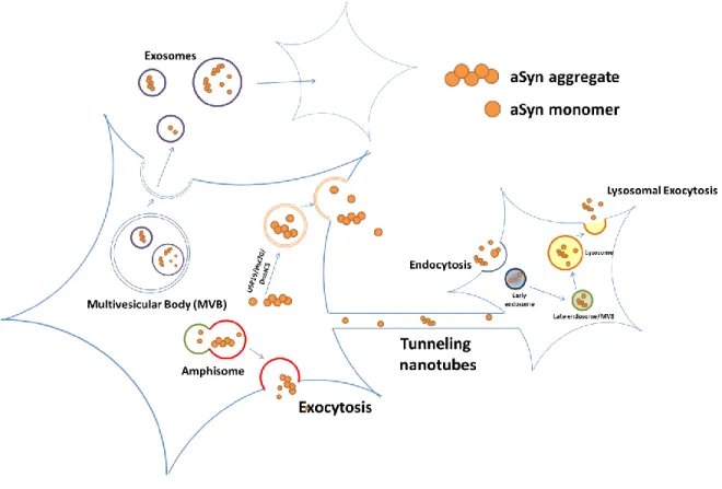
aSyn is thought to be released via a Golgi-independent unconventional pathway (Figure 1) [53-55].
Lee et al. found that under stress condition, aSyn is released outside in a pathway where USP19 acts as a chaperone [54]. Another study showed that the chaperone complex Hsc70/DnaJC5 facilitates removal of aSyn and Tau from neuronal cells [56]. Organelles like multivesicular bodies (MVBs), autophagosomes and amphisomes are also potential means for the secretion of aSyn via exocytosis [57, 58]. However, there are debates over whether MVB-derived exosomes play a part in the propagation of aSyn [59, 60]. Apart from vesicle-based transmission, tunneling nanotubes have also been suggested as an intercellular transmission mechanism of aSyn [61, 62].
Recently, lysosome-mediated exocytosis, or lysosomal exocytosis has emerged to be another possible candidate for protein propagation. Amyloid-β peptides are released via lysosomal exocytosis [63]. Inhibition of lysosomal exocytosis using Vacuolin-1 reduces secretion and transmission of aSyn. Furthermore, knocking-down of RAB27a blocks lysosomal exocytosis and at the same time, decreases the secretion of aSyn (unpublished data). There are not many evidence to date regarding lysosomal exocytosis-mediated protein propagation despite its potential, which makes this pathway a very promising research target.
4
Figure 1. Cellular pathways implicated in the secretion and propagation of aSyn.
Niemann-Pick C (NPC) disease as the model for impaired cholesterol metabolism
In 1955, Christian de Duve discovered the cellular organelles which he called “lysosome” [64, 65].
Lysosomes are acidic organelles (with pH range 4.6-5.0), serving as the terminal compartment responsible for the degradation of endocytic and autophagic substrates to maintain the homeostasis of the cell [66]. It is estimated that about 60 lysosomal acid hydrolases are in charge of performing the catabolism of the cells [67]. Dysfunctions in lysosomal hydrolases, activator protein and cofactor of lysosomal proteins give rise to more than 60 distinct LSDs that occurs collectively in 1 in every 5000 birth worldwide [68]. LSDs are characterized as a group of metabolic diseases of which more than two-thirds patient exhibit neurological symptoms at various extents [69]. The underlying mechanism of neurodegeneration observed in LSDs is not clearly understood, though the
5
accumulation of lysosomal substrates is thought to be the culprit of neuronal loss in LSDs [70-73].
A few examples of LSDs that are associated with central nervous system (CNS) and peripheral nervous system (PNS) pathology include Gaucher’s disease, Krabbe disease, Sandhoff disease (SD), Niemann–Pick type C (NPC), mucolipidoses, and the group of neuronal ceroid lipofuscinoses (NCLs; or Batten disease) [74].
Niemann-Pick type C (NPC) disease is an inherited neurodegenerative LSDs, characterized by accumulation of unesterified cholesterol in lysosomes and late endosomes [75]. NPC disease is caused by an autosomal recessive mutation in one or both of the NPC1 and NPC2 genes. NPC1 is a 1278 amino acid lysosomal membrane protein with 13 transmembrane domains [76]. NPC1 undergoes correct folding in the ER, transport to the Golgi apparatus and proceed through the maturation process like the majority of other membrane proteins [77].. Eventually, NPC1 proteins reach the lysosomes via the dileucine motif in the C terminus [78]. Crystal structure of human NPC1 combined with computational modeling suggested a cholesterol-harboring cavity in the sterol-sensing domain of the protein [79]. Although the N-terminal domain of NPC1 exhibits high affinity binding for cholesterol and side-chain oxysterols in vitro [80], this binding function seems to be dispensable [81]. Both NPC1 and NPC2 proteins are believed to participate in a coordinative transport of cholesterol in the endosome-lysosome system [81]. Furthermore, NPC1 may also have a role in the retrograde transport of lipids from late endosomes to the trans-Golgi network [82].
Up to now, more than 400 mutations in NPC1 gene account for 95% of NPC disease cases [83].
NPC patients’ symptoms include but not limited to ataxia, dystonia vertical gaze palsy, clumsiness, liver dysfunction, and learning difficulties with age onset ranges from infancy to old age [84]. There is an interindividual difference in the pattern lipid builds up. While cholesterol and sphingomyelin storage is more prominent in peripheral neural tissues, glycosphingolipid is more abundant in peripheral tissues [83]. In addition, researchers also observed variations of the severity of cellular
6
cholesterol lesion and biochemical phenotype [85]. The correlation between genetic mutations and clinical symptoms is not clear bur reports showed that variations of NPC1 mutant generate numerous phenotypes with unpredictable functions. Recent studies have identified and characterized the structural features, maturation pathways and subcellular localization of three classes of NPC1 mutants. To be specific, while the first class of NPC1 mutants resembles wild type case, the second and third class of NPC1 mutants reside partly in the endoplasmic reticulum (ER) and entirely in the ER, respectively [86].
Considering increasing evidence of the link between lysosomal diseases and neurodegenerative diseases, LSDs model could be useful in the study of the molecular mechanisms of neurodegenerative diseases. The NPC1 mutant used in this study was generated using the gene- editing system CRISPR-Cas9. Cas9 is directed to its DNA target site by base pairing between the gRNA and DNA. A PAM (protospacer-adjacent motif, sequence of 5’-NGG-3’) motif is required for Cas9 recognition and cleavage. The Cas9/gRNA complex cuts both strands of the target DNA.
The DNA is then left to an error-prone NHEJ pathway of Double strand break (DSB) repair, in which an InDel (insertion and deletion) is introduced into the position of the breakage [87].
Frameshift mutations created in-frame stop codons on both alleles created a homozygous knockout mutant cell line and the result was confirmed with the absence of NPC1 protein in immunoblot analysis. Depletion of NPC1 reduced lysosomal activity and resulted in the accumulation of lysosomes and buildup of substrates targeted for degradation. Moreover, NPC1-deficient cells have increased deposition and secretion of neurological disease-related proteins such as aSyn and Tau.
Cholesterol as PD risk
Cholesterol is an important lipid, especially for the human brain, where 25% of the total body unesterified cholesterol localizes [88]. Due to NPC1’s prominent role in cholesterol metabolism, it
7
is no surprise that cholesterol accumulation is the typical phenotype of NPC disease and that the buildup of cholesterol might be responsible for perturbations in cellular activities [89-92]. Even though there are more evidences linking NPC disease with AD, the implication of NPC1 in PD has been frequently mentioned. Accumulation of aSyn was also found in NPC1 patients [93, 94].
Subsequent studies linked other heterozygous carriers of NPC1 defects with PD [95, 96]. However, since PD is a common neurological disease, correlation like this could be mere coincidence. Since most NPC1 heterozygotes do not show Parkinsonian symptoms, more thorough population genetic studies would be necessary to pinpoint the exact link between the two diseases [95]. Nevertheless, taken the importance of autophagic pathway in PD into account, increased autophagic activity found in both PD and NPC studies proves the value of studying the mutual autolysosomal pathway [97-100].
In cellular models of PD, the neurotoxin 1-methyl-4-methylpyridinium (MPP+), a mitochondrial toxin, triggered lysosomal cholesterol accumulation preceding cell death [101]. This event was ameliorated by preincubation with U18666A (U18), suggesting that neurons respond to early apoptotic stress increasing accumulation of lysosomal cholesterol to preserve the integrity of lysosomal membrane [101]. Studies show data indicating a dual role of cholesterol accumulation in PD, protecting against lysosomal membrane permeabilization and cell death, while stimulating the accumulation and secretion of aSyn. Whether cholesterol accumulates in lysosomes or if the expression of proteins regulating lysosomal cholesterol trafficking is altered in patients with PD remains to be investigated.
Numerous studies have suggested that cholesterol can be an exocytosis facilitator though the exact mechanism is unclear. The ability of cholesterol to modulate membrane curvature could be one explanation [102]. However, we cannot rule out the fact that membrane cholesterol is tightly intertwined with membrane proteins and receptors, which could play important roles in the secretory process [103, 104]. If it is true that exocytosis of disease-related proteins is the direct
8
result of cellular cholesterol level (Figure 2), targeting disease via cholesterol will hold significant therapeutic potential.
Figure 2. NPC1-deficiency causes cholesterol accumulation which can lead to increase in lysosomal exocytosis of disease-related protein.
Manipulation of cellular cholesterol level via pharmacological means
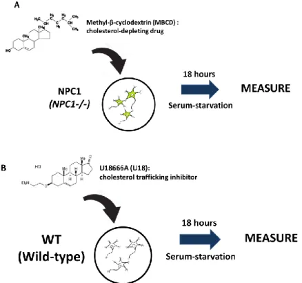
Cyclodextrins (CDs) are cyclic oligosaccharides compound which are commonly used as carriers for hydrophobic drugs [105]. CD exists as hexamers (αCDs), heptamers (βCDs) or octomers (γCDs) among which βCDs have the highest affinity for cholesterol [106]. Due to its low solubility, βCDs normally require modifications to make it viable for use [107]. Methyl-β-cyclodextrins (MBCD) and 2-hydroxyl-β-CD (2OHpβCD) are most widely used for cholesterol depletion. In this study, MBCD is employed as the cholesterol-depleting agent (Figure 3A).
Since we want to focus on the effect of cholesterol accumulation in NPC1-/- cells, U18666A (U18) was chosen as a pharmacological equivalent (Figure 3B). U18 is an androstenolone derivative that inhibits three enzymes in the cholesterol biosynthesis pathway [108]. U18 also inhibits LDL- derived cholesterol egress from lysosomes, which makes it an ideal imitation of NPC1 deficiency [109]. NPC1 was also identified to be one of the binding partners of U18 [110].
9
Figure 3: Experimental scheme of this study. In this study, I investigated (A) NPC1 cells were treated with 200 µM MBCD for 18 hours to deplete cholesterol and (B) WT cells were treated with 5 µM U18 to induce accumulation of cellular cholesterol.
Purpose of the study
Taken the previous studies into consideration, an investigation into the link between cholesterol and PD pathogenesis and progression is needed. Here, I employ two models of cholesterol trafficking defects, a genetic model with NPC1 knockout and a pharmacological model using U18. The data show that modifications of cellular cholesterol contents and cholesterol accumulation in lysosomal pathway can influence the secretion and transmission of aSyn. Also, lowering cholesterol levels with MBCD successfully rescues PD-related phenotypes in NPC1-/- cells. On the other hand, elevated cholesterol level as the result of either genetic or pharmacological tools significantly increases secretion of aSyn and lysosomal enzyme CTSD into the medium.
10
In this study, by using genetic and pharmacological models, I demonstrate that cellular cholesterol contents are crucial for the secretion and transmission of aSyn; the understanding of which will provide an insight into the mechanism of how aSyn contributes to the pathological progress of PD.
11
MATERIALS & METHODS
Compounds and Antibodies
The following chemical compounds and antibodies were used in this study: Purified Mouse Anti-α- Synuclein (Syn-1) (1:1,500 dilution; BD Biosciences; 610787), Anti-Cathepsin D antibody [CTD- 19] (1:2,000 dilution; Abcam; ab6313), Anti-Chromogranin II (SGII) polyclonal antibody (1:250 dilution; Abcam; ab192824), -actin monoclonal antibody (1:10,000 dilution; Sigma; AC-15), Horseradish peroxidase (HRP)-conjugated goat anti-mouse immunoglobulin G (IgG; H+L) (1:3,000 dilution; Bio-Rad Laboratories; 172-1011, Hercules, CA, USA), HRP-conjugated goat anti-rabbit IgG (H+L) (1:3,000 dilution, Bio-Rad Laboratories), Geneticin (Invitrogen; G418); Dextran, Alexa Fluor™ 568 10,000 MW, Anionic, Fixable (Invitrogen, D22912); Methyl--cyclodextrin (MBCD) (Sigma; C-4555); U18666A (U18) (Cayman Chemical Company; Ann Arbor, MI, USA; 10009085);
4-Methylumbelliferyl N-acetyl-β-D-glucosaminide (Sigma; M2133).
Cell culture
Two stable cell lines were previously generated: LAMP1-GFP (WT cells) and NPC1-/- (NPC1 cells). To generate LAMP1-GFP stable cell line, SH-SY5Y human neuroblastoma cells (ATCC CRL-2266; Manassas, VA) were transfected with LAMP1-mGFP plasmid (Addgene Plasmid
#34831) using electroporation. Transfected cells were selected with 500 µg/mL G418 (Invitrogen) and sorted with narrow gate (GFP RFU > 10^4) by FACS. To generate NPC1-/- cell line, LAMP1- GFP stable cells were transfected with sgRNA and Cas9 protein. The target sequence for NPC1 knocout at exon 1 is ACTAAGTCATATCCATCCTTTGG (5’ to 3’). gRNA was designed using http://www.rgenome.net/cas-designer. After transfection, clones were single isolated and the clones
with confirmed deletions and frameshift that introduced in-frame stop codon in both alleles were selected. Selected clones were tested for protein expression using Western blotting. Cells were split every 2 days at 37°C in humidified air with 5% CO2 in Dulbecco’s Modified Eagle’s Medium
12
(SH30243.01, HyClone, Logan, UT, USA) containing 10% Fetal Bovine Serum (SH30396.03, HyClone), 100 units/mL Penicillin and 100 units/mL Streptomycin (15140-122, Gibco, Grand Island, NY, USA). 50 µM all-trans-retinoic-acid (R2625, Sigma-Aldrich) was used for cell differentiation. To overexpress human aSyn protein, differentiated cells were infected with recombinant adenoviral vector (serotype Ad5, CMV promoter) containing human a-synuclein cDNA at a multiplicity of infection of 33. Infected cells were treated with compounds (200 M MBCD; 5 M U18666A) for 18 hours in serum-free condition.
For co-culture, V1S and SV2 stable cells (30,000 cells each) were mixed and culture for 3 days in 96-well plate. The co-culture was maintained with 300 g/mL G418. The fluorescence intensity was measured and analyzed using IN Cell Analyzer 2200 (GE Healthcare).
Preparation of cell extracts
Cells were washed twice with ice-cold PBS and lysed in extraction buffer (1% Triton X-100, 1%(v/v) Protease Inhibitor Cocktail (Sigma, St. Louis, MO, USA) in PBS). Cell lysates were incubated on ice for at least 10 minutes and centrifuged at 16,000 x g for 10 minutes. The Triton X- 100 insoluble fraction was resuspended in 1 x Laemmli Sample Buffer and sonicated briefly.
Medium was collected and centrifuged at 1,000 x g for 10 minutes at 4C and then 16,000 x g for 20 minutes at 4C to further pellet debris. Finally, the medium was concentrated using Amicon Ultra Centrifugal Unit 10K (Millipore; UFC801096) at 3,515 x g for 10 minutes.
Dextran Pulse-Chase Assay
Cells grown on poly-L-Lysine-coated coverslips were incubated with 100 g/mL of Alexa-568- conjugated Dextran (MW. 10,000) (Invitrogen) for 2 hours. After washing with serum-free DMEM, cells were incubated with fresh growth media for 1 hour, then fixed with 4% paraformaldehyde
13
(PFA) solution. The fluorescence intensity was measured using Zeiss 63X (N.A. 1.4) objective on an Axiovert 35 microscope (Zeiss) with an attached MRC1024 laser scanning confocal microscope (LSCM) system (BioRad).
Western blotting
Western blotting was performed as previously described [111]. Protein samples were loaded into 12%
SDS-PAGE gels and transferred to nitrocellulose membrane (0.22 m). Image detection was performed using Amersham Imager 600 (GE Healthcare Life Sciences, Marlborough, MA, USA).
Images were edited and analyzed using Multi Gauge (v3.0) software (Fujifilm, Tokyo, Japan).
-hexosaminidase assay
Enzyme activity was measured using a fluorogenic substrate-based assay. After the treatment, medium samples were incubated at 37°C for 2 hours with Reaction Mix 40 µL of 2mM 4- Methylumbelliferyl N-acetyl-β-D-glucosaminide in 0.1M Sodium Citrate pH 4.5. The reaction was stopped by adding 30 µL mixture of 1.1M Glycine (15 µL) and 2M Na2CO3 (15 µL). The fluorescent product 4-methylumbelliferone was measured in duplicate on a microplate reader using suitable excitation (365 nm) and emission filters (440 nm). The measurements were normalized with the level of SG-II in Western blotting of medium samples.
Amplex Red Cholesterol Assay
The assay was performed in 96-well clear bottom black plate as described in the official protocol (Invitrogen; A12216). Whole cell lysates were diluted 10 times before the assay. Working solution of 300 µM Amplex Red reagent contains 2 U/mL HRP, 2 U/mL cholesterol oxidase, and 0.2 U/mL cholesterol esterase. The assay was performed by adding 50 L of working solution to 50 L of sample. The plate was then incubated at 37C for 30 minutes and measured on a microplate reader
14
using suitable excitation (535nm) and emission filters (590nm). The measurements were normalized with total protein levels in cell lysates.
Quantification and Statistical Analysis
Values shown in the figures are presented as Mean S.E.M. Statistical analyses were performed using Prism 7 software (GraphPad Software Inc., San Diego, CA, USA). The graphs were drawn using Prism 7 software.
15
RESULTS
NPC1 deficiency increases cholesterol accumulation and decreases lysosomal degradation rate
In this study, NPC1 cell line is used as model of defective cholesterol transport. In this cell line, using CRISPR-Cas9, stop codons are introduced on both alleles to generate a homozygous knockout mutant. With the knowledge of NPC1 function in cholesterol metabolism and trafficking, I performed an assay to compare the total cholesterol level in WT and NPC1 cells. Whole cell lysates were collected and diluted to appropriate concentration for the Amplex Red Assay. In agreement with previous studies, knockout of NPC1 remarkably increased total cholesterol content in mutant cells compared to WT (Figure 4A).
Next, I performed the Dextran Pulse-Chase Assay to assess the lysosomal degradation rate in both cell types. Previous studies showed that the rate of endocytosis in cellular models of LSDs was lower than that in wild type cells [112]. Expectedly, the level of dextran uptake in NPC1 cells was decreased compared to WT cells (Figure 4C). While in WT cells dextran signal appeared to be more dispersed, NPC1 cells showed lower dextran intensity and more dextran puncta throughout the cytosol (Figure 4B). Lysosomal degradation rate was also decreased in NPC1 cell line, similar to our previous results (Figure 4D).
16
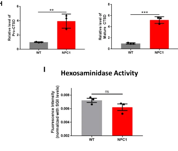
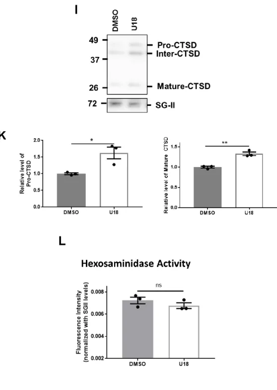
NPC1 deficiency increases secretion of aSyn and lysosomal enzyme CTSD
Catabolites in lysosomes can be secreted into extracellular space via lysosomal exocytosis [113- 115]. The presence of two common lysosomal enzymes, CTSD and β-hexosaminidase, in the extracellular medium can be the indicator of the level of lysosomal exocytosis. Here, I used immunoblot analysis to measure the level of CTSD and fluorometric enzymatic assay to measure the activity level of β-hexosaminidase in the medium samples. Secreted Cathepsin D significantly increased in NPC1 cells compared to WT cells (Figure 4G, H). On the contrary, depletion of NPC1
17
did not change the release of -hexosaminidase (Figure 4I). The data show that knockout of NPC1 gene significantly increases the exocytosis of aSyn alongside with the lysosomal enzyme CTSD, indicating an actual exocytosis of certain lysosomal population (Figure 4E, F, G, H).
18
Figure 4. NPC1 knockout increases cholesterol accumulation, increases secretion of aSyn and CTSD, and decreases lysosomal degradation rate. (A) Amplex Red Cholesterol Assay for total cholesterol level in whole cell lysates of WT and NPC1 cells (n = 3), **** p < 0.0001 by unpaired, two-tailed Student’s t test. (B) Representative images of WT and NPC1 cells showing Alexa 568 intensity from internalized dextran. Cells were incubated with 100 µg/mL Alexa 568-Dextran for 2 hours (0h) then chased for 1 hour in fresh growth media (1h), scale bar: 20 µm. (C) Quantification of dextran uptake by cells, ** p < 0.01 by unpaired, two-tailed Student’s t test. (n = 3, 30 cells were analyzed per experiment),. (D) Degradation rate of internalized dextran calculated from pulse-chase assay, ** p < 0.01 by unpaired, two-tailed Student’s t test. (E) Representative image of immunoblot analysis of secreted aSyn levels in medium samples collected after 18 hours of cell culture in serum-starved condition. (F) Quantification of the levels of total aSyn and aSyn aggregates (High MW aSyn) shown in 1E. ** p < 0.01, *** p < 0.001 by unpaired, two-tailed Student’s t test. (G)
19
Representative image of immunoblot analysis of secreted CTSD levels in medium samples. (H) Quantification of the levels of Pro-CTSD and Mature CTSD shown in 1G ** p < 0.01, *** p <
0.001 by unpaired, two-tailed Student’s t test. (I) β-hexosaminidase assay of medium samples (n = 3), ns: not significant. Data are represented as mean ± SEM.
Depleting cholesterol with MBCD changes distribution of lysosomes and decreases cellular cholesterol in NPC1 cells
Next, I investigated the effects of cholesterol-stripping drug MBCD on NPC1 cells. Cells were treated with 200 µM of MBCD which is adequate to see changes in lysosomal distribution and still preserve the viability of the cells. For the purpose of comparison, similar set of experiments were performed to assess the effects of MBCD on NPC1 cells. Contrary to expectation, stripping cellular cholesterol with MBCD did not bring LAMP1 distribution back to WT levels (Figure 5B). The treatment in effect cleared out peripheral lysosomes and caused perinuclear accumulation of LAMP1-positive compartments (Figure 5A). However, cholesterol assay showed that total cholesterol did decrease after MBCD treatment (Figure 5C).
20
MBCD treatment further decreases lysosomal degradation rate
Dextran Pulse-Chase Assay was performed to assess the protein degradation rate in two conditions.
MBCD-treated cells showed even lower dextran uptake than NPC1 cells in control group (Figure 5E). This may be due to decreased distribution of lysosomes inside the cells. Surprisingly, protein degradation rate in MBCD group also decreased compared to that of control group, which suggests that cholesterol depletion did not rescue disrupted lysosomal function (Figure 5F).
21
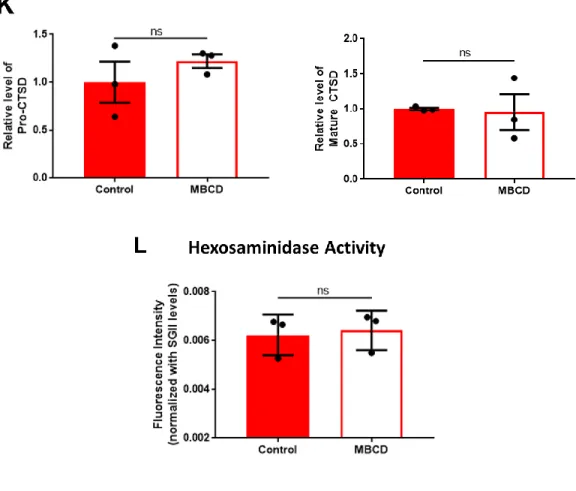
MBCD treatment reverses pathological increase in secretion of aSyn in NPC1 cells via a lysosomal-independent pathway
Immunoblot analysis of medium samples showed significant decrease in the levels of secreted aSyn while secretion of pro-CTSD, Mature CTSD, and -hexosaminidase was not affected (Figure 5E, F, G, H, I). The data suggests that albeit not affecting lysosomal exocytosis and protein degradation, MBCD can still rescue NPC1 knockout-induced increased secretion of aSyn. The data can have great implication in the study the correlation between NPC disease and PD.
22
23
Figure 5. MBCD rescues increased aSyn release in NPC1 cell line via a lysosome-independent pathway. NPC1 cells were treated with 200 µM of MBCD for 18 hours in serum-starved condition.
(A). Representative image of LAMP1 localization in NPC1 cells and MBCD-treated NPC1 cells. (B) Quantification of LAMP1-GFP intensity in NPC1 cells and MBCD-treated NPC1 cells (n = 4, 30 cells were quantified per experiment), ** p < 0.01 by unpaired, two-tailed Student’s test. (C) Amplex Red Cholesterol Assay for total cholesterol level in whole cell lysates of NPC1 and MBCD-treated NPC1 cells (n = 3), * p < 0.05 by unpaired, two-tailed Student’s t test. (D) Representative images of NPC1 and MBCD-treated NPC1 cells showing Alexa 568 intensity from internalized dextran. Cells were incubated with 100 µg/mL Alexa 568-Dextran for 2 hours then chased for 1 hour in fresh growth media, scale bar: 20 µm. (E) Quantification of dextran uptake by cells (n = 3, 30 cells were analyzed per experiment), * p < 0.05 by unpaired, two-tailed Student’s test. (F) Degradation rate of internalized dextran calculated from pulse-chase assay, ** p < 0.01 by
24
unpaired, two-tailed Student’s t test. (G) Representative image of immunoblot analysis of secreted aSyn levels in medium samples collected after 18 hours of cell culture in serum-starved condition.
(H) Quantification of the levels of total aSyn and aSyn aggregates (High MW aSyn) shown in 1E.
ns: not significant, ** p < 0.01 by unpaired, two-tailed Student’s t test. (I) Representative image of immunoblot analysis of secreted CTSD levels in medium samples. (K) Quantification of the levels of Pro-CTSD and Mature CTSD shown in 2I, ns: not significant by unpaired, two-tailed Student’s t test. (L) β-hexosaminidase assay of medium samples (n = 3), ns: not significant. (Data are represented as mean ± SEM.
U18 induces accumulation of cholesterol and lysosomes
Next, I investigated whether induction of cholesterol accumulation using U18 in WT cells can be used as a pharmacological model for cholesterol trafficking defects study. The optimum concentration of U18 at 5 µM was sufficient to induce a NPC1-deficient phenotype and at the same time, preserve the viability of the cells. Similar to previous sections, I will assess the effects of induced cholesterol accumulation in WT cells. Confocal images showed that U18 treatment resulted in increased intensity of LAMP1, especially in the perinuclear region (Figure 6A, B). U18 also increased total cholesterol content in cells compared with non-treated cells (Figure 6C).
25
U18 treatments increases lysosomal degradation rate in WT cells
Dextran Pulse-Chase Assay was performed to assess the protein degradation rate in two conditions.
In U18-treated case, similar to NPC1 cells, dextran signal was not dispersed but rather concentrated in numerous puncta in the cytosol (Figure 6D). Surprisingly, the lysosomal degradation rate was increased upon treatment with U18, opposite what happened in NPC1 knockout (Figure 6F).
26
U18 increased exocytosis of aSyn and CTSD, mimicking NPC1 knockout
Immunoblot analysis showed that there was increase in the levels of secreted aSyn and pro-CTSD and Mature CTSD, similar to what I observed in NPC1 cells (Figure 6G, H, I, K). The degree of increase in this pharmacological imitation was not comparable to what I observed in the complete knock out of NPC1. However, the increase of aSyn secretion was proportionate with the increase of CTSD secretion. β-hexosaminidase level in medium samples post-treatment remained unchanged (Figure 6L). This data shows that it is possible to employ U18 to mimic the effect of knocking out
27
NPC1 in in vitro model. It also emphasizes the discrepancy between the secretion of CTSD and - hexosaminidase under the influence of cholesterol accumulation, despite both proteins being the archetypical lysosomal markers. This observation raises the question whether there exists more than one lysosomal population when lysosomal stress becomes overwhelming for the cellular machinery.
28
Figure 6. U18 partly mimics the NPC1-deficient phenotypes. WT cells were treated with 5 µM of U18 for 18 hours in serum-starved condition. (A). Representative image of LAMP1 localization in WT cells and U18-treated WT cells. (B) Quantification of LAMP1-GFP intensity in WT cells and U18-treated WT cells (n = 3, 30 cells were quantified per experiment), **** p < 0.0001 by unpaired, two-tailed Student’s test. (C) Amplex Red Cholesterol Assay for total cholesterol level in
29
whole cell lysates of WT and U18-treated WT cells (n = 3), ** p < 0.01 by unpaired, two-tailed Student’s t test. (D) Representative images of WT and U18-treated WT cells showing Alexa 568 intensity from internalized dextran. Cells were incubated with 100 µg/mL Alexa 568-Dextran for 2 hours (0h) then chased for 1 hour in fresh growth media (1h), scale bar: 20 µm. (E) Quantification of dextran uptake by cells (n = 3, 30 cells were analyzed per experiment), ns: not significant by unpaired, two-tailed Student’s t test. (F) Degradation rate of internalized dextran calculated from pulse-chase assay, * p < 0.05 by unpaired, two-tailed Student’s t test. (G) Representative image of immunoblot analysis of secreted aSyn levels in medium samples. (H) Quantification of the levels of total aSyn and aSyn aggregates (High MW aSyn) shown in 3G, * p < 0.05 by unpaired, two-tailed Student’s t test. (I) Representative image of immunoblot analysis of secreted CTSD levels in medium samples. (K) Quantification of the the levels of Pro-CTSD and Mature-CTSD shown in 3I,
* p < 0.05, ** p < 0.01 by unpaired, two-tailed Student’s t test (I) β-hexosaminidase assay of medium samples (n = 3), ns: not significant by unpaired, two-tailed Student’s t test. Data are represented as mean ± SEM.
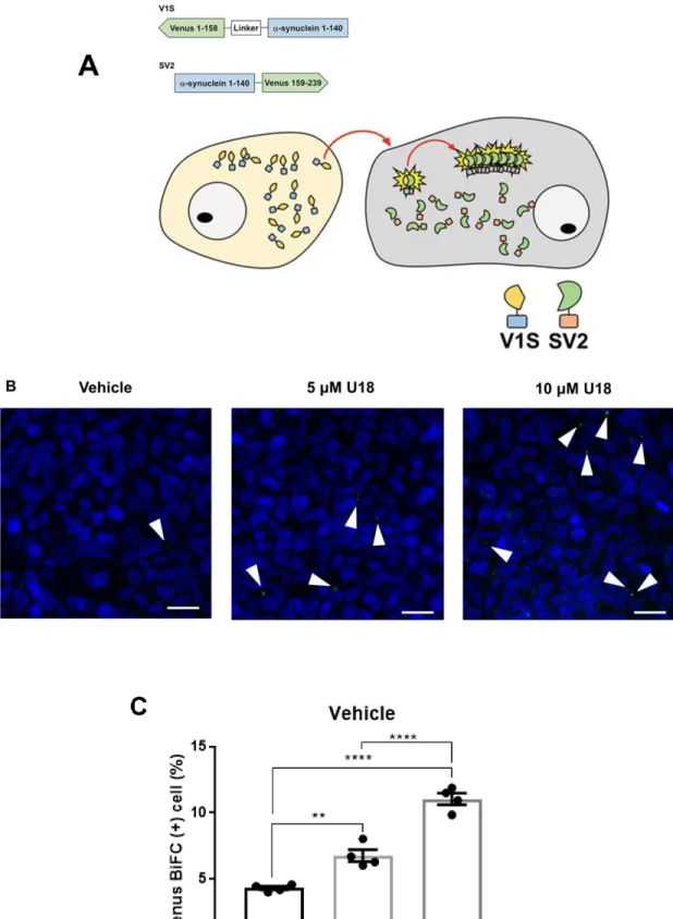
U18 treatment increases cell-to-cell transmission of aSyn aggregates in a dose-dependent manner
Next, using the dual-cell BiFC system, I analyzed the effect of U18 treatment on cell-to-cell transmission of aSyn, a key process in the progression of PD. The working mechanism of BiFC system is described in Figure 7A [116]. Briefly, the transmission of aSyn and formation of aSyn aggregates will be reflected in the intensity Venus fluorescence. Co-culture of V1S and SV2 cells were treated with DMSO (vehicle), 5 µM U18 and 10 µM U18. Results in Figure 7B and 7C showed that Venus fluorescence was significantly increased in 5 µM U18 treatment and further increased to a higher level in 10 µM U18. Therefore, aSyn propagation is affected by the treatment of U18 in a dose-dependent manner.
30
Figure 7. U18 treatment increases cell-to-cell transmission of aSyn aggregates in a dose- dependent manner. (A) Schematic representation of BiFC system using stable cell lines
31
overexpressing V1S (Venus 1-158-α-syn) or SV2 (α-syn-Venus 159-239). Cell-to-cell transmission and aggregation of αSyn can be observed via the reconstitution of green fluorescence. (B) Cell-to- cell transmission in BiFC system. White arrows indicate Venus puncta. Scale bar: 20 μm. (C) Quantification of BiFC-positive cells, approximately 10,000 cells were analyzed per experiment, **
p < 0.01, **** p < 0.0001 by one-way ANOVA with Tukey’s post hoc test.
32
DISCUSSION
Cholesterol is a key player in exocytosis and protein secretion, most likely through changing properties of the plasma membrane [104, 117]. Here, I showed that modification of cholesterol content can greatly affect the secretory mechanism of aSyn. By using cholesterol-modifying agents and cell models for cholesterol trafficking defects, I show that depletion of cholesterol in NPC1 cells with MBCD successfully reversed the increased secretion of aSyn (Figure 3). Complete knockout of NPC1, an important player in cholesterol trafficking and metabolism, shows more aggressive phenotype compared to its pharmacological equivalent using U18 (Figure 4). However, the latter seems to have a graded effect on cell-to-cell transmission and this effect was confirmed in BiFC model (Figure 5).
As previous studies suggest, lysosomal exocytosis may be able to explain how cholesterol accumulation triggers the secretion of aSyn and the lysosomal enzyme CTSD. It would be interesting to address the discrepancy between two lysosomal enzymes used in the study, CTSD and
-hexosaminidase. While CTSD secretion increases accordingly to the increase of aSyn secretion, that is not the case for -hexosaminidase. This puts forward a possibility that lysosomes could be heterogenous and only certain population is responsible for the rescue. Study using exocrine acinar cells presented the evidence that lysosomal morphology and cytochemical contents did vary among lysosomes [118] Further examination into the localization of different lysosomal populations linked organellar distribution with their difference in chemical contents [119]. Lysosomal marker acid phosphatase was shown to be quantitatively different in different fractions of lysosomes in human fibroblasts [120]. The distribution of different lysosomal populations can be the reflection of their distinct functions. Other studies showed that the position of lysosomes could determine lysosomal pH, an important factor for normal proteolytic activity [121]. Perinuclear lysosomes are more acidic and more abundant in Cathepsin L activity [121]. Other papers showed that perinuclear localization of lysosomes also facilitate autophagosome-lysosome fusions and delivery of lysosomal
33
constituents [76, 122]. Nutrient status of the cell can also control where lysosomes localize [123].
From these observations, we can say it is possible that certain lysosomal motility and dynamic could bring about different exocytotic outcomes.
Due to the fact that cyclodextrin depletes cholesterol by spontaneous extraction [124] without a specific target region., it is difficult to pinpoint how this treatment decreases aSyn secretion. There are contradicting evidences regarding the effects of cyclodextrin on exocytosis. Depletion of membrane cholesterol can inhibit vesicle docking and vesicle fusion and thus, prevent exocytosis from occurring [104]. On the contrary, other studies showed treatment with MBCD trigger exocytosis and secretion of a variety of proteins and cytokines [125, 126]. Cyclodextrin is known to have therapeutic effect on NPC patients. In one study using hydropropyl-cyclodextrin (HPCD) on NPC1 models, HPCD trigger endo-lysosomal secretion [127]. MBCD decreases aSyn accumulation in aSyn tg mice and rescues disease phenotype in in vivo models of PD and NPC disease [128, 129].
Demais et al. also showed that reversal of lipid accumulation in models of NCP disease was effective to reduce to release of certain multilamellar structure [130]. We can see that the dose and experimental design can greatly affect experimental outcome. Different cyclodextrins also have different solubility and different potency, which makes it difficult to cross-compare between studies.
While many studies show that increased cholesterol buildup can trigger protein secretion, there is also evidence that it is possible to decrease exocytosis in cells by overloading lysosomes with cholesterol [131]. This method is contradictory with other findings where increased lysosomal stress might trigger lysosomal exocytosis as a rescue track. Lysosomal exocytosis is considered a critical event in aSyn secretion considering that this process has been previously reported to be a protective mechanism in LSD research models [113-115]. Increased lysosomal exocytosis even protects dopaminergic neurons from aSyn toxicity [132].
Taken the intricacy of cholesterol homeostasis into consideration, the question whether or not modifying cholesterol is a promising therapeutic target for aSyn-related diseases is a disputable one.
More studies will be needed to determine the most effective way to make use of cholesterol
34
metabolism for the purpose of reversing the pathological damages caused by numerous detrimental diseases.
35
CONCLUSION
In this study, I investigated the effects of cholesterol on the secretion and transmission of aSyn, a critical step in pathogenesis and disease progress of PD. In the genetic model of impaired cholesterol trafficking, secretion of aSyn significantly increased alongside with other defects in autolysosomal activity caused by knocking out NPC1 gene. In that model, depletion of cholesterol with MBCD was effective to reverse the pathological increase in secretion of aSyn. In addition, pharmacological induction of cholesterol accumulation with U18 also triggered increased exocytosis of aSyn. In brief, this study suggests a therapeutic possibility for PD through controlling the transmission of aSyn via modification of cholesterol metabolism.
36
REFERENCES
1. Nussbaum, R.L. and C.E. Ellis, Alzheimer's disease and Parkinson's disease. N Engl J Med, 2003. 348(14): p. 1356-64.
2. Fearnley, J.M. and A.J. Lees, Ageing and Parkinson's disease: substantia nigra regional selectivity. Brain, 1991. 114 ( Pt 5): p. 2283-301.
3. Poewe, W., et al., Parkinson disease. Nat Rev Dis Primers, 2017. 3: p. 17013.
4. Baba, M., et al., Aggregation of alpha-synuclein in Lewy bodies of sporadic Parkinson's disease and dementia with Lewy bodies. Am J Pathol, 1998. 152(4): p. 879-84.
5. Spillantini, M.G., et al., Alpha-synuclein in Lewy bodies. Nature, 1997. 388(6645): p. 839- 40.
6. Spillantini, M.G., et al., alpha-Synuclein in filamentous inclusions of Lewy bodies from Parkinson's disease and dementia with lewy bodies. Proc Natl Acad Sci U S A, 1998.
95(11): p. 6469-73.
7. Goedert, M., et al., 100 years of Lewy pathology. Nat Rev Neurol, 2013. 9(1): p. 13-24.
8. Maroteaux, L. and R.H. Scheller, The rat brain synucleins; family of proteins transiently associated with neuronal membrane. Brain Res Mol Brain Res, 1991. 11(3-4): p. 335-43.
9. Ueda, K., et al., Molecular cloning of cDNA encoding an unrecognized component of amyloid in Alzheimer disease. Proc Natl Acad Sci U S A, 1993. 90(23): p. 11282-6.
10. Maroteaux, L., J.T. Campanelli, and R.H. Scheller, Synuclein: a neuron-specific protein localized to the nucleus and presynaptic nerve terminal. J Neurosci, 1988. 8(8): p. 2804-15.
11. Kahle, P.J., alpha-Synucleinopathy models and human neuropathology: similarities and differences. Acta Neuropathol, 2008. 115(1): p. 87-95.
12. Abeliovich, A., et al., Mice lacking alpha-synuclein display functional deficits in the nigrostriatal dopamine system. Neuron, 2000. 25(1): p. 239-52.
37
13. Murphy, D.D., et al., Synucleins are developmentally expressed, and alpha-synuclein regulates the size of the presynaptic vesicular pool in primary hippocampal neurons. J Neurosci, 2000. 20(9): p. 3214-20.
14. Cabin, D.E., et al., Synaptic vesicle depletion correlates with attenuated synaptic responses to prolonged repetitive stimulation in mice lacking alpha-synuclein. J Neurosci, 2002.
22(20): p. 8797-807.
15. Larsen, K.E., et al., Alpha-synuclein overexpression in PC12 and chromaffin cells impairs catecholamine release by interfering with a late step in exocytosis. J Neurosci, 2006. 26(46):
p. 11915-22.
16. Fortin, D.L., et al., Neural activity controls the synaptic accumulation of alpha-synuclein. J Neurosci, 2005. 25(47): p. 10913-21.
17. Chandra, S., et al., Alpha-synuclein cooperates with CSPalpha in preventing neurodegeneration. Cell, 2005. 123(3): p. 383-96.
18. Polymeropoulos, M.H., et al., Mutation in the alpha-synuclein gene identified in families with Parkinson's disease. Science, 1997. 276(5321): p. 2045-7.
19. Kruger, R., et al., Ala30Pro mutation in the gene encoding alpha-synuclein in Parkinson's disease. Nat Genet, 1998. 18(2): p. 106-8.
20. Zarranz, J.J., et al., The new mutation, E46K, of alpha-synuclein causes Parkinson and Lewy body dementia. Ann Neurol, 2004. 55(2): p. 164-73.
21. Lesage, S., et al., G51D alpha-synuclein mutation causes a novel parkinsonian-pyramidal syndrome. Ann Neurol, 2013. 73(4): p. 459-71.
22. Proukakis, C., et al., A novel alpha-synuclein missense mutation in Parkinson disease.
Neurology, 2013. 80(11): p. 1062-4.
23. Singleton, A.B., et al., alpha-Synuclein locus triplication causes Parkinson's disease.
Science, 2003. 302(5646): p. 841.
38
24. Miller, D.W., et al., Alpha-synuclein in blood and brain from familial Parkinson disease with SNCA locus triplication. Neurology, 2004. 62(10): p. 1835-8.
25. Chartier-Harlin, M.C., et al., Alpha-synuclein locus duplication as a cause of familial Parkinson's disease. Lancet, 2004. 364(9440): p. 1167-9.
26. Fuchs, J., et al., Phenotypic variation in a large Swedish pedigree due to SNCA duplication and triplication. Neurology, 2007. 68(12): p. 916-22.
27. Maraganore, D.M., et al., Collaborative analysis of alpha-synuclein gene promoter variability and Parkinson disease. JAMA, 2006. 296(6): p. 661-70.
28. Mueller, J.C., et al., Multiple regions of alpha-synuclein are associated with Parkinson's disease. Ann Neurol, 2005. 57(4): p. 535-41.
29. Mizuta, I., et al., Multiple candidate gene analysis identifies alpha-synuclein as a susceptibility gene for sporadic Parkinson's disease. Hum Mol Genet, 2006. 15(7): p. 1151- 8.
30. International Parkinson Disease Genomics, C., et al., Imputation of sequence variants for identification of genetic risks for Parkinson's disease: a meta-analysis of genome-wide association studies. Lancet, 2011. 377(9766): p. 641-9.
31. Al-Chalabi, A., et al., Genetic variants of the alpha-synuclein gene SNCA are associated with multiple system atrophy. PLoS One, 2009. 4(9): p. e7114.
32. Scholz, S.W., et al., SNCA variants are associated with increased risk for multiple system atrophy. Ann Neurol, 2009. 65(5): p. 610-4.
33. Braak, H., et al., Staging of brain pathology related to sporadic Parkinson's disease.
Neurobiol Aging, 2003. 24(2): p. 197-211.
34. Kordower, J.H., et al., Lewy body-like pathology in long-term embryonic nigral transplants in Parkinson's disease. Nat Med, 2008. 14(5): p. 504-6.
35. Desplats, P., et al., Inclusion formation and neuronal cell death through neuron-to-neuron transmission of alpha-synuclein. Proc Natl Acad Sci U S A, 2009. 106(31): p. 13010-5.
39
36. Luk, K.C., et al., Pathological alpha-synuclein transmission initiates Parkinson-like neurodegeneration in nontransgenic mice. Science, 2012. 338(6109): p. 949-53.
37. Luk, K.C., et al., Exogenous alpha-synuclein fibrils seed the formation of Lewy body-like intracellular inclusions in cultured cells. Proc Natl Acad Sci U S A, 2009. 106(47): p.
20051-6.
38. Rodriguez, L., M.M. Marano, and A. Tandon, Import and Export of Misfolded alpha- Synuclein. Front Neurosci, 2018. 12: p. 344.
39. Lee, H.J., et al., Assembly-dependent endocytosis and clearance of extracellular alpha- synuclein. Int J Biochem Cell Biol, 2008. 40(9): p. 1835-49.
40. Holmes, B.B., et al., Heparan sulfate proteoglycans mediate internalization and propagation of specific proteopathic seeds. Proc Natl Acad Sci U S A, 2013. 110(33): p.
E3138-47.
41. Karpowicz, R.J., Jr., et al., Selective imaging of internalized proteopathic alpha-synuclein seeds in primary neurons reveals mechanistic insight into transmission of synucleinopathies.
J Biol Chem, 2017. 292(32): p. 13482-13497.
42. Mao, X., et al., Pathological alpha-synuclein transmission initiated by binding lymphocyte- activation gene 3. Science, 2016. 353(6307).
43. Shrivastava, A.N., et al., alpha-synuclein assemblies sequester neuronal alpha3-Na+/K+- ATPase and impair Na+ gradient. EMBO J, 2015. 34(19): p. 2408-23.
44. Snyder, H., et al., beta-Synuclein reduces proteasomal inhibition by alpha-synuclein but not gamma-synuclein. J Biol Chem, 2005. 280(9): p. 7562-9.
45. Rivero-Rios, P., et al., Targeting the Autophagy/Lysosomal Degradation Pathway in Parkinson's Disease. Curr Neuropharmacol, 2016. 14(3): p. 238-49.
46. Lee, H.J., et al., Clearance of alpha-synuclein oligomeric intermediates via the lysosomal degradation pathway. J Neurosci, 2004. 24(8): p. 1888-96.
40
47. Grabowski, G.A., Phenotype, diagnosis, and treatment of Gaucher's disease. Lancet, 2008.
372(9645): p. 1263-71.
48. Chu, Y., et al., Alterations in lysosomal and proteasomal markers in Parkinson's disease:
relationship to alpha-synuclein inclusions. Neurobiol Dis, 2009. 35(3): p. 385-98.
49. Parnetti, L., et al., Cerebrospinal fluid biomarkers in Parkinson disease. Nat Rev Neurol, 2013. 9(3): p. 131-40.
50. Gegg, M.E., et al., Glucocerebrosidase deficiency in substantia nigra of parkinson disease brains. Ann Neurol, 2012. 72(3): p. 455-63.
51. Murphy, K.E., et al., Reduced glucocerebrosidase is associated with increased alpha- synuclein in sporadic Parkinson's disease. Brain, 2014. 137(Pt 3): p. 834-48.
52. Chiasserini, D., et al., Selective loss of glucocerebrosidase activity in sporadic Parkinson's disease and dementia with Lewy bodies. Mol Neurodegener, 2015. 10: p. 15.
53. Lee, H.J., S. Patel, and S.J. Lee, Intravesicular localization and exocytosis of alpha- synuclein and its aggregates. J Neurosci, 2005. 25(25): p. 6016-24.
54. Lee, J.G., et al., Unconventional secretion of misfolded proteins promotes adaptation to proteasome dysfunction in mammalian cells. Nat Cell Biol, 2016. 18(7): p. 765-76.
55. Rabouille, C., Pathways of Unconventional Protein Secretion. Trends Cell Biol, 2017.
27(3): p. 230-240.
56. Fontaine, S.N., et al., DnaJ/Hsc70 chaperone complexes control the extracellular release of neurodegenerative-associated proteins. EMBO J, 2016. 35(14): p. 1537-49.
57. Tsunemi, T., K. Hamada, and D. Krainc, ATP13A2/PARK9 regulates secretion of exosomes and alpha-synuclein. J Neurosci, 2014. 34(46): p. 15281-7.
58. Hasegawa, T., et al., The AAA-ATPase VPS4 regulates extracellular secretion and lysosomal targeting of alpha-synuclein. PLoS One, 2011. 6(12): p. e29460.
41
59. Fernandes, H.J., et al., ER Stress and Autophagic Perturbations Lead to Elevated Extracellular alpha-Synuclein in GBA-N370S Parkinson's iPSC-Derived Dopamine Neurons. Stem Cell Reports, 2016. 6(3): p. 342-56.
60. Ejlerskov, P., et al., Tubulin polymerization-promoting protein (TPPP/p25alpha) promotes unconventional secretion of alpha-synuclein through exophagy by impairing autophagosome-lysosome fusion. J Biol Chem, 2013. 288(24): p. 17313-35.
61. Abounit, S., et al., Tunneling nanotubes spread fibrillar alpha-synuclein by intercellular trafficking of lysosomes. EMBO J, 2016. 35(19): p. 2120-2138.
62. Dieriks, B.V., et al., alpha-synuclein transfer through tunneling nanotubes occurs in SH- SY5Y cells and primary brain pericytes from Parkinson's disease patients. Sci Rep, 2017. 7:
p. 42984.
63. Annunziata, I., et al., Lysosomal NEU1 deficiency affects amyloid precursor protein levels and amyloid-beta secretion via deregulated lysosomal exocytosis. Nat Commun, 2013. 4: p.
2734.
64. De Duve, C., The lysosome. Sci Am, 1963. 208: p. 64-72.
65. De Duve, C. and R. Wattiaux, Functions of lysosomes. Annu Rev Physiol, 1966. 28: p. 435- 92.
66. Nixon, R.A., D.S. Yang, and J.H. Lee, Neurodegenerative lysosomal disorders: a continuum from development to late age. Autophagy, 2008. 4(5): p. 590-9.
67. Kolter, T. and K. Sandhoff, Principles of lysosomal membrane digestion: stimulation of sphingolipid degradation by sphingolipid activator proteins and anionic lysosomal lipids.
Annu Rev Cell Dev Biol, 2005. 21: p. 81-103.
68. Raben, N., et al., Monitoring autophagy in lysosomal storage disorders. Methods Enzymol, 2009. 453: p. 417-49.
69. Schultz, M.L., et al., Clarifying lysosomal storage diseases. Trends Neurosci, 2011. 34(8):
p. 401-10.
42
70. Kiselyov, K., et al., Autophagy, mitochondria and cell death in lysosomal storage diseases.
Autophagy, 2007. 3(3): p. 259-62.
71. Settembre, C., et al., A block of autophagy in lysosomal storage disorders. Hum Mol Genet, 2008. 17(1): p. 119-29.
72. Matsuda, N. and K. Tanaka, Does impairment of the ubiquitin-proteasome system or the autophagy-lysosome pathway predispose individuals to neurodegenerative disorders such as Parkinson's disease? J Alzheimers Dis, 2010. 19(1): p. 1-9.
73. Tan, C.C., et al., Autophagy in aging and neurodegenerative diseases: implications for pathogenesis and therapy. Neurobiol Aging, 2014. 35(5): p. 941-57.
74. Prada, C.E. and G.A. Grabowski, Neuronopathic lysosomal storage diseases: clinical and pathologic findings. Dev Disabil Res Rev, 2013. 17(3): p. 226-46.
75. Vanier, M.T., Niemann-Pick disease type C. Orphanet J Rare Dis, 2010. 5: p. 16.
76. Li, X., et al., A molecular mechanism to regulate lysosome motility for lysosome positioning and tubulation. Nat Cell Biol, 2016. 18(4): p. 404-17.
77. Alfalah, M., R. Jacob, and H.Y. Naim, Intestinal dipeptidyl peptidase IV is efficiently sorted to the apical membrane through the concerted action of N- and O-glycans as well as association with lipid microdomains. J Biol Chem, 2002. 277(12): p. 10683-90.
78. Watari, H., et al., Mutations in the leucine zipper motif and sterol-sensing domain inactivate the Niemann-Pick C1 glycoprotein. J Biol Chem, 1999. 274(31): p. 21861-6.
79. Li, X., et al., 3.3 A structure of Niemann-Pick C1 protein reveals insights into the function of the C-terminal luminal domain in cholesterol transport. Proc Natl Acad Sci U S A, 2017.
114(34): p. 9116-9121.
80. Infante, R.E., et al., Purified NPC1 protein. I. Binding of cholesterol and oxysterols to a 1278-amino acid membrane protein. J Biol Chem, 2008. 283(2): p. 1052-63.
81. Infante, R.E., et al., Purified NPC1 protein: II. Localization of sterol binding to a 240- amino acid soluble luminal loop. J Biol Chem, 2008. 283(2): p. 1064-75.
43
82. Scott, C. and Y.A. Ioannou, The NPC1 protein: structure implies function. Biochim Biophys Acta, 2004. 1685(1-3): p. 8-13.
83. Lloyd-Evans, E. and F.M. Platt, Lipids on trial: the search for the offending metabolite in Niemann-Pick type C disease. Traffic, 2010. 11(4): p. 419-28.
84. Wraith, J.E., et al., Niemann-Pick type C Suspicion Index tool: analyses by age and association of manifestations. J Inherit Metab Dis, 2014. 37(1): p. 93-101.
85. Vanier, M.T., et al., Type C Niemann-Pick disease: spectrum of phenotypic variation in disruption of intracellular LDL-derived cholesterol processing. Biochim Biophys Acta, 1991. 1096(4): p. 328-37.
86. Shammas, H., et al., Different Niemann-Pick C1 Genotypes Generate Protein Phenotypes that Vary in their Intracellular Processing, Trafficking and Localization. Sci Rep, 2019.
9(1): p. 5292.
87. Mohr, S.E., et al., CRISPR guide RNA design for research applications. FEBS J, 2016.
283(17): p. 3232-8.
88. Dietschy, J.M. and S.D. Turley, Cholesterol metabolism in the brain. Curr Opin Lipidol, 2001. 12(2): p. 105-12.
89. Reid, P.C., et al., A novel cholesterol stain reveals early neuronal cholesterol accumulation in the Niemann-Pick type C1 mouse brain. J Lipid Res, 2004. 45(3): p. 582-91.
90. Mutka, A.L., et al., Secretion of sterols and the NPC2 protein from primary astrocytes. J Biol Chem, 2004. 279(47): p. 48654-62.
91. Liao, G., et al., Allopregnanolone treatment delays cholesterol accumulation and reduces autophagic/lysosomal dysfunction and inflammation in Npc1-/- mouse brain. Brain Res, 2009. 1270: p. 140-51.
92. Karten, B., et al., Cholesterol accumulates in cell bodies, but is decreased in distal axons, of Niemann-Pick C1-deficient neurons. J Neurochem, 2002. 83(5): p. 1154-63.
44
93. Chiba, Y., et al., Niemann-Pick disease type C1 predominantly involving the frontotemporal region, with cortical and brainstem Lewy bodies: an autopsy case. Neuropathology, 2014.
34(1): p. 49-57.
94. Saito, Y., et al., Aberrant phosphorylation of alpha-synuclein in human Niemann-Pick type C1 disease. J Neuropathol Exp Neurol, 2004. 63(4): p. 323-8.
95. Kluenemann, H.H., et al., Parkinsonism syndrome in heterozygotes for Niemann-Pick C1. J Neurol Sci, 2013. 335(1-2): p. 219-20.
96. Josephs, K.A., J.Y. Matsumoto, and N.M. Lindor, Heterozygous Niemann-Pi