저작자표시-비영리-변경금지 2.0 대한민국 이용자는 아래의 조건을 따르는 경우에 한하여 자유롭게
l 이 저작물을 복제, 배포, 전송, 전시, 공연 및 방송할 수 있습니다. 다음과 같은 조건을 따라야 합니다:
l 귀하는, 이 저작물의 재이용이나 배포의 경우, 이 저작물에 적용된 이용허락조건 을 명확하게 나타내어야 합니다.
l 저작권자로부터 별도의 허가를 받으면 이러한 조건들은 적용되지 않습니다.
저작권법에 따른 이용자의 권리는 위의 내용에 의하여 영향을 받지 않습니다. 이것은 이용허락규약(Legal Code)을 이해하기 쉽게 요약한 것입니다.
Disclaimer
저작자표시. 귀하는 원저작자를 표시하여야 합니다.
비영리. 귀하는 이 저작물을 영리 목적으로 이용할 수 없습니다.
변경금지. 귀하는 이 저작물을 개작, 변형 또는 가공할 수 없습니다.
의학석사 학위논문
The Effect of Gastric Acid Suppressants and Prokinetics on
Peritonitis in Peritoneal Dialysis Patients
복막투석 환자에서 위산억제제 및 위장운동촉진제의 사용이 복막염
발생에 미치는 영향
2014 년 2 월
서울대학교 대학원
의과대학 임상의과학과
권 지 은
A thesis of the Degree of Master of Science in Clinical Medical Science
복막투석 환자에서 위산억제제 및 위장운동촉진제의 사용이 복막염
발생에 미치는 영향
The Effect of Gastric Acid Suppressants and Prokinetics on
Peritonitis in Peritoneal Dialysis Patients
Feb. 2014
The Department of Advanced Education for Clinician-Scientists
Seoul National University College of Medicine
Ji Eun Kwon
The Effect of Gastric Acid Suppressants and Prokinetics on
Peritonitis in Peritoneal Dialysis Patients
지도 교수 김 병 관
이 논문을 의학석사 학위논문으로 제출함
2013년 10월
서울대학교 대학원
의과대학 임상의과학과
권 지 은
권지은의 의학석사 학위논문을 인준함
2014년 01월
위 원 장 이 국 래 (인) 부위원장 김 병 관 (인) 위 원 김 지 원 (인)
The Effect of Gastric Acid Suppressants and Prokinetics on
Peritonitis in Peritoneal Dialysis Patients
by
Ji Eun Kwon
A thesis submitted to the Department of Clinical Medical Sciences in partial fulfillment of the requirements for the Degree of Master of Science
in Clinical Medical Science at Seoul National University College of Medicine
Jan. 2014
Approved by Thesis Committee:
Professor Chairman Professor Vice chairman Professor
i
ABSTRACT
Introduction: Peritoneal dialysis (PD) related peritonitis is associated with high morbidity and mortality in end stage renal disease (ESRD) patients. A few studies suggested gastric acid suppressive therapy can increase peritonitis in PD patients and to our knowledge, there was no study to assess the effect of prokinetics on PD related peritonitis. This study was aimed to evaluate the association of gastric acid suppressive therapy or prokinetics treatment and PD related peritonitis in Korea.
Methods: This was a single center, retrospective, case-control design study. Medical records of 398 peritoneal dialysis patients were collected from January 2000 to September 2012 and were analyzed to compare patients with at least one episode of peritonitis (peritonitis group, group A) and patients who never had peritonitis (no peritonitis group, group B). All peritonitis episodes were analyzed to compare between peritonitis caused by enteric organism and peritonitis caused by non-enteric organism.
ii
Results: Among 120 patients who met inclusion criteria, 61 patients had at least one episode of peritonitis and 59 patients never experienced peritonitis. Twenty-four of 61 patients (39.3%) in group A and 15 of 59 patients (25.4%) in group B used gastric acid suppressants. Only the use of H2-bloker (H2B) was a significant risk factor for PD related peritonitis.
The use of proton pump inhibitor (PPI), the other antacids, and prokinetics was not associated with higher PD related peritonitis risk.
A total of 81 episodes of peritonitis among 61 patients (group A) were divided into enteric peritonitis group and non-enteric peritonitis group. Acid suppressive therapy and prokinetics treatment were not associated higher enteric peritonitis risk in peritonitis group.
Conclusions: The use of H2B shows trend to increase the risk of overall PD related peritonitis. Further studies with larger samples are required to evaluate the potential risk of PD related peritonitis with the use of gastric acid suppressants or prokinetics.
iii
--- Keywords: Peritonieal Dialysis, Peritonitis, Proton Pump Inhibitors, Histamine H2 Antagonists, Antacids, Gastrointestinal Motility
Student number: 2012-22677
iv
CONTENTS
Abstract ... i
Contents ... iv
List of tables and figures ... v
List of abbreviations ... vi
Introduction ... 1
Patients and Methods ... 3
Results ... 10
Discussion ... 22
References ... 28
Abstract in Korean ... 34
v
LIST OF TABLES AND FIGURES
Figure 1. Patient inclusion / exclusion chart ... 5 Table 1. Clinical characteristics of the included patients... 11 Table 2. Acid-suppressive therapy and other medications in peritonitis group and no peritonitis group .... 14 Table 3. Variables associated with PD related peritonitis : Multivariable logistic regression analysis ... 15 Table 4. Clinical characteristics of peritonitis patients by the causative pathogen ... 18 Table 5. Isolated microorganisms of the peritonitis episodes by the PD Effluent culture ... 20 Table 6. Acid-suppressive therapy and other medications in enteric peritonitis (EP) group and non- enteric peritonitis (NEP) group
(A) Sterile peritonitis included in NEP group and (B)
Sterile peritonitis excluded in NEP group ... 21
vi
LIST OF ABBREVIATION
PD : peritoneal dialysis
ESRD : end stage renal disease BMI : body mass index
DM : diabetes mellitus HTN : hypertension LC : liver cirrhosis
GERD : gastroesophageal reflux disease PUD : peptic ulcer disease
PPI : proton pump inhibitor H2B : H2-blocker
EP : enteric peritonitis
NEP : non-enteric peritonitis
CAPD : continuous ambulatory peritoneal dialysis
ESI : exit-site infection
1
INTRODUCTION
Peritonitis is common in peritoneal dialysis (PD) patients, and is associated with significant morbidity and mortality (1,2).
Several studies have evaluated the risk factors for PD-related peritonitis. Ethnicity and body mass index have been identified as independent risk factors for PD-related peritonitis in large cohort studies (3,4). Obesity is not a useful predictor of risk for peritonitis in East Asian PD patients because of the lower prevalence of obesity.
Diverticulosis is also a risk factor for PD-related peritonitis (5).
With mucosal thinning and/or microperforation of the colonic diverticulum, microorganisms in the gastrointestinal tract can transmigrate easily into the peritoneal space. The other potential risk factor for PD-related peritonitis is acid suppressive therapy, as several recent studies have shown that a higher pH in the gastrointestinal tract induced by gastric acid suppressants is a good environment for microbial proliferation and growth (6–9). Although these studies showed an association between acid suppression treatment and PD peritonitis, the results remain controversial (9–12).
2
In addition, few studies have examined the effects of acid suppressive therapy in Asian patients with PD. Furthermore, several drugs such as immunosuppressants and prokinetics can influence the development of PD-related peritonitis and have not been evaluated simultaneously, which could cause bias.
Many patients with end-stage renal disease (ESRD) experience gastroparesis or reduced bowel movement. Several studies have identified an association between gastrointestinal dysmotility and spontaneous bacterial peritonitis in cirrhotic patients and animal models of liver cirrhosis (LC ). Decreased intestinal motility and extended transit time can lead to bacterial overgrowth, and bacterial translocation from the gastrointestinal tract (13–16). However, no study has clarified the effects of prokinetics on PD-related peritonitis.
This study aimed to evaluate whether acid suppressive therapy is associated with an increased risk of PD-related peritonitis, especially peritonitis caused by enteric organisms. In addition, we assessed the effects of prokinetics on PD-related peritonitis.
3
PATIENTS AND METHODS
1. Subjects
Data of 398 patients with ESRD who underwent PD at our institution between January 2001 and September 2012 were collected . This study was approved by the Institutional Review Board of the Seoul National University Hospital.
This study included only adult patients aged ≥20 years;
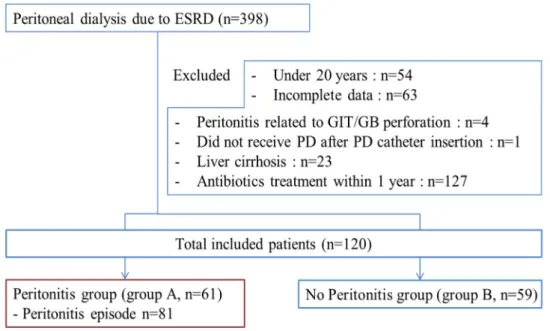
therefore, 54 patients aged <20 years were excluded. Sixty- three patients were excluded because of incomplete data and 4 patients because of peritonitis related to the perforation of the gastrointestinal tract or gallbladder. Twenty-three patients with LC were excluded because there were no standard criteria to distinguish PD-related peritonitis from spontaneous bacterial peritonitis. In addition, 127 patients who were previously administered antibiotics were excluded. One patient who did not receive PD after surgical replacement of the PD catheter was excluded (Figure 1).Peritonitis episodes were identified by a review of the medical records of patients. PD-related peritonitis was diagnosed if at least two the following diagnostic criteria were met: (a) abdominal pain or cloudy PD effluent, (b) leukocytosis in the peritoneal fluid effluent (white
4
blood cells >100/mm3, with at least 50% polymorphonuclear neutrophils), or (c) a positive Gram stain or a positive culture from PD effluent (17). All episodes of peritonitis were initially treated by intraperitoneal administration of first-generation cephalosporin (cefazolin) and third-generation cephalosporin (ceftazidime). Management of peritonitis depended on the clinical course and results of the antibiotic resistance tests for each isolated organism.To minimize potential bias, we excluded individual episodes of recurrent or relapsing peritonitis.
According to the International Society for Peritoneal Dialysis recommendations (2), recurrent peritonitis is defined as newly developed peritonitis caused by a different organism within 4 weeks of completion of therapy for a prior episode, and relapsing peritonitis is defined as newly developed peritonitis caused by the same organism within 4 weeks of completion of therapy for a prior episode. Then, we included only up to the third episode for each patient in the analysis to minimize potential bias.
5
Figure 1. Patient inclusion / exclusion chart.
(Abb : PD, peritoneal dialysis; GIT, gastrointestinal gract; GB, gallbladder; cath, catheter)
2. Data collection and outcome measurement
Medical records were reviewed by a single trained
investigator. Baseline characteristics including age, sex,
cause of ESRD, modality of PD, initial serum albumin level,
and presence of comorbidities such as diabetes mellitus,
hypertension, or diverticulosis were recorded. The
presence of diverticulosis was determined by reviewing
previous findings of abdomen and pelvic contrast-
6
enhanced computed tomography or colonoscopy. Data on
treatment with gastric acid suppressants, prokinetics, and
immunosuppressants were also collected. Data on acid
suppressive therapy with proton pump inhibitors (PPIs),
H2-blockers (H2Bs), and other antacids were recorded
separately. Ribeiro et al. reported that PPI use 48 hours
after the first dose increases and sustains gastric acid
suppression (18). In addition, a single dose of PPI per day
may cause protopathic bias. Therefore, for this study, the
use of acid suppressants was defined as the use of any
PPIs or H2Bs for at least 2 days. PPIs or H2Bs take 5–7
days with daily dosing to achieve their maximum
therapeutic effect level. Therefore, the use of gastric
acid suppressants was classified into four groups: use
within the previous 7 days, use within the previous 30
days but not within the past 7 days, use within the
previous 1 year but not within the last 30 days, and no use
of acid suppressants. Appropriate indications for a gastric
acid suppressant were defined as follows:
7
gastroesophageal reflux disease (GERD), peptic ulcer disease (PUD), Barrett’s esophagus, and concomitant use of a steroid or non-steroidal anti-inflammatory drug.
Prokinetic agents were assumed to be administered to
patients with clinical symptoms associated with decreased
gastrointestinal motility. In most of the cases, the reasons
for prescription were not described in the medical
records.In group A (the peritonitis group), the use of
gastric acid suppressants and the other medications was
determined by electronic medical records from the most
recent clinical visits 1 year prior to the first episode of
peritonitis. In group B (no peritonitis group), the use of
medications was determined from the most recent clinical
visit 1 year prior to the last outpatient visit, renal
transplantation, death, or transfer date to hemodialysis or
other renal replacement modalities. Each peritonitis
episode was classified as enteric peritonitis (EP) or non-
enteric peritonitis (NEP) depending on the isolated
organism from PD effluent culture. Microorganisms known
8
to colonize the gastrointestinal tract, such as Gram- negative bacteria, Enterococcus and Candida species were the causative organisms of EP, whereas all other organisms were pathogens of NEP. For the comparison of mediations used in the EP and NEP groups, the medications used were determined from the records of the most recent clinical visits 1 year prior to the first episode of peritonitis.
3. Statistical analysis
Baseline characteristics are presented as mean ± standard deviation or median and percentage for categorical variables.
Statistical analysis was performed using the Student’s t-test for the comparison of numerical variables, and the chi-square test or Fisher’s exact test for the comparison of categorical variables between groups. All probability values were two- tailed and the significance levels were set at 0.05. Multivariate logistic regression analysis was performed to assess the association of significant variables in univariate analysis with
9
the occurrence of peritonitis. All analyses were performed using SPSS version 19.0 for Windows (IBM, New York, USA).
10
RESULTS
1. Baseline characteristics of the study patients
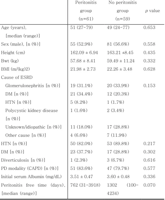
A total of 120 patients were included in the study: 61 patients in group A and 59 patients in group B. Table 1 summarizes the clinical characteristics of the two groups. In both groups, the common causes of renal failure were diabetic nephropathy and glomerulonephritis (GN). GN included immunoglobulin A nephropathy (6/16 in group A, 12/20 in group B), focal segment glomerular sclerosis (4/16 in group A, 3/20 in group B), lupus nephritis (1/16 in group A, 1/20 in group B), hepatitis B virus- associated GN (1/16 in group A, no one in group B), and other types of GN (7/16 in group A, 4/20 in group B). Baseline concentrations of serum albumin were 3.51 ± 0.47 mg/dL in group A and 3.60 ± 0.48 mg/dL in group B. There was no significant difference in the baseline characteristics between the two groups (p = 0.336).
11
Table 1. Clinical characteristics of the included patients
(Abb : Bwt, body weight; BMI, body mass index; DM, diabetes mellitus; HTN, hypertension; LC, liver cirrhosis; PD, peritoneal dialysis; CAPD, continuous ambulatory peritoneal dialysis)
Peritonitis group (n=61)
No peritonitis group (n=59)
p value
Age (years), [median (range)]
51 (27-79) 49 (24-77) 0.653
Sex (male), [n (%)] 55 (52.9%) 81 (56.6%) 0.558
Height (cm) 162.09 ± 6.94 163.21 ±8.45 0.435
Bwt (kg) 57.68 ± 8.41 59.49 ± 11.24 0.332
BMI (m/(kg)2) 21.98 ± 2.73 22.26 ± 3.48 0.628 Cause of ESRD
Glomerulonephritis [n (%)] 19 (31.1%) 20 (33.9%) 0.153
DM [n (%)] 21 (34.4%) 12 (20.3%)
HTN [n (%)] 5 (8.2%) 1 (1.7%)
Polycystic kidney disease [n (%)]
1 (1.6%) 2 (3.4%)
Unknown/idiopathic [n (%)] 11 (18.0%) 17 (28.8%) Other cause [n (%)] 4 (6.6%) 7 (11.9%)
HTN [n (%)] 50 (82.0%) 53 (89.8%) 0.217
DM [n (%)] 23 (37.7%) 17 (28.8%) 0.302
Diverticulosis [n (%)] 1 (2.3%) 3 (6.7%) 0.616 PD modality (CAPD) [n (%)] 51 (83.6%) 47 (79.7%) 0.577 Initial serum Albumin (mg/dL) 3.51 ± 0.47 3.60 ± 0.48 0.336 Peritonitis free time (days),
[median (range)]
762 (31-3918) 1302 (100- 4234)
0.070
12
2. Drug effects on PD-related peritonitis
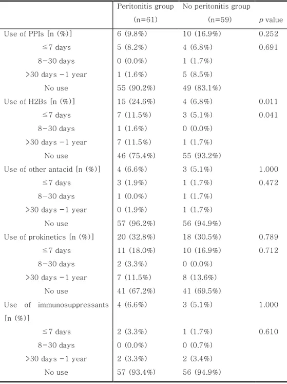
Table 2 summarizes the results of the comparison between group A and group B patients in terms of acid suppressive therapy, prokinetics, and immunosuppressants. PPIs or H2Bs were administered to 20 of the 61 patients (32.8%) in group A and 13 of the 59 patients (22.0%) in group B. Pantoprazole was the most frequently prescribed PPI and famotidine was the most frequently used H2B. The reasons for prescribing PPI or H2B were not recorded in 18 of the 33 patients (54.5%).
Inappropriate indications for the drugs included gastritis (24.2%) and nausea or vomiting (12.1%). The drugs were administered properly in 3 patients, including 1 patient with GERD (3.0%), 1 patient with PUD (3.0%), and 1 patient who concomitantly received steroids (3.0%).
Univariate analysis showed that the use of H2Bs significantly increased the risk of PD-related peritonitis. Using linear-by- linear association analysis, a higher proportion of patients with peritonitis were using H2Bs than those without peritonitis for each time interval of treatment (previous 7 days, 8–30 days prior to peritonitis, and within the last 1 year but not in the previous 90 days). No association was between PPI, other
13
antacid, or prokinetic use and PD-related peritonitis was found.
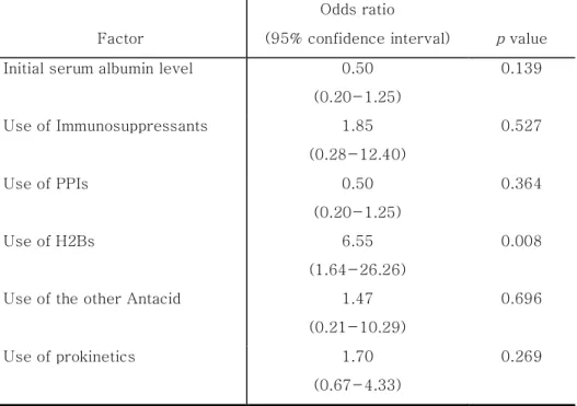
Furthermore, exposure to immunosuppressants was not associated with PD-related peritonitis. On multivariable analysis, we found that H2B use within 1 year prior to development of peritonitis was an independent risk factor for the increased risk of peritonitis development (odds ratio, 6.55;
95% confidence interval, 1.64–26.26; p = 0.008) (Table 3).
14
Table 2. Acid-suppressive therapy and other medications in peritonitis group and no peritonitis group
(Abb : PPIs, proton pump inhibitors; H2B, H2-blockers)
Peritonitis group (n=61)
No peritonitis group
(n=59) p value
Use of PPIs [n (%)] 6 (9.8%) 10 (16.9%) 0.252
≤7 days 5 (8.2%) 4 (6.8%) 0.691
8-30 days 0 (0.0%) 1 (1.7%)
>30 days -1 year 1 (1.6%) 5 (8.5%)
No use 55 (90.2%) 49 (83.1%)
Use of H2Bs [n (%)] 15 (24.6%) 4 (6.8%) 0.011
≤7 days 7 (11.5%) 3 (5.1%) 0.041
8-30 days 1 (1.6%) 0 (0.0%)
>30 days -1 year 7 (11.5%) 1 (1.7%)
No use 46 (75.4%) 55 (93.2%)
Use of other antacid [n (%)] 4 (6.6%) 3 (5.1%) 1.000
≤7 days 3 (1.9%) 1 (1.7%) 0.472
8-30 days 1 (0.0%) 1 (1.7%)
>30 days -1 year 0 (1.9%) 1 (1.7%)
No use 57 (96.2%) 56 (94.9%)
Use of prokinetics [n (%)] 20 (32.8%) 18 (30.5%) 0.789
≤7 days 11 (18.0%) 10 (16.9%) 0.712
8-30 days 2 (3.3%) 0 (0.0%)
>30 days -1 year 7 (11.5%) 8 (13.6%)
No use 41 (67.2%) 41 (69.5%)
Use of immunosuppressants [n (%)]
4 (6.6%) 3 (5.1%) 1.000
≤7 days 2 (3.3%) 1 (1.7%) 0.610
8-30 days 0 (0.0%) 0 (0.7%)
>30 days -1 year 2 (3.3%) 2 (3.4%)
No use 57 (93.4%) 56 (94.9%)
15 Factor
Odds ratio
(95% confidence interval) p value Initial serum albumin level 0.50
(0.20-1.25)
0.139
Use of Immunosuppressants 1.85 (0.28-12.40)
0.527
Use of PPIs 0.50
(0.20-1.25)
0.364
Use of H2Bs 6.55
(1.64-26.26)
0.008
Use of the other Antacid 1.47
(0.21-10.29)
0.696
Use of prokinetics 1.70
(0.67-4.33)
0.269
Table 3. Variables associated with PD related peritonitis : Multivariable logistic regression analysis
(Abb : PPIs, proton pump inhibitors; H2B, H2-blockers)
16
3. Drug effects on enteric peritonitis
Over the study period, 54 of the 61 patients in group A had a single episode of peritonitis, which met the inclusion criteria, 12 patients had two episodes, and one patient had three episodes.
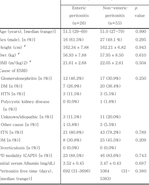
A total of 81 episodes were included in the study, and all episodes were divided into EP group or NEP group depending on the causative organism. Baseline characteristics did not show statistically significant differences between the groups (Table 4).
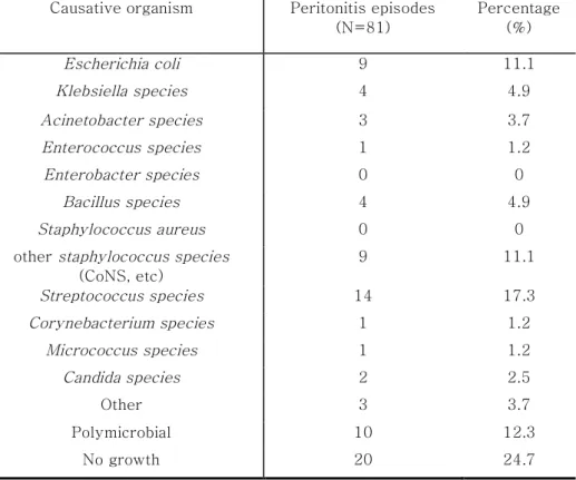
The causative microorganisms were identified from the peritoneal fluid culture in 61 of the 81 episodes. Escherichia coli (9/26, 34.6%) was the most frequently isolated enteric microorganism, and Streptococcus species (14/55, 24.5%) were the most frequently identified non-enteric microorganisms. The peritoneal fluid cultures from 20 episodes were negative (Table 5). The effects of various medications taken within 1 year prior to peritonitis on the development of EP are summarized in Table 6. When culture-negative peritonitis was included in the NEP group, no statistically significant association was found between exposure to gastric acid suppressants and EP. In addition, the use of prokinetics
17
and immunosuppressants did not influence the development of EP (Table 6A). When culture-negative peritonitis was excluded from the NEP group, the overall results were similar to those of the NEP group that included culture-negative peritonitis. The use of PPIs, H2Bs, other antacids, prokinetics, and immunosuppressants did not increase the risk of development of EP (Table 6B).
18
Table 4 Clinical characteristics of peritonitis patients by the causative pathogen.
# : available cases only
Included peritonitis episode (n=81) Enteric
peritonitis (n=26)
Non-enteric peritonitis
(n=55) p value
Age (years), [median (range)] 51.5 (29-69) 51.0 (27-79) 0.990 Sex (male), [n (%)] 16 (61.5%) 27 (49.1 %) 0.295 Height (cm) # 162.34 ± 7.88 162.21 ± 6.82 0.943
Bwt (kg) # 56.93 ± 7.88 57.95 ± 8.50 0.610
BMI (m/(kg)2) # 21.61 ± 2.68 22.05 ± 2.81 0.504 Cause of ESRD
Glomerulonephritis [n (%)] 12 (46.2%) 17 (30.9%) 0.250
DM [n (%)] 7 (26.9%) 20 (36.4%)
HTN [n (%)] 3 (11.5%) 3 (5.5%)
Polycystic kidney disease [n (%)]
0 (0.0%) 1 (1.8%)
Unknown/idiopathic [n (%)] 3 (11.5%) 11 (20.0%) Other cause [n (%)] 1 (3.8%) 3 (5.5%)
HTN [n (%)] 21 (80.8%) 43 (78.2%) 0.789
DM [n (%)] 8 (30.8%) 25 (45.5%) 0.209
Diverticulosis [n (%)] 0 (0.0%) 0 (0.0%)
PD modality (CAPD) [n (%)] 23 (88.5%) 46 (83.6%) 0.743 Initial serum Albumin (mg/dL) 3.52 ± 0.45 3.47 ± 0.43 0.687 Peritonitis free time (days),
[median (range)]
692 (31–3696) 1064 (31- 5383)
0.380
19
(Abb : Bwt, body weight; BMI, body mass index; ESRD, end stage renal disease; DM, diabetes mellitus; HTN, hypertension;
LC, liver cirrhosis; PD, peritoneal dialysis; CAPD, continuous ambulatory peritoneal dialysis)
20
Table 5. Isolated microorganisms of the peritonitis episodes by the PD Effluent culture
(Abb : CoNS, Coagulase negative staphylococcus; etc, et cetera)
Causative organism Peritonitis episodes (N=81)
Percentage (%)
Escherichia coli 9 11.1
Klebsiella species 4 4.9
Acinetobacter species 3 3.7
Enterococcus species 1 1.2
Enterobacter species 0 0
Bacillus species 4 4.9
Staphylococcus aureus 0 0
other staphylococcus species (CoNS, etc)
9 11.1
Streptococcus species 14 17.3
Corynebacterium species 1 1.2
Micrococcus species 1 1.2
Candida species 2 2.5
Other 3 3.7
Polymicrobial 10 12.3
No growth 20 24.7
21 (A)
(B)
Table 6. Acid-suppressive therapy and other medications in enteric peritonitis (EP) group and non-enteric peritonitis (NEP) group
Included peritonitis episode (n=81)
Enteric peritonitis (n=26)
Non-enteric peritonitis (n=55)
p value
Use of PPIs 3 (11.5%) 8 (14.5%) 1.000
Use of H2Bs 6 (23.1%) 15 (27.3%) 0.687
Use of other antacid 1 (3.8%) 5 (9.1%) 0.658
Use of prokinetics 5 (19.2%) 18 (32.7%) 0.209
Use of immunosuppressants 2 (7.7%) 7 (12.7%) 0.501
Included peritonitis episode (n=61)
Enteric peritonitis (n=26)
Non-enteric peritonitis (n=35)
p value
Use of PPIs 3 (11.5%) 5 (14.3%) 1.000
Use of H2Bs 6 (23.1%) 9 (25.7%) 0.818
Use of other antacid 1 (3.8%) 3 (8.6%) 0.629
Use of prokinetics 5 (19.2%) 13 (37.1%) 0.129 Use of immunosuppressants 2 (7.7%) 5 (14.3%) 0.688
22
DISCUSSION
Bacterial overgrowth and proliferation in an intraluminal environment with a high pH is a well-recognized mechanism for the development of peritonitis, especially in LC patients (6,7,10,13). Several studies have investigated if this mechanism causes PD-related peritonitis, especially enteric pathogen infections. However, conflicting results were obtained for the association between acid suppressive therapy and PD- related peritonitis. Caravaca et al. reported that gastric acid suppressive therapy is an independent risk factor for EP (10).
Nessim et al. reported that H2B use was associated with a higher risk of EP in PD patients (11). Conversely, del Peso et al.
did not find any effect of gastric acid suppressants on EP (12).
These studies were valuable, but were limited in design.
Therefore, we carefully excluded patients with liver cirrhosis, prior use of antibiotics, and a history of medication use that might influence peritonitis development to investigate the relationship between acid suppressive therapy and PD-related peritonitis. In the present study, H2B use, but not PPI use, was an independent risk factor for the development of PD-related peritonitis after adjusting for variable factors. Therefore, we
23
suggest an association between H2Bs and PD-related peritonitis.
Keane et al. evaluated the metabolism of PPIs such as rabeprazole, and showed that PPI clearance is not affected by renal failure (19). Conversely, Sica et al. analyzed the pharmacokinetics of H2B using ranitidine, and reported that H2B clearance was decreased in continuous ambulatory PD patients compared with that in individuals with normal kidney function (20). Therefore, acid suppression by H2B might be more powerful than that by PPI in PD patients. However, we showed that neither H2Bs nor PPIs increased the risk of EP development, which suggests that a mechanism independent of acid suppression is involved in the development of PD-related peritonitis. Peritoneal macrophages, mast cells, and migrated leukocytes have crucial roles in the immune response to bacterial invasion. Histamine, which is released by peritoneal mast cells, stimulates vasodilation and encourages leukocyte transmigration. Moreover, histamine induces aggregation of complement and opsonin, which promotes bacterial phagocytosis (21–23). H2Bs block these mechanisms.
Furthermore, H2Bs inhibit inflammation-generated increases in
24
nitric oxide concentrations (24), resulting in reduced phagocytosis and antimicrobial effects. These facts support the hypothesis that H2Bs increase the risk of PD-related peritonitis
Many ESRD patients experience abdominal discomfort, dyspepsia, and constipation because of decreased dietary fiber content, inadequate liquid intake, electrolyte imbalance, use of certain medications such as phosphate binders containing aluminum, and calcium- or iron-replacement therapy. Diabetic nephropathy is the most common cause of ESRD (25,26); most patients also have diabetic gastroparesis and decreased intestinal motility. Although the reasons for prescription were not described in detail, these symptoms might be related to the use of gastric acid suppressants and/or prokinetic drugs.
Previous studies have shown an association between gastrointestinal dysmotility and spontaneous bacterial peritonitis in patients with LC (13–16). Therefore, we investigated the effects of prokinetics on PD-related peritonitis.
Our data showed that prokinetic use is not associated with a reduced risk of PD-related peritonitis. To our knowledge, this is the first study to investigate the association between
25
prokinetic drugs and PD-related peritonitis. Further studies are needed to assess the effects of prokinetics on PD-related peritonitis.
Prognosis of EP is worse in patients with PD-related peritonitis.
Peritonitis caused by enteric organisms is associated with increased rates of PD catheter loss, prolonged hospitalization, and mortality (27,28). In this study, we found no statistically significant variables that increase the risk of EP. However, this result should be interpreted with caution because of the small number of peritonitis episodes that were found with each individual drug use.
This study was superior to previous studies in the following aspects. First, we carefully excluded patients with LC or previous antibiotic use. Furthermore, we performed multiple logistic regression analysis using various clinical factors such as the use of immunosuppressive agents and prokinetics, thus eliminating these potentially confounding factors. Second, a complete history of H2B and PPI use was obtained, since both required a prescription in Korea during the study period. In addition, H2B and PPI use was defined as the use of these
26
drugs for at least 2 days to prevent protopathic bias, which would increase the causality of the results.
There are several limitations in this study. First, this was a retrospective study that lacked clinical data on the reasons for gastric acid suppressant prescription. The study design did not permit analysis of individual drug doses and potential confounding factors such as type of PD and exit-site infection.
Furthermore, only a few patients were using H2Bs or PPIs, so the sample size was small. When we compared the EP and NEP groups, the number of peritonitis episodes treated with H2B or other medications was considerably smaller than that in groups A and B. To overcome these limitations, a prospective, randomized, placebo-control study is needed, which may put patients at unnecessary risk. As an alternative, a well-designed, prospective cohort study might provide further evidence of the association between acid suppressive therapy and PD-related peritonitis.
In conclusion, H2Bs tend to increase the risk of PD-related peritonitis, whereas PPIs and prokinetics do not. Further large prospective cohort studies are required to assess the effects of
27
gastric acid suppressants and prokinetics on the development of peritonitis in PD patients.
28
REFERENCES
1. Brown F, Liu WJ, Kotsanas D, Korman TM, Atkins RC. A quarter of a century of adult peritoneal dialysis-related peritonitis at an Australian medical center. Perit Dial Int J Int Soc Perit Dial. 2007 Oct;27(5):565–74.
2. Li PK-T, Szeto CC, Piraino B, Bernardini J, Figueiredo AE, Gupta A, et al. Peritoneal dialysis-related infections recommendations: 2010 update. Perit Dial Int J Int Soc Perit Dial. 2010 Aug;30(4):393–423.
3. Kidney International - Abstract of article: Race and the risk of peritonitis: An analysis of factors associated with the initial episode. Kidney Int. 1994 Nov;46(5):1392–6.
4. Stephen P McDonald JFC. Obesity is a risk factor for peritonitis in the Australian and New Zealand peritoneal dialysis patient populations. Perit Dial Int J Int Soc Perit Dial.
24(4):340–6.
5. Tranaeus A, Heimbürger O, Granqvist S. Diverticular disease of the colon: a risk factor for peritonitis in continuous peritoneal dialysis. Nephrol Dial Transplant Off Publ Eur Dial
29
Transpl Assoc - Eur Ren Assoc. 1990;5(2):141–7.
6. Lewis SJ, Franco S, Young G, O’Keefe SJ. Altered bowel function and duodenal bacterial overgrowth in patients treated with omeprazole. Aliment Pharmacol Ther. 1996 Aug;10(4):557–61.
7. Thorens J, Froehlich F, Schwizer W, Saraga E, Bille J, Gyr K, et al. Bacterial overgrowth during treatment with omeprazole compared with cimetidine: a prospective randomised double blind study. Gut. 1996 Jul;39(1):54–9.
8. Fried M, Siegrist H, Frei R, Froehlich F, Duroux P, Thorens J, et al. Duodenal bacterial overgrowth during treatment in outpatients with omeprazole. Gut. 1994 Jan;35(1):23–6.
9. Stiefel U, Rao A, Pultz MJ, Jump RLP, Aron DC, Donskey CJ.
Suppression of gastric acid production by proton pump inhibitor treatment facilitates colonization of the large intestine by vancomycin-resistant Enterococcus spp. and Klebsiella pneumoniae in clindamycin-treated mice.
Antimicrob Agents Chemother. 2006 Nov;50(11):3905–7.
10. Del Peso G, Bajo MA, Gadola L, Millán I, Codoceo R,
30
Celadilla O, et al. Diverticular disease and treatment with gastric acid inhibitors do not predispose to peritonitis of enteric origin in peritoneal dialysis patients. Perit Dial Int J Int Soc Perit Dial. 2001 Aug;21(4):360–4.
11. Nessim SJ, Tomlinson G, Bargman JM, Jassal SV. Gastric acid suppression and the risk of enteric peritonitis in peritoneal dialysis patients. Perit Dial Int J Int Soc Perit Dial.
2008 Jun;28(3):246–251; discussion 236–237.
12. Caravaca F, Ruiz-Calero R, Dominguez C. Risk factors for developing peritonitis caused by micro-organisms of enteral origin in peritoneal dialysis patients. Perit Dial Int J Int Soc Perit Dial. 1998 Feb;18(1):41–5.
13. Garcia-Tsao G, Albillos A, Barden GE, West AB.
Bacterial translocation in acute and chronic portal hypertension. Hepatol Baltim Md. 1993 Jun;17(6):1081–5.
14. Sandhu BS, Gupta R, Sharma J, Singh J, Murthy NS, Sarin SK. Norfloxacin and cisapride combination decreases the incidence of spontaneous bacterial peritonitis in cirrhotic ascites. J Gastroenterol Hepatol. 2005 Apr;20(4):599–605.
31
15. Chang CS, Chen GH, Lien HC, Yeh HZ. Small intestine dysmotility and bacterial overgrowth in cirrhotic patients with spontaneous bacterial peritonitis. Hepatol Baltim Md.
1998 Nov;28(5):1187–90.
16. Teltschik Z, Wiest R, Beisner J, Nuding S, Hofmann C, Schoelmerich J, et al. Intestinal bacterial translocation in rats with cirrhosis is related to compromised paneth cell antimicrobial host defense. Hepatology. 2012;55(4):1154–63.
17. Keane WF, Alexander SR, Bailie GR, Boeschoten E, Gokal R, Golper TA, et al. Peritoneal dialysis-related peritonitis treatment recommendations: 1996 update. Perit Dial Int J Int Soc Perit Dial. 1996 Dec;16(6):557–73.
18. Ribeiro TC, Chebli JM, Kondo M, Gaburri PD, Chebli LA, Feldner ACA. Spontaneous bacterial peritonitis: How to deal with this life-threatening cirrhosis complication? Ther Clin Risk Manag. 2008 Oct;4(5):919–25.
19. Keane WF, Swan SK, Grimes I, Humphries TJ.
Rabeprazole: pharmacokinetics and tolerability in patients with stable, end-stage renal failure. J Clin Pharmacol. 1999
32 Sep;39(9):927–33.
20. Sica DA, Comstock T, Harford A, Eshelman F. Ranitidine pharmacokinetics in continuous ambulatory peritoneal dialysis. Eur J Clin Pharmacol. 1987;32(6):587–91.
21. Hall* JC, Heel* KA, Papadimitriou‡ JM, Platell* C. The pathobiology of peritonitis. Gastroenterology. 1998 Jan;114(1):185–96.
22. Heel KA, Hall JC. Peritoneal defences and peritoneum- associated lymphoid tissue. Br J Surg. 1996 Aug;83(8):1031–
6.
23. Carlos D, Spiller F, Souto FO, Trevelin SC, Borges VF, de Freitas A, et al. Histamine h2 receptor signaling in the pathogenesis of sepsis: studies in a murine diabetes model. J Immunol Baltim Md 1950. 2013 Aug 1;191(3):1373–82.
24. Witte MB, Barbul A. General principles of wound healing.
Surg Clin North Am. 1997 Jun;77(3):509–28.
25. Jin DC. Current status of dialysis therapy for ESRD patients in Korea. J Korean Med Assoc. 2013;56(7):562.
33
26. Himmelfarb J, Tuttle KR. New Therapies for Diabetic Kidney Disease. N Engl J Med. 2013 Nov 9;
27. Troidle L, Gorban-Brennan N, Kliger A, Finkelstein F.
Differing outcomes of gram-positive and gram-negative peritonitis. Am J Kidney Dis Off J Natl Kidney Found. 1998 Oct;32(4):623–8.
28. Bunke CM, Brier ME, Golper TA. Outcomes of single organism peritonitis in peritoneal dialysis: gram negatives versus gram positives in the Network 9 Peritonitis Study.
Kidney Int. 1997 Aug;52(2):524–9.
34
국문 초록
서론: 복막투석 환자에서 발생한 복막염은 기타 합병증의 이환율 및 사망률을 높인다. 위산억제제가 복막투석환자에서 복막염의 발생에 미치는 영향에 대한 몇몇 보고가 있었으나, 위장운동촉진제가 복막 염의 발생에 미치는 영향에 대한 연구는 거의 이루어지지 않았다.
저자들은 한국의 복막투석 환자에서 위산억제제와 위장운동촉진제 가 복막염의 발생에 미치는 영향을 평가하고자 하였다.
방법: 단일기관 후향적 연구로, 2000 년 1 월부터 2013 년 9 월까지 서울대학교병원에서 추적관찰중인 복막투석 환자를 대상으로 하였 다. 연구에 포함된 환자들을 복막염이 발생한 병력이 있는 군 (환자 군)과 그렇지 않은 군 (대조군)으로 나누어 분석하였다. 덧붙여, 연 구에 포함된 복막염들을 장내세균에 의한 복막염과 그렇지 않은 복 막염의 두 군으로 나누어 비교하였다.
결과: 총 120 명의 환자가 분석에 적절한 대상으로 분류되어, 61 명 의 환자는 복막염군, 59 명의 환자는 비복막염군으로 분류되었다. 복 막염군의 24 명(39.3%)와 비복막염군의 15 명(25.4%)이 위산억제 제를 사용한 병력이 있었다. H2 수용체 길항제는 유의하게 투석환자
35
의 복막염을 증가시켰다. 반면, 양성자 펌프 억제제, 기타 위산억제 제 그리고 위장운동촉진제는 복막투석 환자에서 복막염의 발생을 유의하게 증가시키지 않았다. 복막염군에서 복막염은 총 81 례가 분 석 대상이 되었고, 이들을 각각 원인균주에 따라 장내세균에 의한 복막염과, 그 외 원인균주에 의한 복막염으로 나누어 비교하였다.
위산억제제와 위장운동촉진제는 모두 장내세균에 의한 복막염의 발 생을 증가시키지 않았다.
결론: 결론적으로, H2 수용체 길항제만이 복막투석 환자에서 복막염 의 발생률을 유의하게 증가시키는 경향을 보였다. 여전히 이론적으 로는 잠재적인 위험인자인 위산억제제와 위장운동촉진제와 복막염 과의 상관관계를 평가하기 위해서는 대규모 복막투석 환자를 대상 으로 한 연구가 필요하겠다.
--- 주요어 : 복막투석, 복막염, 양성자 폄프 억제제, H2 수용체 길항제, 위산억제제, 위장관 운동
학 번 : 2012-22677