저작자표시-비영리-변경금지 2.0 대한민국 이용자는 아래의 조건을 따르는 경우에 한하여 자유롭게
l 이 저작물을 복제, 배포, 전송, 전시, 공연 및 방송할 수 있습니다. 다음과 같은 조건을 따라야 합니다:
l 귀하는, 이 저작물의 재이용이나 배포의 경우, 이 저작물에 적용된 이용허락조건 을 명확하게 나타내어야 합니다.
l 저작권자로부터 별도의 허가를 받으면 이러한 조건들은 적용되지 않습니다.
저작권법에 따른 이용자의 권리는 위의 내용에 의하여 영향을 받지 않습니다. 이것은 이용허락규약(Legal Code)을 이해하기 쉽게 요약한 것입니다.
Disclaimer
저작자표시. 귀하는 원저작자를 표시하여야 합니다.
비영리. 귀하는 이 저작물을 영리 목적으로 이용할 수 없습니다.
변경금지. 귀하는 이 저작물을 개작, 변형 또는 가공할 수 없습니다.
藥學碩士學位論文
Structure and Functional Study on HigBA from Streptococcus pneumoniae
Streptococcus pneumoniae 에서 유래한 HigBA 단백질의 구조 및 기능 연구
2017年 8月
서울대학교 대학원
약학과 물리약학전공金 成 龍
Structure and Functional Study on HigBA from Streptococcus pneumoniae
Streptococcus pneumoniae 에서 유래한 HigBA 단백질의 구조 및 기능 연구
指導敎授 李 奉 振
이 論文을 藥學碩士學位 論文으로 提出함 2017年 8月
서울大學校 大學院
藥學科 物理藥學專攻金 成 龍
金 成 龍의 碩士學位 論文을 認准함 2017年 8月
委 員 長 (인)
副委員長 (인)
Abstract
Structure and Functional Study on HigBA from Streptococcus pneumoniae
JIN CHENGLONG, Physical Pharmacy, the Graduate School,
Seoul National University
Streptococcus pneumoniae is a gram-positive strain that causes diseases mainly through respiratory infections. Diseases caused by pneumococcal species are meningitis, bacteremia, pneumonia, otitis and sinusitis. Patients infected with s.pneumonia have developed antibiotic resistant strains, so new antibiotics need to be developed.
Ultimately, developing novel antibiotic candidates through structural analysis and functional studies of proteins is a major goal.
The target TA complex protein present in the s.pneumoniae TIGR4 strain is a type II toxin-antitoxin system and is classified under HigBA family. The toxin protein HigB is predicted to be a ribosome-dependent mRNA interferases, which cleave mRNAs at the
ribosomal A site. The antitoxin protein HigA regulates transcription through binding of palindromic sequence to its operator region.
In order to obtain the tertiary structure of the protein, the target protein was obtained by recombinant process and was over-expressed in Rosetta (DE3) pLysS of escherichia coli. Affinity chromatography was used to purify the hexa-histidine tagged protein. Further, higher purity of target protein was obtained by ion-exchange chromatography and size-exclusion chromatography. The structure of target protein was obtained using X-ray crystallography techniques.
Key words
Streptococcus pneumoniae, Toxin-antitoxin system, HigBA, X-ray crystallography, EMSA, RNase activity assay
Student Number: 2015-22388
Contents
I. Introduction ··· 1
1.1. Streptococcus pneumoniae ··· 1
1.2. Toxin-Antitoxin system ··· 2
1.3. HigBA family in type II TA system ··· 3
1.4. Purpose of Study ··· 4
II. Materials and Methods ··· 5
2.1. Materials ··· 5
2.1.1. Reagents ···5
2.1.2. Apparatus ···5
2.2. Methods ··· 6
2.2.1. Gene cloning ···6
2.2.2. Over-expression and purification ···7
2.2.3. Crystallization ···8
2.2.4. X-ray data collection and structure determination ···8
2.2.5. EMSA (Electrophoretic Mobility Shift Assay) ··· 9
2.2.6. Ribonuclease activity assay ···9
III. Results ··· 11
3.1. Over-expression and purification ··· 11
3.1.1. Over expression ···11
3.1.2. Purification ···12
3.1.2.1. Affinity chromatography ···12
3.1.2.2. Ion exchange chromatography ···12
3.1.2.3. Size exclusion chromatography ···13
3.2. Crystallization ··· 14
3.3. EMSA (Electrophoretic Mobility Shift Assay) ··· 15
3.4. I n vitro ribonuclease activity assay ··· 17
V. Discussion ··· 19
VI. References ··· 23
국문초록 ··· 28
Ⅰ. Introduction
1.1. Streptococcus pneumoniae
Streptococcus pneumoniae is a gram-positive strain that causes diseases mainly through respiratory infections. Diseases caused by pneumococcal species are classified into invasive diseases and non-invasive diseases.
Invasive diseases include meningitis, bacteremia, and pneumonia.
Non-invasive diseases include otitis and sinusitis.
Among the infections caused by s.pneumonia, 1.6 million deaths occur every year, of which 900,000 are children under 5 years of age. Patients infected with s.pneumonia bacteria are mainly treated with antibiotics such as penicillin, macrolide, clindamycin, and cephalosporin. However, about 30% of infected patients have developed antibiotic resistant strains for several reasons, so new antibiotics need to be developed.
Fig. 1. The incidence of invasive pneumococcal Fig. 2. Pneumococcal mortality rate disease varies according to age group
1.2. Toxin-Antitoxin system
The toxin–antitoxin (TA) systems were discovered in the 1980s as plasmid genes that guarantee inheritance of a plasmid in daughter cells.
Homologues of TA plasmid system modules were later found in the chromosomes of most bacteria, and their roles in bacterial cells have been intensively investigated.
Recent advances in genome sequencing and bioinformatics have revealed a high prevalence of toxin–antitoxin (TA) systems in prokaryotes, which has served as a starting point for diverse and intensive studies of TA systems.
TA systems are typically encoded in operons that are located on bacterial plasmids and chromosomes. The operons consist of adjacent toxin and antitoxin genes. Toxins have diverse cellular functions such as inhibitions of protein synthesis, DNA replication, and cell wall synthesis in response to unfavorable growth conditions. Many toxins are ribonucleases that degrade mRNA in a specific or nonspecific fashion, and some toxins act as gyrase inhibitors and kinases.
TA systems are usually classified into type I, type II and type III TA systems. These three major groups have been subdivided into six groups, each of which has a different mechanism. The antitoxin in the type I TA system are antisense RNAs that bind to toxins, which are mRNAs. Type I antitoxins inhibit the translation of toxins and cause gradual toxin degradation. In the type II TA system, the toxins and antitoxins are proteins that interact with each other to form a non-toxic TA complex. In the type III TA system, antitoxins act as RNAs that bind directly to toxin proteins.
In the most widely distributed type II TA systems, the toxin is thermodynamically stable, whereas the antitoxin is unstable and is cleaved by some cellular proteases because its C-terminal region adopts a flexible conformation that is susceptible to proteolysis.
Fig. 3. Mechanism of type Ⅱ TA system
1.3. HigBA family in type II TA system
The higBA (host inhibition of growth) was originally identified on the Rts1 plasmid conferring plasmid maintenance in Proteus spp. Subsequently, chromosomal higBA homologues have been reported from several pathogens such as Vibrio cholera, proteus vulgaris, Streptococcus pneumonia, Mycobacterium tuberculosis, and two virulent E. coli species (CFT073, and
O157:H7). All the higBA genes are bicistronic arrayed on genome in an unusual reverse way with the toxin higB locus preceding the antitoxin gene.
The toxin protein HigB is a ribosome-dependent mRNA interferases belonging to RelE family, which cleave mRNAs at the ribosomal A site.
Although HigB contains a complete RNase fold, HigB does not possess ribonuclease activity against naked RNA in vitro. The HigB-mediated cleavage sites were observed and confined to the coding frames of the translating RNAs on ribosome. This HigB cleavage elicited a global effect on all cellular mRNA in vivo and ceased cell growth. The antitoxin HigA possesses DNA binding activity within its helix-turn-helix fold (HTH) and modulates the HigBA expression.
1.4. Purpose of the study
The main objective of the study is to determine the tertiary structure of HigBA complex from Streptococcus pneumoniae, to demonstrate the function of the target protein, and to discover a new antimicrobial agent of S. pneumoniae. To achieve the goal, higBA gene was selected and cloned through the known genome sequence. Then, they were produced by E.coli expression system and the expressed proteins were carried out solubility test to optimize the best soluble condition of the target proteins. Through several procedures of purification, we can get the purified proteins. Finally, crystallization was performed to get the crystal of HigBA complex and the three-dimensional structure of target protein was determined by X-ray crystallography.
Ⅱ. Materials and Methods
2.1. Materials
2.1.1. Reagents
The Streptococcus pneumoniae genomic DNA (strain TIGR4) and
polymerase chain reaction (PCR) primers, which are used for target gene amplification, were purchased from ATCC and BIONEER, respectively. Luria Broth (LB), Kanamycin and Ampicillin were bought from Sigma Aldrich.
Restriction enzymes (NdeI and XhoI) were purchased from New England Biolabs (NEB). The expression vector pET 21a(+) and pET 28b(+) were obtained from Novagen.
2.1.2. Apparatus
PCR reaction was carried out by Perkin-Elmer PCR system 9600 (Perkin-Elmer, U.S.A.). Cell lysis was performed by the sonic oscillator, Sonifier 450 produced by Branson Ultrasonic Corporation (Connecticut, U.S.A.). J2-MC and the fraction collector were purchased from Bio-Rad Laboratories Inc. (California, U.S.A.). Centricon, Centriprep were bought from Millipore Corporation. Affinity chromatography, ion exchange chromatography were purchased from GE healthcare (Germany). Specimens
were observed in Fast Protein Liquid Chromatography (FPLC) (AKTA, U.S.A.). Size exclusion chromatography was performed by Hiload 16/60 Superdex 200 prep-grade column (GE Health care, U.S.A.). Results of EMSA were visulized using a printgraph 2M (ATTO). Fluorescence spectroscopy was performed by SPECTRAmax GEMINI XS spectrofluorometer.
2.2. Methods
2.2.1. Gene cloning
The genes encoding S. peumoniae (strain TIGR4) higBA were amplified by polymerase chain reaction (PCR) using the S. peumoniae genomic DNA (strain TIGR4) as templete. The forward and reverse of gene encoding higA were 5’- GGAATTCCATATGATGAAAAATAATGCTATTGG -3’ and 5’- CCGCTCGAGTTAAACCTGCTCATGCTCTAATGGT- 3’, respectively. Also, the forward and reverse of gene encoding higB were designed as followed.
The forward was 5’- GGAATTCCATATGATGCATAATATCTATTTTTA -3’
and reverse was 5’- CCGCTCGAGTTATTTTTCATTGTCTAAAC -3’. The underlined sequences are NdeI and XhoI digestion sites, respectively. The amplified gene encoding higB was cloned into the expression vector pET 28b(+) (Novagen) to enabled the production of HigB with an N-terminal hexa-histidine tag. The gene encoding higA was cloned into the expression vector pET 21a(+) (Novagen) with no hexa-histidine tag. Then they was transformed into competent cell of E. coli DH5α. After validation of DNA sequence of target protein, the plasmid of higA and higB were
co-transformed into competent cell of E. coli Rosetta (DE3) pLysS to perform the over expression test.
2.2.2. Over-expressionand purification
The recombinant protein was over expressed in E.coli Rosetta (DE3) pLysS cells. Cells were grown in LB culture medium at 310 K, 180 rpm shaking condition up to OD 600 of 0.5 in LB culture medium containing 50 mg/ml ampicillin and 30 mg/ml kanamycin, and the over-expression of the target proteins were induced by isopropyl-β-D-thiogalactopyranoside (IPTG). The cells continued to be incubated at 310 K, 180rpm for additional 4h after IPTG induction and were harvested by centrifugation at 6,000 rpm for 10min at 277K.
The cell pellets were lysed by sonication in a buffer A (20 mM Tric-HCl, 500 mM NaCl, pH 7.9) containing 10% (v/v) glycerol. The entire lysate was centrifuged at 18,000 rpm for 1h at 277 K.
The cell debris was discarded and the supernatant was loaded into the HIS-tag affinity column. The regenerated column was previously washed with buffer A. Then target proteins sticking to the resin was eluted with the imidazole gradient (50 mM, 100 mM, 150 mM, 200 mM, 250 mM, 300 mM, 400 mM, 500 mM, 600 mM, 700 mM of imidazole). Purified fractions containing target protein were pooled and concentrated. Then it was diluted by the buffer (50 mM Mes, 500 mM Nacl, pH 6.0) at 1:100 ratio. The diluted protein was applied to ion exchange chromatography on Hitrap SP column (GE Heathcare). The protein was eluted with a linear gradient of 0.0-1.5M Nacl. As a final step, gel filtration was performed on a Hiload 16/60 Superdex 200 prep-grade column (GE Heathcare), which was
preciously equilibrated with final buffer (50 mM Mes, 500 mM Nacl, pH6.0). All the purified proteins were assessed by sodium dodecyl sulfate-polyarylamide gel electrophoresis (SDS-PAGE) and concentrated by ultrafiltration (Centriconm Millipore Corp.).
2.2.3. Crystallization
Crystallization was performed at 293 K by the sitting-drop vapor-diffusion method using 96-well crystallization plates. Two kinds of crystals were obtained. They are Native and SeMet, respectively. Each sitting drop was prepared by mixing 0.8µl of protein solution (0.45 mM protein concentration for Native, 0.6mM protein concentration for SeMet) and 0.8µl of reservoir solution, and was placed over 100µl reservoir solution. Initial crystallization conditions were established using screening kits from Hampton Research Inc. (IndexⅠ,Ⅱ), Molecular Dimentions Inc.
(Structure Screening) and Rigaku inc. (Wizard Ⅰ,Ⅱ). Consequently, the best hits were occurred in the crystallization solution of Index 39 and were optimized by made buffer in the same condition.
2.2.4. X-ray data collection and structure determination
X-ray diffraction data was collected using synchrotron radiation on ADSC Q315r detector at beamline PAL-5C(SBII) (Pohang, South Korea) at λ= 0.97944. Crystal of HigBA complex belonged to space group P21, with unit cell parameters of a = 74.903 Å, b =74.403 Å, c = 98.321 Å,α = γ = 90.00˚, β=90.08˚ for the native HigBA crystal and a = 74.575 Å, b = 67.038 Å, c=87.717 Å, α = γ = 90.00˚, β= 94.22˚ for the SeMet-labeled
HigBA crystal. Raw data was processed and scaled using HKL2000 program package (Otwinowski and Minor, 2002). The structure was phased by single-wavelength anomalous dispersion (SAD) and was refined by molecular replacement (MR) using the phase program in CCP4 suite of programs and Phenix (Adams et al., 2010). Coot (Emsley and Cowtan, 2004) was used for manual model.
2.2.5 EMSA (Electrophoretic Mobility Shift Assay)
Both HigA and (HigBA)2 have been estimated that they would bind to palindromic sequence in their own operator region. Three DNA duplexes were generated by annealing three pairs of complementary oligonucleotides.
Reaction mixtures for EMSA containing 10µM of each DNA duplex and a defined concentration of different proteins were assembled in binding buffer (50mM MES, 500mM Nacl, pH 6.0) and incubated for 30min at room temperature. EMSA reactions were electrophoresed on a 0.5% agarose gel with 0.5x TBE for 20 min. The results were visualized using a Printgraph 2M (ATTO).
2.2.6. Ribonuclease activity assay
There is evidence that HigB toxins are structurally similar to RelE and YoeB, which have RNase folds for interacting with the 50S ribosomal subunits . That is to say that HigB toxins are known as sequence-specific endo-ribonucleases that impede the global translations of cellular mRNAs.
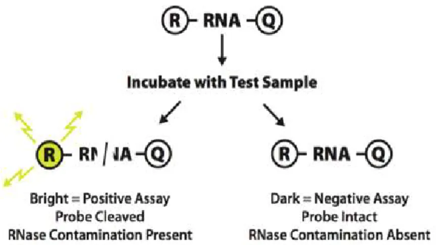
We have confirmed the ribonuclease activity of HigB using an RNase Alert kit(IDT). We conducted the fluorescence quenching assay according to the protocol provided by the manufacturer. In this system, a fluorophore is covalently attached to one end of a synthetic RNA strand and is quenched by a quencher group at the other end of the RNA strand. If synthetic RNA containing a fluorophore-quencher pair interacts with ribonuclease, the synthetic RNA is digested and the quencher is released. The released fluorophore emits fluorescence at 520 nm upon excitation at 490 nm. The relative fluorescence units (RFU) were observed on a SPECTRAmax GEMINI XS spectrofluorometer.
Ⅲ. Results
3.1. Over-expression and purification
3.1.1 Over expression
The HigBA protein consists of HigA antitoxin and HigB toxin. Two proteins were co-expressed in E.coli Rosetta (DE3) pLysS and over expressed after induction by adding IPTG. The recombinant proteins were over expressed to a high level respectively and nearly almost of the over expressed HigBA complex protein was in the soluble fraction.
Fig. 4. Expression Test Fig. 5. Solubility Test
3.1.2 Purification
3.1.2.1. Affinity chromatography
The first step utilized the N-terminal hexa-histidine tag by affinity chromatography on a IMAC column, which was previously charged with 50 mM NiSO4 and equilibrated with buffer A. Upon eluting with a gradient of imidazole in the same buffer, the target complex protein was eluted at 100-700 mM imidazole concentration. Most contaminants were removed at this step.
Fig. 6. The result of purification using affinity chromatography
3.1.2.2. Ion exchange chromatography
In order to separate the target protein from DNA contaminant protein, ion exchange chromatography on Hitrap SP column (GE Heathcare) was performed. The protein was eluted with a linear gradient of 0.0-1.5 M Nacl.
Consequently, the target protein was obtained at about 0.8-1.2 M Nacl concentration.
Fig. 7. The result of purification using ion exchange chromatography
3.1.2.3. Size exclusion chromatography
In order to strip the rest impurities and increase the purity of the target protein, size exclusion chromatography was performed. Before loading the target proteins, the column was equilibrated by the final buffer (pH 6.0, 50 mM MES, 500 mM Nacl). As a result, we obtained a single peak.
Fig. 8. The result of purification using size exclusion chromatography
3.2. Crystallization
Crystals of HigBA complex were grown using sitting-drop vapor diffusion at 293 K. The optimized crystallization solution for HigBA was 100 mM HEPES, 30% v/v JeffamineED-2001 (pH 7.0). X-ray diffraction data of native and SeMET were collected to 2.8Å resolution and 3.2Å resolution, respectively. The structure of HigBA complex was firstly phased at 3.2Å resolution by single-wavelength anomalous dispersion using SeMET.
The whole structure could not be determined because of the relatively low resolution. Then the diffraction data of native come to rescue. The structure of HigBA complex was refined using 2.8Å resolution native diffraction data by molecular replacement based on SeMET protein model. Crystals of HigBA complex belong to the space group P21, with unit cell parameters of a = 74.903 Å, b = 74.403 Å, c = 98.321 Å, α = γ = 90.00˚, β= 90.08˚
for the native HigBA crystal and a = 74.575 Å, b = 67.038 Å, c=87.717 Å, α = γ = 90.00˚, β= 94.22˚ for the SeMet-labeled HigBA crystal. All raw data were scaled and processed using the HKL2000.
Fig. 9. Crystal of HigBA native state Fig. 10. Crystal of HigBA SeMET state
Fig. 11. Overall structure of HigBA complex
3.3. EMSA (Electrophoretic Mobility Shift Assay)
In HigBA family, the antitoxin alone or in complex with toxin typically interacts with its corresponding operator DNA, repressing the transcription of the TA operon. Therefore, the DNA-binding properties of the antitoxin and complex are important for the regulation of TA systems. We selected three pairs of palindromic sequence (P1, P2 and P3) from own operon region. As a result, electrophoretic mobility shift assays were shown that HigBA complex specifically bound to P1, P2, and P3, but HigA were only binding to P3. It indicated that P1, P2, and P3 would be the HigBA operator regions which were corresponding with target protein. In addition, HigBA
displayed higher affinity than HigA, given that HigBA produced more intensity shifted band. It demonstrated that the DNA-binding modes of HigA and the HigBA complex might be distinct, implying that HigA and HigBA auto-regulate differentially in vivo.
Palindromic
Sequence Forward Reverse
P1 5’-TGCTTTTTATTTTAATA
ACTTAAA-3’
3’-ACGAAAAATAAAATTAT TGAATTT-5’
P2 5’-CAAACAATAACTTTTAG
GTTATAATTGT-3’
3’-GTTTGTTATTGAAAATCC AATATTAACA-5’
P3 5’-TATTTTAATAACTTAAA
AGT-3’
3’-ATAAAATTATTGAATTT TCA-5’
Form 1. Palindromic sequence from higBA operator DNA
Fig. 12. Results of EMSA using 3 pairs of palindromic sequence (P1, P2, P3)
3.4. In vitro ribonuclease activity assay
We performed assays of the RNase activity of HigB toxin and HigBA complex using fluorescent RNA substrates. When these substrates are cleaved, they emit fluorescence in proportion to the amount of the substrate cleaved by HigB or HigBA. The fluorescence data are shown in Figure14.
Relative fluorescence units (RFU) was significantly increasing as the concentrate of HigB increased from 1uM to 2uM, whereas the RFU of HigBA was negligible. It illustrated that HigB had the capacity for cleaving the RNA molecules, when HigBA did not have. Based on the result of in vitro RNase activity assay, we confirm that HigB toxins are sequence-specific endo-ribonucleases that impede the global translations of cellular mRNAs. Also, the HigA antitoxin wraps around the toxin, forming a large complex structure that inhibits the entrance of the toxin into the ribosomal A-site.
Fig. 13. Mechanism of RNase activity assay
Fig. 14. In vitro ribonuclease activity of Streptococcus pneumoniae
HigBA complex and HigB
Ⅳ. Discussion
The main objective of this study was the structural determination of the HigBA complex. In order to achieve the goal, cloning of the target gene, over-expression and purification of the target protein, and crystallization were performed. With a lot of trials, we established purification methods for target protein and optimized the condition of crystallization solution. Then, we obtained several crystals of SeMET and Native state, which were used for collecting X-ray diffraction data. Consequently, X-ray diffraction data of native and SeMET were collected to 2.8 Å resolution and 3.2 Å resolution, respectively. The structure of HigBA complex was firstly phased by single-wavelength anomalous dispersion (SAD) using SeMET. Then we refined the HigBA structure using native diffraction data by molecular replacement (MR) based on SeMET protein model.
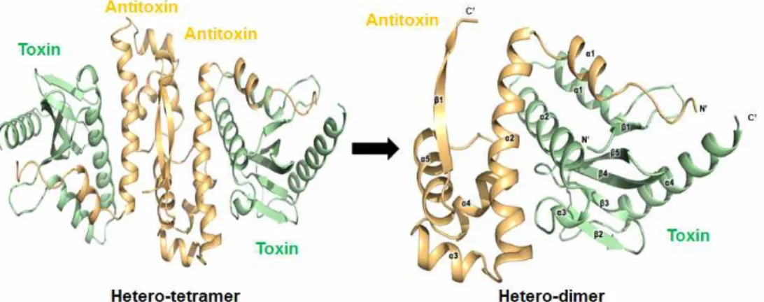
The asymmetric unit of the crystal of the HigBA complex contains four HigA antitoxins and four HigB toxins in a hetero-octameric assembly. Four of the heterodimeric HigBA complexes are included in the asymmetric unit.
The HigA dimer binds two HigB monomers, and two of the HigB₂A₂ complexes are related to each other by a dyad axis. The flexible hinge loop of the antitoxin wraps the toxin as a hook, known as a looped arm. The structure of the HigB toxin is composed of five β-strands and four α-helices in the following order: β1 (residues 4–6), α1 (residues 15–25), α2 (residues 29–48), α3 (residues 49–52), β2 (residues 57–61), β3 (residues 64–67), β4 (residues 72–78), β5 (residues 84–91) and α4 (residues 99–115). Also, the HigA antitoxin contains five α-helices and one β-strand. The six secondary structure elements correspond to residues 10–17 (α1), residues 20–42 (α2),
residues 47–54 (α3), residues 58–66 (α4), residues 73–83 (α5) and residues 85–91 (β1). HigA consists of the following two domains: an N-terminal toxin-binding domain and a C-terminal DNA-binding domain (RHH). The two antitoxins interact with each other through their C-terminal β-strands to form a homodimer.
Fig. 15. Overall structure of HigBA and its subunits
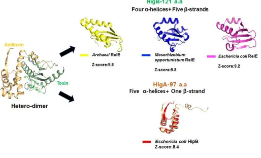
A search for structural homologues of HigBA using the DALI server was conducted to analyze their structural similarities, and obvious structural differences between the HigBA and its structural homologues were observed.
Interestingly, HigA antitoxin and HigB toxin share structure similarity with other TA sysem families from different bacteria strains. HigA antitoxin shares structural similarity with HipB from Eschericia coli (Z-scores of 8.4) when HigB toxin has high homology to several RelE toxin proteins from different bacteria strains (Z-scores-9.8, 9.8 and 9.2, respectively). The structure similarities of target protein are shown in Figure 16. In conclusion,
it adds a lot of value to further research that toxin and antitoxin of HigBA shares structure similarity with other TA system families.
Fig. 16. Comparison of the structure of the HigB and HigA with their homologs
After electrophoretic mobility shift assay (EMSA) , two results were drawn. Firstly, In HigBA family, the antitoxin alone or in complex with toxin typically interacts with its corresponding operator DNA, repressing the transcription of the TA operon. Secondly, HigBA displayed higher affinity than HigA, given that HigBA produced more intensity shifted band. It demonstrated that the DNA-binding modes of HigA and the HigBA complex might be distinct, implying that HigA and HigBA auto-regulate differentially in vivo. Furthermore, advanced experiment will be performed to conclusively approve the results of EMSA. For example, Isothermal titration calorimetry (ITC) will be performed to estimate the binding affinity of HigA to operator
DNA when DNA titration is performed using NMR technique to reveal the key residues which interact with DNA.
Based on the RNase activity assay, we confirm that HigB toxins are sequence-specific endo-ribonucleases that impede the global translations of cellular mRNAs. Also, the HigA antitoxin wraps around the toxin, forming a large complex structure that inhibits the entrance of the toxin into the ribosomal A-site. To reveal key residues of HigB which affect the RNase activity, mutation of residues of HigB will be performed. By comparison of RNase activity between the wild-type and mutant, the designed objective will be achieved.
Ⅴ. References
R.B.Jensen, K. Gerdes, Programmed cell death in bacteria: proteic plasmidstabilization systems, Mol. Microbiol., 17 (1995) 205-210.
Q.B.Tian,M.Ohnishi, T.Murata, K.Nakayama, Y.Terawaki, T.Hayashi, Specific protein-DNAand protein-protein interaction in the hig gene system, a plasmid-borne proteickiller gene system of plasmid Rts1, Plasmid, 45 (2001) 63-74.
C.F.Schuster,R.Bertram, Toxin-antitoxin systems are ubiquitous and versatile modulators ofprokaryotic cell fate, FEMS Microbiol.Lett., 340 (2013) 73-85.
R.Leplae, D. Geeraerts, R. Hallez, J. Guglielmini, P. Dreze, L. Van Melderen,Diversity of bacterial type II toxin-antitoxin systems: a comprehensive searchand functional analysis of novel families, NucleicAcids Res., 39 (2011) 5513-5525.
N.Goeders,L.VanMelderen, Toxin-antitoxin systems as multilevel interaction systems, Toxins (Basel), 6 (2014) 304-324.
W.T.Chan, M. Espinosa, C.C. Yeo, Keeping the wolves at bay: antitoxins ofprokaryotic type II toxin-antitoxin systems, Front. Mol. Biosci., 3 (2016).
M.Overgaard, J. Borch, M.G. Jorgensen, K. Gerdes, Messenger RNA
interferase RelEcontrols relBE transcription by conditional cooperativity, Mol.
Microbiol., 69(2008) 841-857.
D.Andreev, V. Hauryliuk, I. Terenin, S. Dmitriev, M. Ehrenberg, I. Shatsky, Thebacterial toxin RelE induces specific mRNA cleavage in the A site of theeukaryote ribosome, RNA, 14 (2008) 233-239.
A.Boggild, N. Sofos, K.R. Andersen, A. Feddersen, A.D. Easter, L.A.
Passmore,D.E. Brodersen, The crystal structure of the intact E. coli RelBE toxin-antitoxin complex provides the structural basisfor conditional cooperativity., Structure, 20 (2012) 1641-1648.
V.Zorzini,L.Buts,E.Schrank, Y.G.Sterckx, M.Respondek, H.Engelberg-Kulka, R. Loris, K.Zangger, N.A. van Nuland, Escherichiacoli antitoxin MazE as transcription factor: insights into MazE-DNA binding, Nucleic Acids Res., 43 (2015) 1241-1256.
S.Feng, Y. Chen, K. Kamada, H. Wang, K. Tang, M. Wang, Y.G. Gao, YoeB- ribosomestructure: a canonical RNase that requires the ribosome for its specificactivity, Nucleic Acids Res., 41 (2013) 9549-9556.
M.Christensen-Dalsgaard, K. Gerdes, Two higBA loci in the Vibrio cholerae superintegron encode mRNA cleaving enzymes and canstabilize plasmids, Mol. Microbiol., 62 (2006) 397-411.
J.M.Hurley, N.A. Woychik, Bacterial toxin HigB associates with ribosomes andmediates translation-dependent mRNA cleavage at A-rich sites, J. Biol.
Chem., 284 (2009) 18605-18613.
W.T.Chan, I.Moreno-Cordoba, C.C.Yeo, M.Espinosa, Toxin-antitoxin genes of theGram-positive pathogen Streptococcuspneumoniae: so few and yet so many, Microbiol. Mol. Biol. Rev., 76(2012) 773-791.
D.L.Schuessler, T. Cortes, A.S. Fivian-Hughes, K.E. Lougheed, E. Harvey, R.S.Buxton, E.O. Davis, D.B. Young, Induced ectopic expression of HigB toxin inMycobacterium tuberculosis results in growth inhibition, reduced abundance of asubset of mRNAs and cleavage of tmRNA, Mol.Microbiol., 90 (2013) 195-207.
M.A. Schureck, A. Repack, S.J. Miles, J. Marquez,C.M. Dunham, Mechanism of endonuclease cleavage by the HigB toxin, Nucleic Acids Res.
(2016).
M. Christensen-Dalsgaard, M.G. Jorgensen, K.Gerdes, Three new RelE- homologous mRNA interferases of Escherichia coli differentially induced by environmental stresses, Mol. Microbiol., 75 (2010) 333-348.
Shao,Y., Harrison,E.M., Bi,D., Tai,C., He,X.,Ou,H.Y., Rajakumar,K. and Deng,Z. TADB: a web-based resource for Type 2toxin-antitoxin loci in bacteria and archaea. Nucleic Acids Res., 39 (2011) D606–D611.
Pandey,D.P. and Gerdes,K., Toxin-antitoxin loci are highly abundant in free-living but lost from host-associated prokaryotes, NucleicAcids Res., 33 (2005) 966–976.
Van Melderen, L., Toxin-antitoxin systems: why somany, what for? Curr.
Opin. Microbiol., 13 (2010) 781–785.
Hayes,F. and Van Melderen, L., Toxins-antitoxins:diversity, evolution and function. Crit. Rev. Biochem. Mol. Biol., 46(2011) 386–408.
Wang, X. and Wood, T.K., Toxin-antitoxin systemsinfluence biofilm and persister cell formation and the general stress response.Appl. Environ.
Microbiol., 77 (2011) 5577–5583.
Park,S.J., Son,W.S. andLee,B.J., Structural overview of toxin-antitoxin systems in infectious bacteria:a target for developing antimicrobial agents.
Biochim. Biophys. Acta, 1834(2013) 1155–1167.
Lioy,V.S., Rey,O., Balsa,D., Pellicer,T. andAlonso,J.C., A toxin-antitoxin module as a target for antimicrobialdevelopment. Plasmid, 63 (2010) 31–39.
Adams,P.D., Afonine,P.V., Bunkoczi,G., Chen,V.B., Davis,I.W., Echols,N., Headd,J.J., Hung,L.W., Kapral,G.J., Grosse-Kunstleve,R.W. et al., PHENIX: a comprehensive Python-basedsystem for macromolecular structure solution.
Acta Crystallogr. D Biol.Crystallogr., 66 (2010) 213–221.
Emsley,P., Lohkamp,B., Scott,W.G. and Cowtan,K., Features and development of Coot. Acta Crystallogr. D Biol. Crystallogr., 66 (2010) 486–
501.
Schreiter,E.R. and Drennan,C.L., Ribbon-helix-helix transcription factors:
variations on a theme. Nat. Rev. Microbiol., 5 (2007) 710–720.
Chou,P.Y. and Fasman,G.D., Secondary structural prediction of proteins from their amino-acidsequence. Trends Biochem. Sci., 2 (1977) 128–131.
Black,D.S., Kelly, A.J.,Mardis, M.J. and Moyed, H.S., Structure and organization of hip, an operon thataffects lethality due to inhibition of peptidoglycan or DNA-synthesis. J.Bacteriol., 173 (1991) 5732–5739.
Ahmad,S., Keskin, O.,Sarai, A. and Nussinov,R., Protein-DNA interactions:
structural, thermodynamicand clustering patterns of conserved residues in DNA-binding proteins. NucleicAcids Res., 36 (2008) 5922–5932.
Chopra,N., Agarwal, S.,Verma, S., Bhatnagar,S. and Bhatnagar,R., Modeling of the structure andinteractions of the B. anthracis antitoxin, MoxX: deletion mutant studieshighlight its modular structure and repressor function. J.
Comput. AidedMol. Des., 25 (2011) 275–291.
Williams,J.J. and Hergenrother, P.J., Artificial activation of toxin-antitoxin systems as anantibacterial strategy. Trends Microbiol., 20 (2012) 291–298.
국문초록
폐렴연쇄상구균(streptococcus pneumoniae)은 그램양성 균주로서 주로 호흡기를 통한 감염으로 질병을 야기한다. 폐렴상구균종의 질병은 감염방 식에 의해 침입성(Invasive)질병과 비침입성(Noninvasive)질병으로 구분이 되는데 침입성 질병으로는 뇌막염, 균혈증, 폐렴이 있고 비침입성 질병으 로는 중이염, 축농증이 있다. 폐렴연쇄상구균에 의한 감염환자 중 매년 160만 명의 사망환자가 발생하는데 이중 90만 명은 5세미만의 어린이 환 자이다. 폐렴연쇄상구균종의 질병환자는 주로 항생제로 치료하며 쓰이는 약물로는 페니실린, 마크로라이드계, 클린다마이신, 세팔로스포린계 등이 있다. 그러나 현재 전체 감염환자 중 30% 정도가 항생제 내성 폐렴연쇄상 구균에 의한 것으로 파악되고 있다. 따라서 기존 치료제와 다른 새로운 항 생제 개발이 필요하다.
S.pneumoniae TIGR4 균주에는 7개의toxin-antitoxin 쌍이 존재한다. 이 미 밝혀진 바에 의하면 toxin-antitoxin system 은 박테리아의 기능, 성장 및 사멸에 영향을 미칠 뿐만 아니라 박테리아의 항생제의 내성과도 관련이 있다고 추정된다. 따라서toxin-antitoxin system 관련 단백질의 구조분석 및 기능연구를 통하여 새로운 항생제 후보물질을 개발하는 것이 최종목적이 다.
S.pneumoniae TIGR4 균주에 존재하는 target TA complex 단백질은
Type II toxin-antitoxin system 이며 HigBA 계열 단백질로 명명이 되어 있 다. 이 단백질의 3차 구조를 구하기 위하여 target gene을 유전자 재조합 과정을 통하여 얻었으며, 이를 대장균의 Rosetta (DE3) pLysS에 형질전환 하여 과발현 시켰다.
Affinity chromatography을 이용하여 Hexa-histidine tag이 붙어 있는 단 백질을 정제하였고, ion-exchange chromatography, size-exclusion chromatography 과정을 거쳐 순도 높은 HigBA complex 단백질을 얻을 수 있었다. 이 단백질은 X-ray crystallography법을 이용하여 3차 구조를 얻을 수 있었다.
HigBA 계열 단백질은 complex 상태와 antitoxin이 단독으로 존재하는 상태에서 자신의 operator region의 palindromic sequence와 binding을 하여 transcription regulation 작용을 하는 것으로 추정되기 때문에 EMSA (Electrophoretic Mobility Shift Assay)을 진행하였다.
또한 toxin인 HigB는 ribosome A-site에서 mRNA cleavage 작용을 하 는 것으로 추정되기 때문에 in vitro RNase assay도 진행하였다.
주요어: Streptococcus pneumoniae, Toxin-antitoxin system, HigBA, X-ray crystallography, EMSA, RNase activity assay
학번: 2015-22388