저작자표시-비영리-변경금지 2.0 대한민국 이용자는 아래의 조건을 따르는 경우에 한하여 자유롭게
l 이 저작물을 복제, 배포, 전송, 전시, 공연 및 방송할 수 있습니다. 다음과 같은 조건을 따라야 합니다:
l 귀하는, 이 저작물의 재이용이나 배포의 경우, 이 저작물에 적용된 이용허락조건 을 명확하게 나타내어야 합니다.
l 저작권자로부터 별도의 허가를 받으면 이러한 조건들은 적용되지 않습니다.
저작권법에 따른 이용자의 권리는 위의 내용에 의하여 영향을 받지 않습니다. 이것은 이용허락규약(Legal Code)을 이해하기 쉽게 요약한 것입니다.
Disclaimer
저작자표시. 귀하는 원저작자를 표시하여야 합니다.
비영리. 귀하는 이 저작물을 영리 목적으로 이용할 수 없습니다.
변경금지. 귀하는 이 저작물을 개작, 변형 또는 가공할 수 없습니다.
의학박사 학위논문
비용종을 동반한 호산구성 비부비동염에 대한 resveratrol 의 치료효과
Resveratrol: a potential new drug for the treatment of eosinophilic
rhinosinusitis with nasal polyps
2012 년 8 월
서울대학교 대학원 의학과 이비인후과학 전공
김 상 욱
비용종을 동반한 호산구성 비부비동염에 대한 resveratrol 의 치료효과
지도교수 민 양 기
이 논문을 의학과 박사 학위논문으로 제출함 2012 년 4 월
서울대학교 대학원 의학과 이비인후과학 전공
김 상 욱
김상욱의 박사 학위논문을 인준함 2012 년 6 월
위원장 한 성 구 (인) 부위원장 민 양 기 (인) 위원 조 상 헌 (인) 위원 이 재 서 (인) 위원 동 헌 종 (인)
Resveratrol: a potential new drug for the treatment of eosinophilic
rhinosinusitis with nasal polyps
by
Sang-Wook Kim
A Thesis Submitted to the Department of Otorhinolaryngology in Partial Fulfillment of the Requirements for the Degree of Doctor of Philosophy in
Medicine (Otorhinolaryngology) at the Seoul National University College of Medicine
June, 2012
Approved by thesis committee:
Chairman Sung Koo Han
Vice chairman Yang-Gi Min
Member Sang-Heon Cho
Member Chae-Seo Rhee
Member Hun-Jong Dhong
학위논문 원문제공 서비스에 대한 동의서
본인의 학위논문에 대하여 서울대학교가 아래와 같이 학위논문 저작물을 제공하는 것에 동의합니다.
1. 동의사항
① 본인의 논문을 보존이나 인터넷 등을 통한 온라인 서비스 목적으로 복제할 경우 저작물의 내용을 변경하지 않는 범위 내에서의 복제를 허용합니다.
② 본인의 논문을 디지털화하여 인터넷 등 정보통신망을 통한 논문의 일부 또는 전부의 복제․배포 및 전송 시 무료로 제공하는 것에 동의합니다.
2. 개인(저작자)의 의무
본 논문의 저작권을 타인에게 양도하거나 또는 출판을 허락하는 등 동의 내용을 변경하고자 할 때는 소속대학(원)에 공개의 유보 또는 해지를 즉시 통보하겠습니다.
3. 서울대학교의 의무
①서울대학교는 본 논문을 외부에 제공할 경우 저작권 보호장치(DRM)를 사용하여야 합니다.
②서울대학교는 본 논문에 대한 공개의 유보나 해지 신청 시 즉시 처리해야 합니다.
논문제목: 비용종을 동반한 호산구성 비부비동염에 대한 resveratrol 의 치료효과 (Resveratrol: a potential new drug for the treatment of eosinophilic rhinosinusitis with nasal polyps)
학위구분 : 석사 □ 박사 ■
학 과(부) : 의학과 (이비인후과학 전공) 학 번 : 2010-30501
연 락 처 :
저 작 자 : 김 상 욱 (인) 제 출 일: 2012 년 7 월 31일 서울대학교총장 귀하
i
Abstract
Resveratrol: a potential new drug for the treatment of eosinophilic rhinosinusitis with
nasal polyps
Sang-Wook Kim Department of Otorhinolaryngology The Graduate School Seoul National University
Background: Patients with chronic rhinosinusitis often suffer from nasal stuffiness, olfactory dysfunction, and the decline of quality of life. Chronic rhinosinusitis is frequently accompanied by nasal polyps. Although systemic administration of corticosteroids is efficient medical treatment for nasal polyps, it cannot be used for long-term periods because of its detrimental side effects such as osteoporosis, depression and diabetes mellitus. Therefore, search for safe and effective novel drugs for nasal polyps is needed. Recently, a murine model of eosinophilic rhinosinusitis with nasal polyps was established by using Staphylococcus aureus enterotoxin B. Using the same protocols, therapeutic effects of resveratrol on eosinophilic rhinosinusitis with nasal polyps were examined, and the mechanism of actions were investigated in this study.
ii
Materials and Methods: Mice in the experimental groups were sensitized with ovalbumin whereas those in the control group received phosphate-buffered saline. Experimental groups were challenged intranasally with Staphylococcus aureus enterotoxin B weekly during the late 8 weeks of experiments, and divided into 4 subgroups according to the administered drugs: a mixture of dimethyl sulfoxide and phosphate- buffered saline, triamcinolone acetonide, low-dose and high-dose resveratrol. Histopathologic changes were observed using hemotoxylin and eosin for overall inflammation and polyp-like lesions, Sirius red for eosinophils, Giemsa for mast cells, Masson’s trichrome for collagen, and alcian blue staining for secretory cells. The expression of cyclooxygense- 2 and 5-lipooxygense were evaluated by immunohistochemical staining and Western blot analysis. The levels of interleukin (IL)-4, IL-5, prostaglandin D synthase, and leukotriene C4 synthase transcripts were determined by quantitative real-time PCR. The differences in histologic and immunologic findings were compared between groups.
Results: The degree of eosinophilic infiltration and subepithelial fibrosis, the proportion of eosinophils in total inflammatory cells were significantly decreased by administration of high-dose resveratrol, and its potency was similar to that of triamcinolone acetonide. By means of immunohistochemical staining and Western blot analysis, it was identified that 5-lipooxygenase expression was strongly inhibited by high-dose resveratrol. The gene expression of IL-4, IL-5, prostaglandin D synthase, and leukotriene C4 synthase was highest in mice with eosinophilic
iii
rhinosinusitis with nasal polyps, and administration of low- or high-dose resveratrol lowered their expression significantly. The number of polyp- like lesions also decreased, but this change was not statistically significant. Although low-dose resveratrol did not show definite anti- inflammatory effects, it reduced the proportion of eosinophils in total inflammatory cells and the degree of subepithelial fibrosis.
Conclusion: Resveratrol, particularly in a high dose, exhibited apparent anti-inflammatory and polyp-reducing effects in a murine model of eosinophilic rhinosinusitis with nasal polyps, and a key mechanism of its action is believed to be the inhibition of the lipooxygenase pathway.
Resveratrol appears to be a new potential drug for the treatment of eosinophilic rhinosinusitis with nasal polyps although a further human study is needed to confirm it.
Keywords: resveratrol, rhinosinusitis, nasal polyp, mouse model, eosinophil, lipooxygenase.
Student Number: 2010-30501
iv
Contents
Abstract --- i
Contents --- iv
List of Figures --- v
List of Abbreviations --- vii
Introduction ---1
Materials and Methods ---3
Results ---10
Discussion ---16
References ---25
Figures ---34
Abstract (Korean) ---48
Acknowledgement ---- ---50
v
List of Figures
Figure 1. Protocols for the development of eosinophilic rhinosinusitis with nasal polyps in mice.---34
Figure 2. Comparison of the count of polyp-like lesions between groups (Hematoxylin and eosin stain, ×400). ---35
Figure 3. Comparison of the eosinophil count between groups (**P < 0.01, Sirius red stain, ×400). ---36
Figure 4. Differences in the proportion of eosinophils in total inflammatory cells between groups. ---38
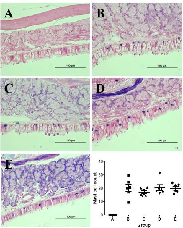
Figure 5. Comparison of the mast cell count between groups (Giemsa stain, ×400). ---39
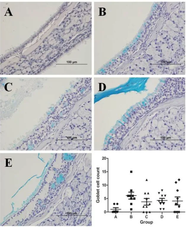
Figure 6. Comparison of the secretory cell count between groups (Alcian blue stain, ×400). ---40
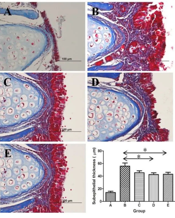
Figure 7. Comparison of the subepithelial thickness between groups (*P <
0.05, Masson’s trichrome stain, ×400). ---41
vi
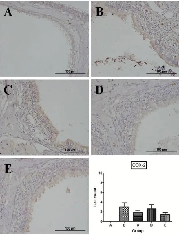
Figure 8. Representative microphotographs of immunohistochemistry and quantitative analysis of positive cells for cyclooxygenase-2. ---42
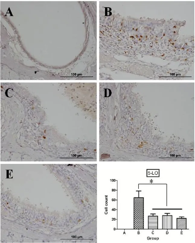
Figure 9. Representative microphotographs of immunohistochemistry and quantitative analysis of positive cells for 5-lipooxygenase (*P < 0.05,
×400). ---44
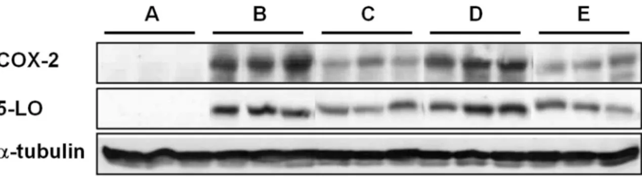
Figure 10. Western blot analysis of cyclooxygenase-2 and 5- lipooxygenase expression. ---46
Figure 11. mRNA expression of key cytokines IL-4, IL-5, and eicosanoid pathway-related enzymes PGDS and LTC4S in the murine sinonasal mucosa. ---47
vii
List of Abbreviations
SAEs: Staphylococcus aureus exotoxins SEB: Staphylococcus aureus enterotoxin B OVA: Ovalbumin
COX-2: cyclooxygenase-2 5-LO: 5-lipooxygenase DMSO: dimethyl sulfoxide
PBS: phosphate-buffered saline IHC: Immunohistochemistry
qPCR: quantitative real-time PCR IL: interleukin
PGDS: prostaglandin D synthase LTC4S: leukotriene C4 synthase Sir2: silent information regulator 2 NO: nitric oxide
PMN: polymorphonuclear leukocytes DMBA: 7,12-dimethylbenz(α)anthracene TGF-β: transforming growth factor-β
1
Introduction
The prevalence of chronic rhinosinusitis is known to reach approximately 12.5% in general population and it is frequently accompanied by nasal polyps which occur in 1% - 4% of general population.1,2 Nasal polyps often bring about nasal stuffiness and olfactory dysfunction resulting in the decline of quality of life. Furthermore, it has propensity to recur after surgical removal. In addition, nasal polyp-related medical cost is increasing along with the increase of allergic diseases and air pollution.3 Systemic administration of corticosteroids has been the most efficient medical treatment thus far, and its crucial mode of action is known to be an anti-inflammatory effect. However, it cannot be used for long-term periods because prolonged use of them result in detrimental side effects such as diabetes mellitus, osteoporosis, depression, and peptic ulcer.4 Thus, it is mandatory to search for safe and effective novel drugs for the treatment of rhinosinusitis with nasal polyps.
Understanding the pathomechanism of rhinosinusitis with nasal polyps is of paramount importance in order to discover a new potential drug for it.
Previous studies elucidated that nasal polyps are not simple mucosal edema, but benign inflammatory growth consisting of cystically dilated glands which are totally different from the seromucinous glands of normal turbinal mucosa.5 Numerous factors have been reported to be associated with nasal polyp formation, including infection,6,7 allergy,8-11 superantigens,12-15 and genetic predisposition,16,17 yet the precise mechanism of nasal polyp formation is still unknown. Since infiltration of
2
inflammatory cells, particularly eosinophils, and tissue remodeling are consistently found in nasal polyps, nontheless, the molecules which have anti-inflammatory and/or remodeling-preventing effects are assumed to inhibit nasal polyp formation.18-21
Resveratrol was first extracted from a nonedible Peruvian legume, Cassia quinquangulata Rich. Although resveratrol is ubiquitous in nature, it is found in only a few edible substances, most notably the grape. 22 Resveratrol has been found to have various beneficial effects thus far:
strong inhibition of cyclooxygenase (COX), cancer chemoprevention, and even life extension. The molecular targets of resveratrol, which mediate its diverse cellular effects, are the subject of ongoing investigations.22,23 In terms of safety, several animal studies have been performed, and no significant toxicity such as genotoxicity and carcinogenicity was identified.
Moreover, nephrotoxicity was found only at very high dosages (2,000 - 3000 mg/kg/day). On the basis of these studies, an acceptable daily intake of resveratrol in food has been defined as 450 mg/day.24
Since a mouse model of eosinophilic rhinosinusitis with nasal polyps was established recently, research for the development of potential new drugs and elucidation of the mechanism of their effect has been feasible.25 In brief, the model was induced by systemic sensitization with ovalbumin (OVA) followed by intranasal instillatioin of OVA and Staphylococcus aureus enterotoxin B (SEB). Using this murine model, therapeutic effects of resveratrol on eosinophilic rhinosinusitis with nasal polyps, and the mechanism of actions were investigated in this study.
3
Materials and Methods
Experimental Animals
BALB/c mice (4 weeks of age) were purchased from Koatech Laboratory Animal, Inc. (Pyeongtaek, Korea). These animals were kept in a special pathogen-free biohazard containment facility maintained at 22℃ to 24℃ and 50% to 60% humidity. All experimental protocols complied with the Guidelines of the National Institute of Health and the Declaration of Helsinki, and were approved by the Committee on the Use and Care of Animals at Gyeongsang National University.
Experimental Protocols
Mice were categorized into a single control and 4 experimental groups, and eosinophilic inflammation in the nasal and sinus mucosa was induced in each mouse according to the protocols which were previously established.25 In brief, OVA (Grade V; Sigma, St. Louis, MO, USA) was used to obtain systemic sensitization and local stimulation, followed by intranasal instillation of SEB in experimental groups (Figure 1). Instead, in the control group (group A), phosphate-buffered saline (PBS) was applied systemically as well as locally. The experimental groups were designated as follows: intraperitoneal injection of vehicle, which means a mixture of dimethyl sulfoxide (DMSO) and PBS with a ratio of 1:9 (group B), intraperitoneal injection of triamcinolone acetonide with a concentration of 1 mg/kg (group C), intraperitoneal injection of resveratrol with a
4
concentration of 0.5 mg/kg (group D) and 5 mg/kg (group E). Mice in the experimental groups were systemically sensitized with 25 µg OVA dissolved in 300 µL PBS in the presence of 2 mg aluminum hydroxide gel as adjuvants by intraperitoneal injection on days 0 and 5. One week after the second intraperitoneal injection, mice were challenged intranasally with 3% OVA diluted in 40 µL of PBS daily for 1 week. Thereafter, continual local stimulation was maintained in the same fashion three times a week for 4 consecutive weeks. Finally, 3% OVA diluted in 40 µL of PBS was applied intranasally accompanied by intraperitoneal injection of drugs including triamcinolone acetonide and resveratrol with the same intervals for eight consecutive weeks. During that period, 10ng SEB diluted in 20 µL of PBS was challenged intranasally, subsequent to the instillation of OVA once a week. Twenty-four hours after the final nasal challenge, mice were euthanized and decapitated. Each experimental and control group contained 20 and 10 mice, respectively; half were prepared for histologic examination while nasal mucosa was obtained in the remaining half for Western blot analyses and quantitative real-time PCR (qPCR). Since a single mouse in group B died during the experiments, nine mice were finally prepared for histologic analysis.
Histologic Analyses
The skin and soft tissues of the mice were removed from the skull.
Heads were immediately fixed in 2% paraformaldehyde and decalcified in 5% nitric acid for 4 - 5 days at 4°C. The specimens were excised from the
5
second palatal ridge to the first upper molar teeth. The tissues were dehydrated and processed according to standard paraffin-embedding procedures, and then were cut in coronal sections with a thickness of 4 μm. Several stains were conducted in order to compare the characteristics between groups: hematoxylin and eosin (H&E) for overall inflammation, Sirus red for eosinophils, Giemsa for mast cells, alcian blue for secretory cells, and Masson’s trichrome staining for collagen in the subepithelial layer.
An atlas of normal murine sinonasal anatomy was used to standardize the anatomic locations being examined. First, the vomeronasal organ was identified. The superior and inferior maxillary turbinelles were identified for anatomic orientation. The true maxillary sinus and ethmoidal labyrinths were identified at the lesions posterior to the two maxillary turbinelles.
Three coronal sections which were similar to the sinus cavity were chosen for evaluation, according to the previous study. The number of polyp-like lesions, inflammatory and secretory cells was counted under high-power fields (10 X 40) by an examiner blinded to the groups. Polyp- like lesions were defined as distinct mucosal bulges with eosinophilic infiltration and microcavities. The thickness of the subepithelial layer was measured by using an image analysis system (NIS-Elements BR 3.0 system; Nikon Eclipse, Tokyo, Japan). Three consecutive slides were reviewed in order to exclude processing errors.
Immunohistochemistry (IHC)
6
Other sections were immunostained for COX-2 and 5-lipooxygenase (LO) using the avidin-biotinylated-horseradish peroxidase-complex kits (ABC; Vector Laboratories, Burlingame, CA). After deparaffinization in xylene, sections were rehydrated with ethanol. After washing in PBS, the sections were blocked with 1% normal goat serum and then treated with each primary polyclonal antibody for COX-2 (Cell Signaling Tech, Beverly, MA) and 5-LO (Cell Signaling Tech) at 4°C overnight in a humidified chamber. After washing in PBS, they were incubated for 90 minutes at room temperature with secondary antibody (biotin-conjugated goat anti- rabbit immunoglobulin G, 1:200). Finally, the sections were incubated with ABC for 60 minutes at room temperature, rinsed in PBS, and then developed by 0.027% 3, 3-diaminobenzidine tetrahydrochloride (Sigma) with 0.003% hydrogen peroxide. Finally, the sections were counterstained with hematoxylin (Sigma). Each section was examined under high-power field (10 × 40) by an independent researcher blinded to the experiments.
Cells containing positive signals were counted at the transition zone of the olfactory and respiratory epithelium.
Protein Extraction and Western blot
Protein was obtained from the nasal mucosa of each mouse 1 day after the final nasal challenge and homogenized in lysis buffer (1% Triton X- 100 and 1mM EDTA in 1x PBS [pH 7.4]) which contained 10µM leupeptin and 200 µM phenylmethylsulfonyl fluoride. Cell lysates were sonicated several times for 3-5 minutes each and centrifuged at 12,000 rpm for 20
7
minutes at 4 . Following the collection of super℃ natant, protein concentration of each lysate was determined by using bicinchoninic acid kit (Pierce, Rockford, IL) using bovine serum albumin as standard in accordance with the manufacturer’s instructions. Equal amount of protein (60 µg) was loaded to 10% to 12% sodium dodecylsulfate-polyacrylamide gel. After electrophoresis, proteins in the gel were transferred to a nitrocellulose membrane (Schleicher & Schuell, Dassel, Germany), and the membranes were washed in Tris-buffered saline containing 0.1%
Tween-20. Subsequent to the incubation with each primary rabbit polyclonal antibody for COX-2 (Cell Signaling Tech) and 5-LO (Cell Signaling Tech), the membrane was incubated with secondary antibodies (horseradish peroxidase-conjugated goat anti-rabbit immunoglobulin G, 1:10,000; Pierce). Then, the blots were visualized by using ECL kit (Amersham Pharmacia Biotech, Piscataway, NJ).
Quantitative Real-time PCR
The levels of key cytokines interleukin (IL)-4, IL-5, and eicosanoid pathway-related enzymes prostaglandin D synthase (PGDS), and leukotriene C4 synthase (LTC4S) transcripts were determined by qPCR.
Nasal mucosa was resuspended in 1ml of Trizol Reagent (Invitrogen Life Technologies, Carlsbad, CA) and total RNA was prepared according to the manufacturer’s instructions. Ten microgram of purified RNA was subsequently reverse-transcribed into cDNA using an iScript cDNA synthesis kit (Bio-Rad Laboratories, Hercules, CA) and oligo-dT primers.
8
After reverse transcription, total DNA was diluted by ddH2O for quantitative real-time PCR. Quantitative cDNA amplification was performed using a CFX-96TM Real-Time System (Applied Biosystems Inc., Foster City, CA), and PCR conditions as specified by the manufacturer.
Each of the reaction mixtures contained: 9 μl of template cDNA, 10 μl of universal KAPA probe fast qPCR master mix (KAPA Biosystems, Woburn, MA), and 1 μl of 20× TaqMan gene expression Assay Mix for the genes of interest (Applied Biosystems Inc.) to a final volume of 20 μl. Samples were normalized using mouse GAPDH expression. Thermal cycle conditions were as follows: denaturation at 95°C for 3 minutes, followed by 50 cycles of denaturation at 95°C for 10 seconds, and annealing and extension at 60°C for 30 seconds.
Ct values representing the number of cycles at which the fluorescence value for each sample exceeded the threshold value were recorded with Bio-Rad CFX Manger 2.0 (Applied Biosystems Inc.). The expression level in each sample was compared with a calibrator. The following formula was applied: gene expression = 2−ΔΔCt
In brief, relative quantification was performed using 2−ΔΔCt method.
Reference gene for GAPDH was used. GAPDH as internal control was used for the normalization of the quantity of RNA. For normalization of the results Ct value for GAPDH was subtracted from Ct value for each of target genes (IL-4, IL-5, PGDS, and LTCS).
ΔCt = Average CtTarget gene-Average CtGAPDH
The obtained difference (ΔCt) was then used to calculate ΔΔCt with the
9 formula:
ΔΔCt = ΔCtexperimental group - ΔCtcontrol group
The relative gene expression for cytokines normalized to an endogenous reference and relative to a calibrator was expressed as:
Gene expression level = 2-ΔΔCt
Relative gene expression for targeted genes in group A equals 1 (since 20 = 1). Relative gene expression for measured cytokines and enzymes was presented as the fold change comparing to the control group.
Statistical Analyses
Data were expressed as mean ± SEM (standard error of the mean). The Mann-Whitney U-test and Kruskal-Wallis test generated by SPSS version 18.0 (SPSS, Chicago, IL) were used to determine the difference in the number of polyp-like lesions, inflammatory and secretory cells, the proportion of eosinophils in total inflammatory cells, the thickness of the subepithelial layer, the production of COX-2 and 5-LO, and the gene expression level of IL-4, IL-5, PGDS and LTC4S between groups. A value of P < 0.05 was considered significant.
10
Results
Histologic Analyses
Polyp-like lesions
Polyp-like lesions were not observed in the nasal or sinus mucosa in group A. Sixteen polyp-like lesions were noted in seven out of nine mice in group B. Similarly, 12 polyp-like lesions were shown in seven out of ten mice in group D. However, only five polyp-like lesions were demonstrated in 5 out of 10 mice in group C, which seemed definitely fewer than in group B. Nontheless, statistical significance could not be obtained presumably due to the lack of number of polyp-like lesions. Similarly, the number of polyp-like lesions was also reduced in mice in group E compared to that in group B, but the difference was not statistically significant (Figure 2).
Eosinophilic inflammation
The number of eosinophils was largest in group B whereas mice in group A showed no apparent infiltration of inflammatory cells in the nasal and sinus mucosa. The degree of eosinophilic infiltration seemed less severe in group D than that in group B, yet the difference was not statistically significant. In contrast, mice in both groups C and E demonstrated much lower number of eosinophils than in group B (P <
11
0.001 and P = 0.003, respectively). Likewise, group E showed fewer eosinophilic infiltration in the nasal mucosa than group D (P = 0.006).
Finally, the degree of eosinophilic inflammation was similar between group C and E (Figure 3).
The percentage of eosinophils in total inflammatory cells (mean ± SEM) was also compared between groups: 55.0 ± 1.9%, 42.0 ± 3.5%, 41.6 ± 2.7%, and 41.5 ± 3.3% in groups B, C, D, and E, respectively. The proportion of eosinophils was significantly smaller in groups C, D, and E compared with that in group B (P = 0.017, P < 0.001, and P = 0.006, respectively; Figure 4).
Distribution of mast cells
Mast cells were scarcely observed in the nasal mucosa even in group B while they were easily found in the mucosa of the maxillary sinus. Thus, mast cells were counted (mean ± SEM) in the maxillary sinus and compared between groups: 20.2 ± 2.8, 17.0 ± 1.0, 20.3 ± 2.2, and 19.9 ± 1.4 in groups B, C, D, and E, respectively. In group A, mast cells were not noted in the mucosa of maxillary sinus. The degree of mast cell infiltration seemed less severe in group C compared to other groups, but there was no significant difference between experimental groups (P = 0.39; Figure 5).
Secretory cell hyperplasia
The number of secretory cells (mean ± SEM) was calculated at the
12
transition zone of olfactory and respiratory epithelium: 1.0 ± 0.6, 6.1 ± 1.3, 3.8 ± 1.2, 4.3 ± 0.8, and 4.1 ± 1.5 in groups A, B, C, D, and E, respectively.
A limited number of secretory cells were found in group A. Their number was largest in group B, and smaller in groups C, D, and E compared to that in group B. However, the difference was statistically insignificant (P = 0.43; Figure 6).
Subepithelial fibrosis
The thickness of subepithelial layer (mean ± SEM) was measured at the inferior end of nasal septum: 14.1 ± 2.1 µm, 55.8 ± 5.1 µm, 45.4 ± 3.1 µm, 42.6 ± 2.8 µm, and 43.1 ± 3.1 µm in groups A, B, C, D, and E, respectively. The subepithelial layer was considerably thick in group B compared to that in group A. It was apparently thinner in groups D and E than in group B (P = 0.02 and P = 0.024, respectively). Similarly, group C demonstrated thinner subepithelial layer than group B, but the difference was not statistically significant (P = 0.09; Figure 7).
Immunohistochemistry for COX-2 and 5-LO
To elucidate the mechanism of anti-inflammatory effects by high-dose resveratrol, IHC was performed for COX-2 and 5-LO. In group A, no definite positive signals were observed for COX-2 in the nasal mucosa.
They were also scarcely noted in experimental groups B - E. Cells containing positive signals for COX-2 were counted at the transition zone of the olfactory and respiratory epithelium: 3.0 ± 0.8, 1.8 ± 0.5, 2.6 ± 0.9,
13
and 1.4 ± 0.4 in groups B, C, D, and E, respectively. It appeared that the number of positive signals is smaller in groups C and E than in groups B and D. However, there was no statistically significant difference between groups probably due to the scarcity of positive signals in all groups (P = 0.36; Figure 8).
For 5-LO, no definite positive signals were observed in the nasal mucosa in group A. However, there was strong expression of 5-LO in group B.
The number of cells with positive signals was as follows: 64.6 ± 13.8, 26.6
± 4.8, 28.0 ± 4.3, and 22.0 ± 3.1 in groups B, C, D, and E, respectively.
The degree of positive signals was apparently reduced in groups C, D, and E compared with that in group B (P = 0.03, P = 0.014, and P = 0.01, respectively). Finally, there was no definite difference in 5-LO expression among groups C, D, and E (Figure 9).
Western blot Analysis
The protein levels of COX-2 and 5-LO were measured using Western blot analysis. The expression level of COX-2 was strongest in group B while it was undetectable in group A. Mice in group C exhibited the weakest expression level of COX-2, which was similar in group E. In contrast, there was no apparent decrease in COX-2 expression in group D.
Similar inter-group differences were identified in the expression level of 5- LO. Groups B and D exhibited strong expression of 5-LO, but it was weakened in groups C and E (Figure 10).
14 Quantitative Real-time PCR
The qPCR was used to determine the mRNA expression of key cytokines IL-4, IL-5 in the murine sinonasal mucosa. The relative expression level of IL-4 (mean ± SEM) was calculated: 1.0 ± 0.1, 79.4 ± 8.1, 40.3 ± 8.2, 48.4
± 12.8, and 35.8 ± 6.8 in groups A, B, C, D, and E, respectively. Group B showed highest expression level of IL-4, which was significantly reduced in group C, D, and E compared with that in group B (P = 0.009, P = 0.035, and P = 0.002, respectively). There was no apparent difference in the expression level of IL-4 among groups C, D, and E (Figure 11). The relative expression level of IL-5 (mean ± SEM) was as follows: 1.0 ± 0.1, 2.4 ± 0.2, 0.7 ± 0.1, 0.5 ± 0.1, and 0.5 ± 0.2 in groups A, B, C, D, and E, respectively. Group B showed highest expression level of IL-5, which was considerably diminished in group C, D, and E compared with that in group B (P < 0.001, P < 0.001, and P = 0.002, respectively). There was no definite difference in the expression level of IL-5 among groups C, D, and E (Figure 11).
The mRNA expression of eicosanoid pathway-related enzymes PGDS and LTC4S was also determined using the qPCR. The relative expression level of PGDS (mean ± SEM) was as follows: 1.0 ± 0.1, 4.7 ± 1.5, 4.1 ± 0.9, 0.6 ± 0.2, and 0.6 ± 0.1 in groups A, B, C, D, and E, respectively.
Group B showed highest expression level of PGDS, which was significantly reduced in groups D and E compared with that in group B (P
= 0.001 and P < 0.001, respectively). Groups D and E also showed significantly lower expression level of PGDS than that in group C (P <
15
0.001 and P < 0.001, respectively). There was no apparent difference in the expression level of PGDS between groups B and C (P = 0.91; Figure 11). The relative expression level of LTC4S (mean ± SEM) was as follows:
1.0 ± 0.1, 2.1 ± 0.2, 1.2 ± 0.1, 0.5 ± 0.1, and 0.9 ± 0.2 in groups A, B, C, D, and E, respectively. Group B showed highest expression level of LTC4S, which was considerably decreased in group C, D, and E compared with that in group B (P = 0.009, P < 0.001, and P < 0.001, respectively). Group D showed significantly lower expression of LTC4S than group C (P = 0.02), while there was no definite difference between groups C and E (P = 0.32;
Figure 11).
16
Discussion
Animal models: Useful tools for the exploration of new drugs Animal models are instrumental for the clarification of pathomechanisms of the disease, the investigation into the biomarkers, and the exploration of novel treatment modalities. Thus, animal models have been widely used in the various fields in medicine such as allergy, cancer, cardio- or cerebro-vascular diseases, and psychiatric diseases.27-31 Several trials have been made to establish an animal model of rhinosinusitis accompanied by nasal polyps. First, an agent containing bacteria including Streptococcus pneumoniae serotype 3, Bacteroides fragilis NCTC 9343, or Staphylococcus aureus V8 was applied to New Zealand White rabbits, and unilateral sinusitis with mucosal polyps was identified irrespective of inducing agent.6 Thereafter, the same researchers identified that, besides bacterial infection, the deposition of agarose in the sinus cavity or a chemotactic peptide such as N-formyl-methionyl-leucyl- phenylalanine can also contribute to the formation of nasal polyps.7 Additionally, they analyzed detailed structural changes: epithelial disruption and the migration of immature branching epithelium were key features of polyp formation. Some branches of migrating epithelia eventually covered the mucosal defect, or spread into the intraepithelial microcavities which lied in the connective tissue, resulting in the separation of polyp body from the adjacent mucosa. Although those reports provided useful information on the histologic features of nasal
17
polyp formation, the tissues obtained from the rabbits did not show characteristic eosinophilic infiltration, which are typically noted in human nasal polyps. In recent years, there were two reports on an animal model of nasal polyps associated with eosinophilic infiltration. In the preceding study, rabbits received valine-glycine-serine-glutamine or poly-L-arginine in their maxillary sinuses after repeated exposure to OVA.9 Consequently, apparent eosinophilic infiltration, thickened lamina propria, and polyp formation were identified. Poly-L-arginine is a synthetic cationic polypeptide which is known to increase vascular permeability and induce airway hyperresponsiveness.32,33 Given that human nasal polyps are closely related to the eosinophilic inflammation, and eosinophilic cationic protein is one of the major components in eosinophils, it seems reasonable to make an animal model of nasal polyps by using a cationic protein. Nontheless, there are only limited data on the relationship between poly-L-arginine and nasal polyps. On the other hand, the latest study utilized a mouse for the establishment of an animal model of nasal polyps.25 The reason why mice were chosen was as follows: mice can be readily genetically manipulated, and a wide array of murine reagents is available and their housing costs are lower than other animals.
Additionally, SEB was used to induce the formation of eosinophilic nasal polyps. Numerous previous studies have provided the information on a link between Staphylococcus aureus exotoxins (SAEs) and nasal polyps;
SEB is one of the SAEs commonly detected in nasal polyps.15,34-38 It was added on the mice with pre-existing allergic inflammation caused by OVA.
18
Serial changes in histology were examined monthly and it was found that prolonged stimulation with OVA/SEB is mandatory to induce definite nasal polyps. The current study was conducted based on the protocols established in that study and similar findings were confirmed: the proliferation of secretary cells, the amplification of inflammatory cells such as eosinophils and mast cells, increased subepithelial fibrosis, and characteristic findings of nasal polyps including elevated lesions with eosinophilic infiltration and microcavities. Using these findings as parameters for the degree of inflammation and polyp formation, the therapeutic effect of resveratrol on eosinophilic rhinosinusitis with nasal polyps was investigated.
Versatile actions of resveratrol
Mediterranean diets are known to be rich in resveratrol. In a previous population-based study named ‘Lyon Diet Heart Study’, patients who suffered from first myocardial infarction were followed up for an average of 46 months. Interestingly, Mediterranean dietary pattern significantly reduced the rate of recurrence even after the adjustment of traditional risk factors such as high blood cholesterol and blood pressure.39 Subsequently, a number of studies were reported on the cardioprotective effect of resveratrol and its mechanism.23 Based on the structural similarity of resveratrol to diethylstilbestrol, resveratrol is characterized as a phytoestrogen.40 Given this structural similarity, the cardioprotective benefits of resveratrol was first assumed to be modulated by activation of
19
the estrogen receptor.23 Yet, more recently there have been a number of studies that suggest that the estrogen receptor is not the main cellular target of resveratrol in the vasculature. Instead, SIRT 1, a mammalian homolog of the Saccharomyces cerevisae silent information regulator 2 (Sir2) protein, has been recognized as the main target of resveratrol.41,42 Resveratrol is also known to induce major cellular anti-oxidant enzymes such as glutathione peroxidase and superoxide dismutase.43,44 In a previous study, resveratrol prevented H2O2-mediated apoptotic cell death in cultured aortic segments of rat, and its effect was attenuated by inhibition of glutathione peroxidase and heme oxygenase-1.43 Furthermore, resveratrol treatment upregulated the expression of glutathione peroxidase, catalase, and heme oxygenase-1 in cultured arteries. In another study, resveratrol increased nitric oxide (NO) production by enhancing endothelial NO synthase expression, and reduced O2- production by inhibiting NAD(P)H oxidase activity in mice.
Additionally, the anti-oxidant effects of resveratrol led to improved cardiac function by increasing the left ventricular diastolic peak filling rate.44 Of note, some studies showed that resveratrol has a lifespan extension effect although it was not proven in the upper vertebrates.45,46 In a prior study using Caenorhabditis elegans and Drosophila melanogaster, resveratrol extended the lifespan of these animals without reducing fecundity by activating sirtuins, a family of NAD+-dependent deacetylases conserved from Escherichia coli to humans.45 In another study, resveratrol mimicked calorie restriction by stimulating Sir2, leading to
20
increased DNA stability and extended lifespan by 70% in yeast.46
Anti-inflammatory effects of resveratrol
In the present study, resveratrol showed definite anti-inflammatory effects, particularly in a high dose. The formation of nasal polyp-like lesions was reduced along with the decrease in the overall thickness of nasal mucosa.
The degree of eosinophilic infiltration was also declined by administration of high-dose resveratrol, which was similar to the effect of triamcinolone acetonide. In the present study, tissue inflammation in mice was initiated by OVA, which increases total and OVA-specific IgE production through elevating IL-4 production.47 Thus, IL-4 can be a good marker for evaluating OVA-induced tissue inflammations. In the current study, the gene expression level of IL-4 was approximately 80 times higher in mice with eosinophilic rhinosinusitis with nasal polyps than in control group, and a marked decrease in the IL-4 expression level was observed in mice treated with low- or high-dose resveratrol. Their effect was similar with that of triamcinolone acetonide. Furthermore, the gene expression level of PGDS and LTC4S was remarkably inhibited by low- or high-dose resveratrol. PGDS catalyzes the isomerization of the 9,11-endoperoxide group of PGH2, a common precursor of various prostanoids, to produce PGD2 which is an end product of the COX pathway.48 LTC4S conjugates LTA4 with reduced glutathione to form LTC4, the parent compound of the cysteinyl leukotrienes.49 Accordingly, the decline of gene expression of PGDS and LTC4S by administration of resveratrol indicates that
21
resveratrol can exert an inhibitory action on both the COX and LOX pathway. The inhibitory effect of resveratrol on the LOX pathway, particularly in a high dose, was reconfirmed by IHC and Western blot analysis of 5-LO production. 5-LO is highly expressed in leukocytes such as neutrophils, eosinophils, and mast cells.50 Considering that eosinophils highly express 5-LO for the pro-inflammatory action, blockage of the LOX pathway by resveratrol is believed to a key mechanism of inhibition of eosinophilic inflammation. The inhibitory action of resveratrol on the LOX pathway was also identified in some previous studies.51-54 In a prior study, human polymorphonuclear leukocytes (PMN) were isolated from venous blood of healthy subjects and the effects of resveratrol on arachidonate metabolism were investigated. Resveratrol was found to inhibit several 5- LO products such as 5-hydroxy-6,8,11,14-eicosatetraenoic acid (5-HETE), 5,12-dihydroxy-6,8,10,14-eicosatetraenoic acid (5,12-diHETE) and LTC4.52 Similar effects of resveratrol were confirmed by some ensuing papers. In a study which examined anti-apoptotic activity of resveratrol using human erythroleukemia K562 cells, resveratrol was found to act as a competitive inhibitor of purified 5-LO and 15-LO and PDH synthase; as a consequence, LTB4 and PGE2 was reduced.53 In another study on the anti-cancer effect of resveratrol, it potently inhibited the 5-LO expression and LTB4 production in 7,12-dimethylbenz(α)anthracene (DMBA)-induced mammary cancer developments in rats. Moreover, resveratrol normalized the expression of transforming growth factor (TGF)-β1 which had been down-regulated in DMBA-challenged rat mammary tissue.51 In addition to
22
the anti-5-LO effect, resveratrol was found to have strong inhibitory effects on LTA4 hydrolase activity, which stimulates the production of pro- inflammatory cytokines and mediators by catalyzing the hydrolysis of LTA4
to LTB4. By means of small hairpin RNA-mediated knockdown of LTA4
hydrolase, the reduction of inhibitory effects of resveratrol was identified.54
In addition to the inhibitory actions on the LOX pathway, anti-inflammatory effects of resveratrol were ascertained in each type of leukocytes.55-57 In a previous study, PMN were isolated from venous blood of healthy volunteers, and stimulated with formyl methionyl leucyl phenylalamine or calcium ionophore subsequent to the treatment with resveratrol.
Resveratrol not only inhibited the production of reactive oxygen species, but also prevented the release of inflammatory mediators including elastase, β-glucuronidase, and LTB4 from PMN.55 In another in vitro study, eosinophils were obtained from venous blood of healthy non-atopic volunteers, and following reactions were significantly inhibited by resveratrol: eosinophil peroxidase release after activation with IL-5 or C5a, the production of LTC4 following stimulation with calcium ionophore, and eosinophil chemotaxis in response to eotaxin.56 In the current study, high- dose resveratrol not only reduced overall eosinophilic inflammation, but also decreased the proportion of eosinophils in total inflammatory cells.
Additionally, the expression level of IL-5 mRNA was remarkably decreased by administration of a low- or high-dose resveratrol, which had similar effects with triamcinolone acetonide. It is well established that IL-5
23
induces terminal maturation of eosinophil precursors, prolongs eosinophil survival, possesses eosinophilic chemotactic activity, and enhances eosinophilic effector function.58 Moreover, using the anti-IL-5 antibody, named mepolizumab, a significant decrease in blood eosinophil counts was ascertained.59 Taken together, it appears that resveratrol may inhibit the production and activation of eosinophils not only by inhibiting the LOX pathway, but also by suppressing the production of IL-5. On the other hand, inhibitory actions of resveratrol on mast cells were also reported.
Bone marrow-derived murine mast cells were triggered by IgE or calcium ionophore, and the effect of resveratrol was identified. The release of inflammatory mediators including histamine, tumor necrosis factor-α, LTs and PGD2 was inhibited.57 In histological analyses in the present study, however, no definite inhibitory action of resveratrol was observed. A further study will be needed to confirm the effect of resveratrol on mast cells.
Although low-dose resveratrol did not show a definite inhibitory action on 5-LO in Western blot analysis, on the other hand, it decreased the proportion of eosinophils in total inflammatory cells, and the subepithelial fibrosis. Subepithelial fibrosis is known to be prominent in patients with asthma, in particular, accompanying tissue eosinophilia.60 In addition, multiple cytokines, growth factors, and adhesion molecules, including IL- 13 and TGF-β, are implicated in the pathophysiology of subepithelial fibrosis in asthma.61 Several studies have been conducted regarding the inhibition of fibrosis by resveratrol. In a prior study using ex vivo human
24
lung fibroblasts, resveratrol prevented TGF-β-induced proliferation and differentiation of fibroblasts into myofibroblasts.62 In another study, resveratrol not only inhibited the production of pro-fibrogenic factors such as IL-6 and TGF-β, but also strongly activated the nuclear factor erythroid 2-related factor 2 which is known as a critical regulator of cellular defense against oxidative stress.63 Although the anti-oxidant effect of resveratrol is deemed to decrease subepithelial fibrosis, a further study will be needed to elucidate the precise mechanism of anti-fibrogenic effects of resveratrol.
In summary, it was ascertained that resveratrol exerts anti-inflammatory effects, particularly on eosinophils, and prevents subepithelial fibrosis in a murine model of eosinophilic rhinosinusitis accompanied by polyp-like lesions. Inhibition of 5-LO appears to be one of the key mechanisms of anti-inflammatory effects of resveratrol. Consequently, resveratrol may be a new potential drug for the treatment of eosinophilic rhinosinusitis with nasal polyps although a further human study is needed to confirm it.
25
References
1. Hamilos DL. Chronic rhinosinusitis: epidemiology and medical management. J Allergy Clin Immunol 2011;128:693-707; quiz 708- 699.
2. Klossek JM, Neukirch F, Pribil Cet al. Prevalence of nasal polyposis in France: a cross-sectional, case-control study. Allergy 2005;60:233- 237.
3. DeMarcantonio MA, Han JK. Nasal polyps: pathogenesis and treatment implications. Otolaryngol Clin North Am 2011;44:685-695, ix.
4. Bachert C, Watelet JB, Gevaert P, Van Cauwenberge P.
Pharmacological management of nasal polyposis. Drugs 2005;65:1537-1552.
5. Pawankar R, Nonaka M. Inflammatory mechanisms and remodeling in chronic rhinosinusitis and nasal polyps. Curr Allergy Asthma Rep 2007;7:202-208.
6. Norlander T, Fukami M, Westrin KM, Stierna P, Carlsoo B. Formation of mucosal polyps in the nasal and maxillary sinus cavities by infection. Otolaryngol Head Neck Surg 1993;109:522-529.
7. Norlander T, Westrin KM, Fukami M, Stierna P, Carlsoo B.
Experimentally induced polyps in the sinus mucosa: a structural analysis of the initial stages. Laryngoscope 1996;106:196-203.
26
8. Larsen K. The clinical relationship of nasal polyps to asthma. Allergy Asthma Proc 1996;17:243-249.
9. Sejima T, Kajiwara D, Kikuchi H, Imayoshi S, Yamauchi T, Ichimura K. Experimentally induced eosinophilic polyps in rabbit sinuses. Am J Rhinol Allergy 2010;24:341-347.
10. Settipane GA, Chafee FH. Nasal polyps in asthma and rhinitis. A review of 6,037 patients. J Allergy Clin Immunol 1977;59:17-21.
11. Bachert C, Gevaert P, Holtappels G, Johansson SG, van Cauwenberge P. Total and specific IgE in nasal polyps is related to local eosinophilic inflammation. J Allergy Clin Immunol 2001;107:607-614.
12. Gevaert P, Holtappels G, Johansson SG, Cuvelier C, Cauwenberge P, Bachert C. Organization of secondary lymphoid tissue and local IgE formation to Staphylococcus aureus enterotoxins in nasal polyp tissue. Allergy 2005;60:71-79.
13. Perez Novo CA, Jedrzejczak-Czechowicz M, Lewandowska-Polak Aet al. T cell inflammatory response, Foxp3 and TNFRS18-L regulation of peripheral blood mononuclear cells from patients with nasal polyps-asthma after staphylococcal superantigen stimulation.
Clin Exp Allergy 2010;40:1323-1332.
14. Ryan MW, Davis LS. T cells in chronic rhinosinusitis with nasal polyposis. Curr Opin Otolaryngol Head Neck Surg 2010;18:200-205.
27
15. Seiberling KA, Conley DB, Tripathi Aet al. Superantigens and chronic rhinosinusitis: detection of staphylococcal exotoxins in nasal polyps.
Laryngoscope 2005;115:1580-1585.
16. Robertson JM, Friedman EM, Rubin BK. Nasal and sinus disease in cystic fibrosis. Paediatr Respir Rev 2008;9:213-219.
17. Tos M, Mogensen C, Thomsen J. Nasal polyps in cystic fibrosis. J Laryngol Otol 1977;91:827-835.
18. Allen JS, Eisma R, LaFreniere D, Leonard G, Kreutzer D.
Characterization of the eosinophil chemokine RANTES in nasal polyps. Ann Otol Rhinol Laryngol 1998;107:416-420.
19. Bachert C, Wagenmann M, Hauser U, Rudack C. IL-5 synthesis is upregulated in human nasal polyp tissue. J Allergy Clin Immunol 1997;99:837-842.
20. Elovic A, Wong DT, Weller PF, Matossian K, Galli SJ. Expression of transforming growth factors-alpha and beta 1 messenger RNA and product by eosinophils in nasal polyps. J Allergy Clin Immunol 1994;93:864-869.
21. Liu CM, Hong CY, Shun CTet al. Matrix metalloproteinase-1 and tissue inhibitor of metalloproteinase-1 gene expressions and their differential regulation by proinflammatory cytokines and prostaglandin in nasal polyp fibroblasts. Ann Otol Rhinol Laryngol 2001;110:1129-1136.
22. Pezzuto JM. The phenomenon of resveratrol: redefining the virtues of promiscuity. Ann N Y Acad Sci 2011;1215:123-130.
28
23. Csiszar A. Anti-inflammatory effects of resveratrol: possible role in prevention of age-related cardiovascular disease. Ann N Y Acad Sci 2011;1215:117-122.
24. Edwards JA, Beck M, Riegger C, Bausch J. Safety of resveratrol with examples for high purity, trans-resveratrol, resVida((R)). Ann N Y Acad Sci 2011;1215:131-137.
25. Kim DW, Khalmuratova R, Hur DGet al. Staphylococcus aureus enterotoxin B contributes to induction of nasal polypoid lesions in an allergic rhinosinusitis murine model. Am J Rhinol Allergy 2011;25:255-261.
26. Jacob A, Chole RA. Survey anatomy of the paranasal sinuses in the normal mouse. Laryngoscope 2006;116:558-563.
27. Diaz JA, Obi AT, Myers DD, Jr.et al. Critical review of mouse models of venous thrombosis. Arterioscler Thromb Vasc Biol 2012;32:556- 562.
28. Herreros-Villanueva M, Hijona E, Cosme A, Bujanda L. Mouse models of pancreatic cancer. World J Gastroenterol 2012;18:1286- 1294.
29. Kumar RK, Herbert C, Foster PS. The "classical" ovalbumin challenge model of asthma in mice. Curr Drug Targets 2008;9:485- 494.
30. O'Collins VE, Macleod MR, Donnan GA, Howells DW. Evaluation of combination therapy in animal models of cerebral ischemia. J Cereb Blood Flow Metab 2012;32:585-597.
29
31. Tye KM, Deisseroth K. Optogenetic investigation of neural circuits underlying brain disease in animal models. Nat Rev Neurosci 2012;13:251-266.
32. Coyle AJ, Uchida D, Ackerman SJ, Mitzner W, Irvin CG. Role of cationic proteins in the airway. Hyperresponsiveness due to airway inflammation. Am J Respir Crit Care Med 1994;150:S63-71.
33. Strek ME, Williams FS, Gleich GJ, Leff AR, White SR. Mechanisms of smooth muscle contraction elicited by cationic proteins in guinea pig trachealis. Am J Physiol 1996;270:L133-140.
34. Bachert C, Zhang N, Holtappels Get al. Presence of IL-5 protein and IgE antibodies to staphylococcal enterotoxins in nasal polyps is associated with comorbid asthma. J Allergy Clin Immunol 2010;126:962-968, 968 e961-966.
35. Tripathi A, Conley DB, Grammer LCet al. Immunoglobulin E to staphylococcal and streptococcal toxins in patients with chronic sinusitis/nasal polyposis. Laryngoscope 2004;114:1822-1826.
36. Tripathi A, Kern R, Conley DBet al. Staphylococcal exotoxins and nasal polyposis: analysis of systemic and local responses. Am J Rhinol 2005;19:327-333.
37. Van Zele T, Gevaert P, Holtappels G, van Cauwenberge P, Bachert C. Local immunoglobulin production in nasal polyposis is modulated by superantigens. Clin Exp Allergy 2007;37:1840-1847.
38. Van Zele T, Vaneechoutte M, Holtappels G, Gevaert P, van Cauwenberge P, Bachert C. Detection of enterotoxin DNA in
30
Staphylococcus aureus strains obtained from the middle meatus in controls and nasal polyp patients. Am J Rhinol 2008;22:223-227.
39. De Lorgeril M, Salen P, Martin JL, Monjaud I, Delaye J, Mamelle N.
Mediterranean diet, traditional risk factors, and the rate of cardiovascular complications after myocardial infarction: Final report of the Lyon Diet Heart Study. Circulation 1999;99:779-785.
40. Gehm BD, McAndrews JM, Chien PY, Jameson JL. Resveratrol, a polyphenolic compound found in grapes and wine, is an agonist for the estrogen receptor. Proc Natl Acad Sci U S A 1997;94:14138- 14143.
41. Danz ED, Skramsted J, Henry N, Bennett JA, Keller RS. Resveratrol prevents doxorubicin cardiotoxicity through mitochondrial stabilization and the Sirt1 pathway. Free Radic Biol Med 2009;46:1589-1597.
42. Baur JA, Sinclair DA. Therapeutic potential of resveratrol: the in vivo evidence. Nat Rev Drug Discov 2006;5:493-506.
43. Ungvari Z, Orosz Z, Rivera Aet al. Resveratrol increases vascular oxidative stress resistance. Am J Physiol Heart Circ Physiol 2007;292:H2417-2424.
44. Zhang H, Morgan B, Potter BJet al. Resveratrol improves left ventricular diastolic relaxation in type 2 diabetes by inhibiting oxidative/nitrative stress: in vivo demonstration with magnetic resonance imaging. Am J Physiol Heart Circ Physiol 2010;299:H985- 994.
31
45. Wood JG, Regina B, Lavu Set al. Sirtuin activators mimic caloric restriction and delay ageing in metazoans. Nature 2004;430:686-689.
46. Howitz KT, Bitterman KJ, Cohen HYet al. Small molecule activators of sirtuins extend Saccharomyces cerevisiae lifespan. Nature 2003;425:191-196.
47. Spiegelberg HL. The role of interleukin-4 in IgE and IgG subclass formation. Springer Semin Immunopathol 1990;12:365-383.
48. Urade Y, Hayaishi O. Biochemical, structural, genetic, physiological, and pathophysiological features of lipocalin-type prostaglandin D synthase. Biochim Biophys Acta 2000;1482:259-271.
49. Penrose JF. LTC4 synthase. Enzymology, biochemistry, and molecular characterization. Clin Rev Allergy Immunol 1999;17:133- 152.
50. Okunishi K, Peters-Golden M. Leukotrienes and airway inflammation.
Biochim Biophys Acta 2011;1810:1096-1102.
51. Chatterjee M, Das S, Janarthan M, Ramachandran HK. Role of 5- lipoxygenase in resveratrol mediated suppression of 7,12- dimethylbenz(alpha)anthracene-induced mammary carcinogenesis in rats. Eur J Pharmacol 2011.
52. Kimura Y, Okuda H, Kubo M. Effects of stilbenes isolated from medicinal plants on arachidonate metabolism and degranulation in human polymorphonuclear leukocytes. J Ethnopharmacol 1995;45:131-139.
32
53. MacCarrone M, Lorenzon T, Guerrieri P, Agro AF. Resveratrol prevents apoptosis in K562 cells by inhibiting lipoxygenase and cyclooxygenase activity. Eur J Biochem 1999;265:27-34.
54. Oi N, Jeong CH, Nadas Jet al. Resveratrol, a red wine polyphenol, suppresses pancreatic cancer by inhibiting leukotriene Ahydrolase.
Cancer Res 2010;70:9755-9764.
55. Rotondo S, Rajtar G, Manarini Set al. Effect of trans-resveratrol, a natural polyphenolic compound, on human polymorphonuclear leukocyte function. Br J Pharmacol 1998;123:1691-1699.
56. Tan Y, Lim LH. trans-Resveratrol, an extract of red wine, inhibits human eosinophil activation and degranulation. Br J Pharmacol 2008;155:995-1004.
57. Baolin L, Inami Y, Tanaka H, Inagaki N, Iinuma M, Nagai H.
Resveratrol inhibits the release of mediators from bone marrow- derived mouse mast cells in vitro. Planta Med 2004;70:305-309.
58. Takatsu K, Nakajima H. IL-5 and eosinophilia. Curr Opin Immunol 2008;20:288-294.
59. Leckie MJ, ten Brinke A, Khan Jet al. Effects of an interleukin-5 blocking monoclonal antibody on eosinophils, airway hyper- responsiveness, and the late asthmatic response. Lancet 2000;356:2144-2148.
60. Berry M, Morgan A, Shaw DEet al. Pathological features and inhaled corticosteroid response of eosinophilic and non-eosinophilic asthma.
Thorax 2007;62:1043-1049.
33
61. Brewster CE, Howarth PH, Djukanovic R, Wilson J, Holgate ST, Roche WR. Myofibroblasts and subepithelial fibrosis in bronchial asthma. Am J Respir Cell Mol Biol 1990;3:507-511.
62. Fagone E, Conte E, Gili Eet al. Resveratrol inhibits transforming growth factor-beta-induced proliferation and differentiation of ex vivo human lung fibroblasts into myofibroblasts through ERK/Akt inhibition and PTEN restoration. Exp Lung Res 2011;37:162-174.
63. He X, Wang L, Szklarz G, Bi Y, Ma Q. Resveratrol inhibits paraquat- induced oxidative stress and fibrogenic response by activating the Nrf2 pathway. J Pharmacol Exp Ther 2012.
34
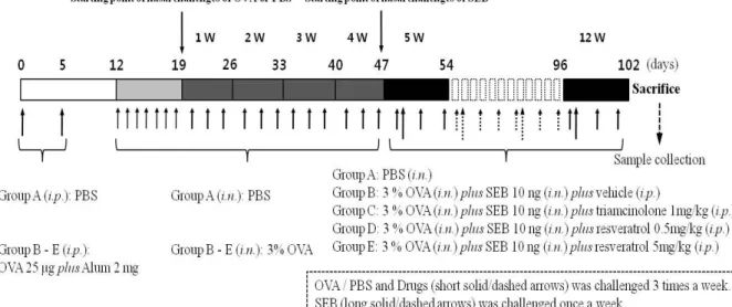
Figure 1. Protocols for the development of eosinophilic rhinosinusitis with nasal polyps in mice. Ovalbumin was used to obtain systemic sensitization and local stimulation, followed by intranasal instillation of Stayphylococcus aureus enterotoxin B (SEB) in experimental groups B - E. Instead, in the control group (group A), phosphate-buffered saline (PBS) was administered via a local or systemic route. The experimental groups were designated as follows: intraperitoneal injection of vehicle, which means a mixture of dimethyl sulfoxide (DMSO) and PBS with a ratio of 1:9 (group B), intraperitoneal injection of triamcinolone acetonide with a concentration of 1 mg/kg (group C), intraperitoneal injection of resveratrol with a concentration of 0.5 mg/kg (group D), and 5 mg/kg (group E). i.p., intraperitoneal injection; i.n., intranasal instillation.
35
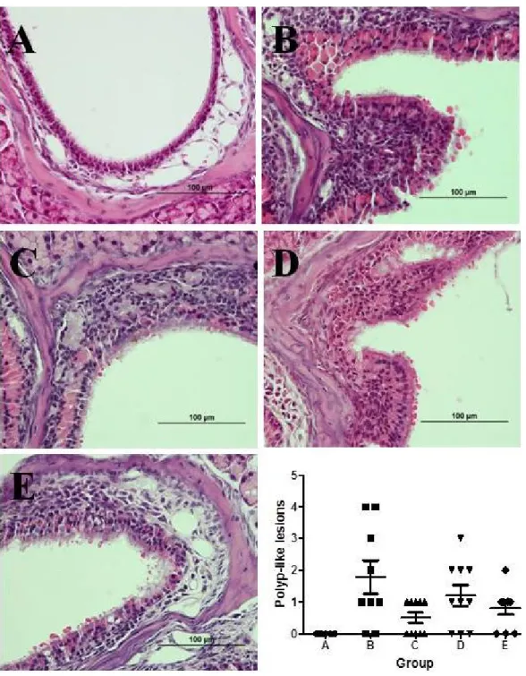
Figure 2. Comparison of the count of polyp-like lesions between groups.
The same experiments were performed twice, and the representative data are depicted. The number of polyp-like lesions was highest in group B, followed by group D. Group C showed lowest number of polyp-like lesions, and group E demonstrated similar outcomes. However, these differences between groups were not statistically significant (Hematoxylin and eosin stain, ×400).
36
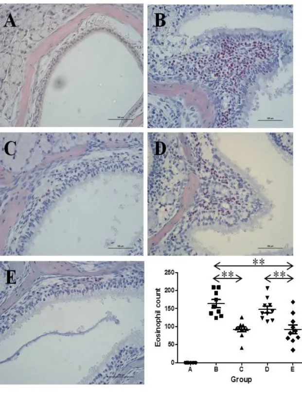
Figure 3. Comparison of the eosinophil count between groups. The same experiments were performed twice, and the representative data are depicted. The number of eosinophils was highest in group B whereas mice in groups A showed no definite infiltration of inflammatory cells in the nasal and sinus mucosa. Both groups C and E demonstrated much lower
37
number of eosinophils than group B. The degree of eosinophilic inflammation was similar between group C and E (**P < 0.01, Sirius red stain, ×400).
38
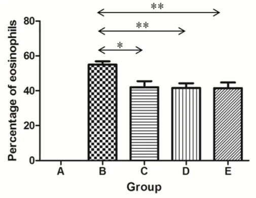
Figure 4. Differences in the proportion of eosinophils in total inflammatory cells between groups. Eosinophils were not found in group A. The percentage of eosinophils in total inflammatory cells was significantly diminished in groups C, D, and E compared with that in group B (*P <
0.05, **P < 0.01).
39
Figure 5. Comparison of the mast cell count between groups. The same experiments were performed twice, and the representative data are depicted. In group A, mast cells were rarely distributed in the sinus
mucosa. The degree of mast cell infiltration seemed less severe in group C, yet there was no significant difference between experimental groups (Giemsa stain, ×400).
40
Figure 6. Comparison of the secretory cell count between groups. The same experiments were performed twice, and the representative data are depicted. A limited number of secretory cells were found in group A. Their number was largest in group B, and smaller in groups C, D, and E compared to that in group B. However, the difference was statistically insignificant (Alcian blue stain, ×400).
41
Figure 7. Comparison of the subepithelial thickness between groups.
The same experiments were performed twice, and the representative data are depicted. The subepithelial layer was considerably thick in group B compared to that in group A. It was apparently thinner in groups D and E than in group B. Similarly, group C demonstrated thinner subepithelial layer than group B, but the difference was not statistically significant (*P <
0.05, Masson’s trichrome stain, ×400).
42
Figure 8. Representative microphotographs of immunohistochemistry and quantitative analysis of positive cells for cyclooxygenase-2. In group A, no definite positive signals were observed for COX-2 in the nasal mucosa. They were also scarcely noted in experimental groups B - E. The number of positive signals appeared to be smaller in groups C and E than
43
in groups B and D, but there was no statistically significant difference between groups.
44
Figure 9. Representative microphotographs of immunohistochemistry and quantitative analysis of positive cells for 5-lipooxygenase. No definite positive signals were observed in the nasal mucosa in group A. However, there was strong expression of 5-LO in group B. The degree of positive signals was apparently reduced in groups C, D, and E compared with that in group B. There was no definite difference in 5-LO expression among
45 groups C, D, and E (*P < 0.05, ×400).