pISSN: 1011-8942 eISSN: 2092-9382
© 2013 The Korean Ophthalmological Society
This is an Open Access article distributed under the terms of the Creative Commons Attribution Non-Commercial License (http://creativecommons.org/licenses /by-nc/3.0/) which permits unrestricted non-commercial use, distribution, and reproduction in any medium, provided the original work is properly cited.
Original Article
In Vitro Effects of Preservative-free and Preserved Prostaglandin Analogs on Primary Cultured Human Conjunctival Fibroblast Cells
Eun Joo Kim1, Yeoun-Hee Kim1, Sun-Hee Kang2, Kyoo Won Lee1, Young Jeung Park1
1Cheil Eye Reserch Institute, Cheil Eye Hospital, Daegu, Korea
2Developmental Biology Laboratory, Department of Biology, College of Natural Sciences, Kyungpook National University, Daegu, Korea
Purpose: Long-term use of topical medication is needed for glaucoma treatment. One of the most commonly pre- scribed classes of hypotensive agents are prostaglandin analogs (PGs) used as both first-line monotherapy; as well as in combination therapy with other hypotensive agents. Several side effects of eye drops can be caused by preservatives. The purpose of this study was to evaluate the effects of PGs with varying concentrations of benzal- konium chloride (BAC), alternative preservatives, or no preservatives on human conjunctival fibroblast cells.
Methods: Primary human conjunctival fibroblast cells were used in these experiments. Cells were exposed to the fol- lowing drugs: BAC at different concentrations, bimatoprost 0.01% (with BAC 0.02%), latanoprost 0.005% (with BAC 0.02%), tafluprost 0.0015% with/without 0.001% BAC and travoprost 0.004% (with 0.001% Polyquad) for 15 and 30 minutes. Cell cytotoxicity was evaluated by phase-contrast microscopy to monitor morphological changes of cells, Counting Kit-8 (CCK-8) assay to cell viability, and fluorescent activated cell sorting (FACS) analysis to measure apoptosis.
Results: BAC caused cell shrinkage and detachment from the plate in a dose-dependent manner. Morphological changes were observed in cells treated with bimatoprost 0.01% and latanoprost 0.005%. However, mild cell shrink- age was noted in cells treated with tafluprost 0.0015%, while a non-toxic effect was noted with travoprost 0.004%
and preservative-free tafluprost 0.0015%. CCK-8 assay and FACS analysis showed all groups had a significantly decreased cell viability and higher apoptosis rate compared with the control group. However, travoprost 0.004%
and preservative-free tafluprost 0.0015% showed lower cytotoxicity and apoptosis rate than other drugs.
Conclusions: This in vitro study revealed that BAC-induced cytotoxicity is dose-dependent, although it is important to emphasize that the clinical significance of toxicity differences observed among the different PGs formulations has not yet been firmly established. Alternatively preserved or preservative-free glaucoma medications seem to be a reasonable and viable alternative to those preserved with BAC.
Key Words: Benzalkonium compounds, Conjunctiva, Pharmaceutical preservatives, Synthetic prostaglandins
Long-term use of topical medication is needed to treat for glaucoma. Chronic use of topical anti-glaucomatic
agents induce side effects, such as ocular inflammation, al- lergy, dry eye syndrome, and failure of filtration surgery [1-9]. The side effects of eye drops are caused by compo- nents such as preservatives. Currently, benzalkonium chlo- ride (BAC) is the most commonly used preservative in ophthalmic preparations. BAC, a quaternary ammonium compound, is a highly effective antimicrobial agent that
Received: March 4, 2013 Accepted: May 28, 2013
Corresponding Author: Young Jeung Park, MD. Cheil Eye Hospital, #1 Ayang-ro, Dong-gu, Daegu 701-820, Korea. Tel: 82-53-959-1751, Fax: 82- 53-959-1758, E-mail: [email protected]
acts by denaturing proteins and disrupting cytoplasmic membranes [10]. In the eyes, BAC turnover is very slow and is retained in ocular tissues up to 48 hours after ad- ministering a single drop [11].
One of the most commonly prescribed classes of hypo- tensive agents are prostaglandin analogs (PGs), which are used as both first-line monotherapy as well as in combina- tion therapy with other hypotensive agents [3]. PGs are su- perior to beta-adrenoceptor antagonists in terms of lower- ing intraocular pressure (IOP), and they have no severe systemic side effects during long-term clinical use [12,13].
New PG formulations without BAC or with alternative preservatives are currently in development. Several studies have revealed that alternative preservatives to BAC, for example sofZia (Alcon, Fort Worth, TX, USA), Purite (a stabilized oxychloro complex), and Polyquad, produce few- er corneal changes and less conjunctival inflammation than BAC [1-3,14,15]. Preservative-free PGs are currently commercially available, and their low toxicity to the corne- al and conjunctival surfaces has been confirmed [4].
The purpose of this study was to evaluate the effects of PGs with varying concentrations of BAC or alternative preservatives on human conjunctival fibroblast cells.
Materials and Methods
Preparation of human tissues and cell culture
Normal human conjunctival tissues were obtained from two patients during cataract surgery. All tissues were re- moved after obtaining informed consent from the donor.
Immediately after excision, the tissue was placed into ei- ther Hank’s balanced salt solution or phosphate buffered saline (PBS) containing a penicillin (5,000 U/mL)/strepto- mycin (5,000 μg/mL) mixture (Gibco BRL, Gaithersburg, MD, USA). Tissues were finely minced into 1-mm3 pieces and placed in six-well culture plates containing 1 mL of Dulbecco’s modified Eagle medium (DMEM)/F12 (1:1 vol/
vol) containing 10% fetal bovine serum (Gibco BRL), ITS (5 mg/mL insulin, 30 nM selenium, 25 mg/mL human transferrin; Sigma, St. Louis, MO, USA), and a penicillin (5,000 U/mL)/streptomycin (5, 000 μg/mL) mixture. Seed- ed tissues were incubated at 37℃ in a humidified air atmo- sphere with 5% CO2 and fed daily. When cells began to form a monolayer, tissue pieces were removed. When con-
junctival fibroblast cultures reached confluence, they were detached from the dishes with trypsin-ethylenediaminete- traacetic acid and replated into new dishes at a ratio of 1:3.
Primary human conjunctival fibroblast cells (passages four and five) were used in these experiments.
Drug treatment
After dividing the cells (6 × 10³ cells/well of a 96-well culture plate, 3 × 10⁵ cells/well of a six-well culture plate), they were allowed to attach for 24 hours, and the media was removed. Cells were washed with D-PBS, and then DMEM:F-12 serum depletion media was replaced with 0.001%, 0.005%, 0.01%, or 0.02% BAC, bimatoprost 0.01%
(Lumigan; Allergan, Irvine, CA, USA), latanoprost 0.005%
(Xalatan; Pfizer, New York, NY, USA), travoprost 0.004%
(Travatan, Alcon), or tafluprost 0.0015% with/without 0.001% BAC (Taflotan/Taflotan-s; Santen, Osaka, Japan).
The cells were exposed to the drugs for 15 or 30 minutes [2,16,17]. The stimuli were removed after incubation. The cells were washed again with D-PBS and then transferred to a serum-free DMEM:F-12 media. Cells were incubated for an additional six hours to stabilize. The control solution was the same volume of PBS added into the cells.
Cell morphology analysis
Treated cells were observed after 30 minutes of drug treatment and upon removing the stimuli. Cells were washed again with PBS and then transferred to serum-free DMEM:F-12 media. Cells were incubated for an additional six hours to stabilize. Finally, the morphological features of the cultured cells in six-well plates were observed by phase-contrast microscopy [18].
Cell viability assay
The Cell Counting Kit-8 (CCK-8) assay was used as a qualitative index of cell viability. The CCK-8 assay was used to measure cytotoxicity under starved conditions, which were based on the conversion of a water-soluble tetrazolium salt, 2-(2-methoxy-4-nitrophenyl)-3-(4-nitro- phenyl)-5-(2,4-disulfophenyl)-2H-tetrazolium, monosodi- um salt (WST-8), to a water-soluble formazan dye upon re- duction by dehydrogenases in the presence of an electron carrier [19,20]. Cells were plated in 96-well microtiter
plates at a density of 6 × 10³ cells per well, then the live cell count was obtained using CCK-8 (Dojindo, Sunnyvale, CA, USA) according to the manufacturer’s protocol. In brief, 10 μL of CCK-8 solution was added to each well, and the samples were incubated for two hours before the absor- bance was measured at 450 nm.
Fluorescent activated cell sorting analysis
Cells (1 × 10⁶) were suspended in 100 μL of PBS, fol- lowed by 200 μL of 95% ethanol while vortexing. Then, the cells were incubated at 4˚C for 1 hour, washed with PBS, and resuspended in 250 μL of 1.12% sodium citrate buffer (pH 8.4) together with 12.5 μg of RNase. Incubation proceeded at 37˚C for 30 minutes. The nuclei were then stained by applying 250 μL of propidium iodide (50 μg/
mL) for 30 minutes at room temperature. Nuclei displaying hypodiploid sub-G1 DNA contents were identified as apop- totic. The stained cells were analyzed by fluorescent acti- vated cell sorting (FACS) on a FACScan flow cytometer (FACScalibur; BectonDickinson Biosciences, San Jose, CA, USA) to determine relative DNA content based on fluores- cence. The sample of each group included with more than 10,000 individual cells [21].
Statistical analysis
Statistical comparisons were performed with SPSS ver.
11.0 (SPSS Inc., Chicago, IL, USA). The cell viability and apoptosis rates were analyzed by Mann-Whitney U-test.
Results
Morphological change of cells
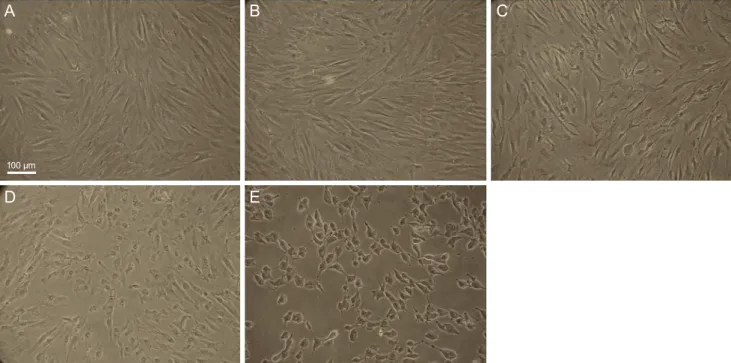
To investigate the cytotoxicity of BAC at different con- centrations in cultured human conjunctival fibroblast cells, we observed morphological changes by phase-contrast mi- croscopy. Cells were treated with BAC at different concen- trations (0.001%, 0.005%, 0.01%, and 0.02%) for 30 min- utes. BAC caused apoptotic characteristics, such as cell shrinkage and detachment from the plate, in a dose-depen- dent manner (Fig. 1).
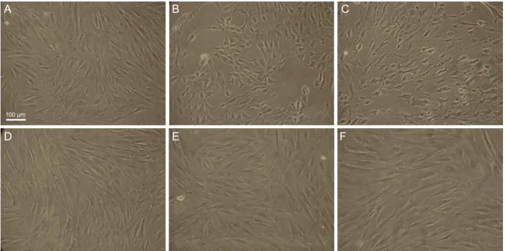
We investigated whether the treatment with different prostaglandin formulations affected the induction of apop- tosis in cultured human conjunctival fibroblast cells. Mor- phological changes, including apoptosis, were observed in cultured cells treated with bimatoprost 0.01% (with BAC 0.02%) and latanoprost 0.005% (with BAC 0.02%). Also, in
Fig. 1. The morphological features of cultured human conjunctival fibroblast cells with different concentrations of benzalkonium chlo- ride (BAC) in six-well plates were observed by phase-contrast microscopy (×100). (A) Control, (B) 0.001% BAC, (C) 0.005% BAC, (D) 0.01%
BAC, and (E) 0.02% BAC. Cell shirnkage increased dose dependently with BAC concentration. Scale bar, 200 μm.
A
D
B
E
C
100 µm
A
D
B
E
C
100 µm
the tafluprost 0.0015% group (with BAC 0.001%), mild cell shrinkage was visible. However, cultured conjunctival fi- broblast cells showed a non-toxic effect to travoprost 0.004% (with 0.001% Polyquad) and preservative-free ta- fluprost 0.0015% treatment, e.g., cell shrinkage and detach- ment from the plate were comparable with those of the control group (Fig. 2).
Cell viability
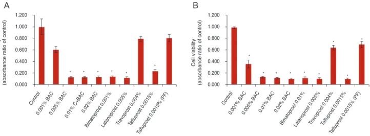
To investigate whether BAC or prostaglandin formula- tions reduce cell viability, the cells were exposed to the drugs for 15 or 30 minutes and then analyzed by CCK-8 assay. The sample absorbance was measured at 450 nm.
CCK-8 assay showed that cell viability of cultured con- junctival fibroblast cells was decreased by treatment with BAC alone or BAC-containing anti-glaucoma drugs. In 15 minutes, the absorbance rates (of control) were 62.8% to 12.8% upon BAC treatment at different concentrations, 13.1% and 11.5% in PGs with BAC (bimatoprost, latano- prost), 22.6% in tafluprost with a small amount of BAC, 78.8% in preservative-free tafluprost, and 78.8% in alterna- tively preserved PG (travoprost with Polyquad). BAC at
different concentrations (0.005% to 0.02%), PGs with BAC (bimatoprost, latanoprost) and tafluprost with a small amount of BAC significantly decreased cell viability com- pared with the control group (p < 0.05). In contrast, 0.001%
BAC, alternatively preserved PG (travoprost with Poly- quad), and preservative-free tafluprost groups had slightly decreased cell viability compared to the control group (Fig.
3A). In 30 minutes, the absorbance rates (of control) were 36.2% to 10.3% upon BAC treatment at different concen- trations, 11.8% and 11.1% in PGs with higher BAC concen- trations (bimatoprost, latanoprost), 10.2% with tafluprost and a small amount of BAC, 70.1% with preservative-free tafluprost, and 64.8% with alternatively preserved PG (travoprost with Polyquad). All groups showed a signifi- cantly decreased cell viability compared with the control group (p < 0.05). However, the preservative-free tafluprost and travoprost with Polyquad groups showed a lower cyto- toxicity than the other drugs (Fig. 3B).
Apoptosis analysis
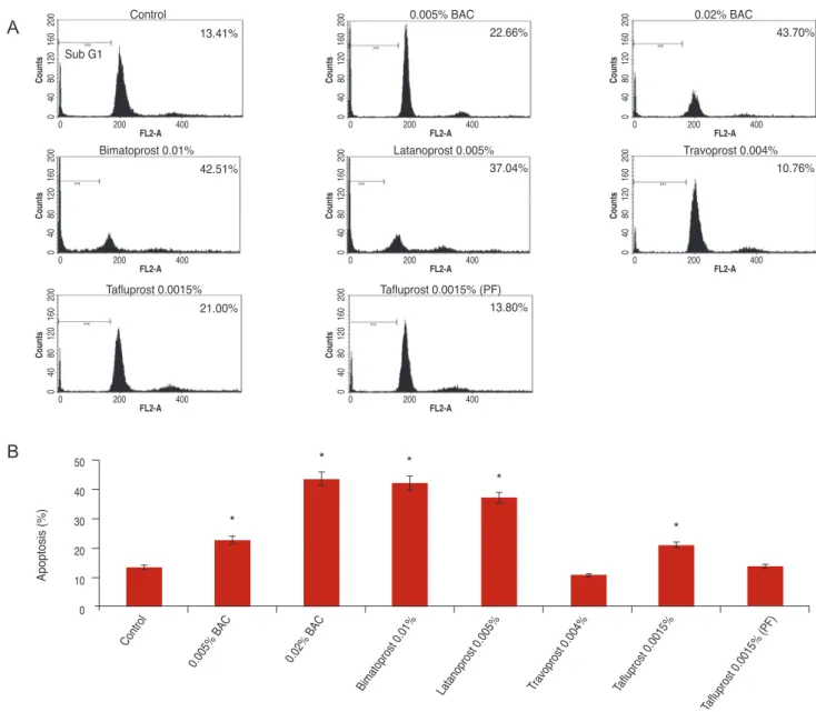
Flow cytometric and quantitative analyses of apoptotic cells were performed after culture for 30 minutes. Apopto-
A
D
B
E
C
F
100 µm
Fig. 2. The morphological features of cultured human conjunctival fibroblast cells with different prostaglandin formulations in six-well plates were observed by phase-contrast microscopy. (A) Control, (B) bimatoprost 0.01% (benzalkonium chloride [BAC] 0.02%), (C) latanoprost 0.005%
(BAC 0.02%), (D) travoprost 0.004% (0.001% Polyquad), (E) tafloprost 0.0015% (BAC 0.001%), and (F) tafluprost 0.0015% (preservative-free).
Compared with the control group, severe cell shrinkage was observed in bimatoprost 0.01% (BAC 0.02%) and latanoprost 0.005% (BAC 0.02%) groups. In the tafluprost 0.0015% group (BAC 0.001%), mild cell shrinkage was visible. In contrast, the travoprost 0.004% (0.001% Polyquad) and preservative-free tafluprost 0.0015% groups showed relatively similar cell morphology to that observed with the control group. Scale bar, 200 μm.
A
D
B
E
C
100 µm
F
sis was analyzed as a sub-G1 fraction by FACS analysis.
The apoptosis rates were 13.4% in control, 22.7% in 0.005%
BAC, 43.7% in 0.02% BAC, 42.5% and 37.0% in PGs with more BAC (bimatoprost, latanoprost), 21.0% in tafluprost with a small amount of BAC, 13.8% in preservative-free tafluprost, and 10.8% in alternatively preserved PG (travo- prost with Polyquad) (Fig. 4A). The groups treated with 0.005% BAC, 0.02% BAC, PGs with more BAC (bimato- prost, latanoprost) and tafluprost with small amount of BAC showed significantly higher apoptosis rates than the control group (p < 0.05). However, preservative-free tafluprost and alternatively preserved PG (travoprost with Polyquad) groups showed similar rates to the control group (Fig. 4B).
Discussion
BAC is the most commonly used preservative for anti- microbial action. Several studies revealed ocular surface toxicity resulting from BAC present in ophthalmic agents [1-11,17,22-28]. Also, in this study, we observed increased cellular shrinkage and apoptosis, and reduced cell viability in BAC-containing agents compared to BAC-free agents.
Upon increasing the concentrations of BAC (0.001%, 0.005%, 0.01% and 0.02%), the cytotoxicity was also in- creased, as noted in other studies (Fig. 1) [3,5,11].
We next determined whether these toxicity were due to the toxicity of BAC or the toxicity of the drug itself. We
investigated a the newly released drug, bimatoprost 0.01%
(with BAC 0.02%), at a low concentration but with a rela- tively high concentration of BAC (with BAC 0.05%) to re- duce the conjunctival hyperemic side effect. The cell via- bility of bimatoprost 0.01% (with BAC 0.02%) was 13.1%
of the control value in 15 minutes and 11.8% of the control value in 30 minutes, while latanoprost 0.005% (with BAC 0.02%) was 11.5% of the control in 15 minutes and 11.1% of the control in 30 minutes. However, that of BAC 0.02%
only was 12.8% of the control in 15 minutes and 10.3% of the control in 30 minutes. So, we can presume there is minimal effect of bimatoprost and latanoprost on conjunc- tival fibroblast cells. In the case of tafluprost 0.0015% (with BAC 0.001%), the cell viability was 22.6% of the control in 15 minutes and 10.2% of the control in 30 minutes, but that of BAC 0.001% showed 62.8% viability of the control in 15 minutes and 36.2% of the control in 30 minutes. As a re- sult, we can presume the relative toxicity of tafluprost.
We also studied preservative-free PGs using tafluprost 0.0015%. The minimum concentration of BAC 0.001% was more cytotoxic than tafluprost 0.0015% (preservative-free) as shown in Fig. 3. Even the small amount of BAC showed cytotoxicity; therefore, considering the long term effects on the ocular surface when using anti-glaucomatic agents and artificial tears, preservative-free agents are of great in- terest.
There is increased interest for alternative preservatives to reduce the cytotoxicity of BAC. Several studies have re-
1.200 1.000 0.800 0.600 0.400 0.200 0.000
Cell viability (absorbance ratio of control) Cell viability (absorbance ratio of control)
Control
0.001% BAC0.005% BAC0.01% C+BAC0.02% BAC
Bimatoprost 0.001%Latanoprost 0.005%Travoprost 0.004%Tafluprost 0.0015%
Tafluprost 0.0015% (PF)
*
* * * *
*
1.200 1.000 0.800 0.600 0.400 0.200 0.000
Control
0.001% BAC0.005% BAC 0.01% BAC 0.02% BAC
Bimatoprost 0.01%Latanoprost 0.005%Travoprost 0.004%Tafluprost 0.0015%
Tafluprost 0.0015% (PF)
*
*
*
* * * *
* *
Fig. 3. Analysis of cell viability by Cell Counting Kit-8 assay. The absorbance was measured at 450 nm. The cells were exposed to the drugs for 15 minutes (A) or 30 minutes (B). *p < 0.05 vs. corresponding value for control by Mann-Whitney U-test. BAC = benzalkonium chloride; PF = preservative-free.
A B
ported that alternative preservatives, such as Sofzia, Purite and Polyquad are less toxic than BAC [1-3,14,17,26,27].
Also, in this study, Fig. 3 shows the viability of cells treat- ed with travoprost 0.004% containing Polyquad was 78.8%
of that of the control in 15 minutes and 64.8% of that of the control in 30 minutes, while that of tafluprost 0.0015%
(preservative-free) was 78.8% of the control in 15 minutes and 70.1% of the control in 30 minutes. The quaternary ammonium compound Polyquad has been used by Alcon as a preservative in artificial tears since 1987 and acts as a surfactant, disrupting bacterial cell membranes and ulti-
mately leading to bacterial cell death. Polyquad is a cation- ic polymer of many quaternary ammonium structures with a 27-fold higher molecular weight than BAK. It lacks a hydrophobic region and, as a result, has no surfactant/de- tergent properties. Due to these two chemical properties, Polyquad is unable to penetrate mammalian cells and cause cytotoxic effects [22]. Ammar et al. [3] reported the cytotoxicity of travoprost 0.004% with BAC, travoprost 0.004% with Polyquad, and travoprost 0.004% with Sof- Zia. The observed relative cytotoxicity values were Poly- quad < SofZia < BAC, and each drug offered more protec-
50 40 30 20 10 0
Control
0.005% BAC 0.02% BAC
Bimatoprost 0.01% Latanoprost 0.005% Travoprost 0.004% Tafluprost 0.0015%
Tafluprost 0.0015% (PF)
*
* *
*
*
Apoptosis (%)
Control
13.41%
Sub G1
04080120200
200
0 400
FL2-A Counts 160
0.005% BAC 22.66%
04080120200
200
0 400
FL2-A Counts 160
0.02% BAC
43.70%
04080120200
200
0 400
FL2-A Counts 160
Bimatoprost 0.01%
42.51%
04080120200
200
0 400
FL2-A Counts 160
Latanoprost 0.005%
37.04%
04080120200
200
0 400
FL2-A Counts 160
Travoprost 0.004%
10.76%
04080120200
200
0 400
FL2-A Counts 160
Tafluprost 0.0015% Tafluprost 0.0015% (PF)
21.00%
04080120200
200
0 400
FL2-A
Counts 160 13.80%
04080120200
200
0 400
FL2-A Counts 160
Fig. 4. Flow cytometric and quantitative analyses of apoptotic cells after culture for 30 minutes. Apoptosis was analyzed as a sub-G1 fraction by fluorescent activated cell sorting analysis (A) and columns (B). *p < 0.05 vs. corresponding value for control by Mann-Whit- ney U-test. BAC = benzalkonium chloride; PF = preservative-free.
A
B
tion to live conjunctival cells than preservatives only, e.g., Travatan 0.004% has a protective effect on cells. We also studied only travoprost 0.004% with Polyquad in the travo- prost 0.004% group, where showed a lower cytotoxicity than other drugs. However, it has the limitation that it was not compared in combination with other preservatives.
The benefits of preservative-free eye drops are already known from several studies, but they also have drawbacks.
The biggest drawback is the inconvenience of use for older people who often suffer from glaucoma and dry eye. Also, preservative-free eye drops are more expensive than preser- vative-containing eye drops. Long-term use of these agents is also a burden. In addition, the action of preservatives for improving corneal penetration is also limited in preserva- tive-free eye drops. Further study is needed regarding alter- native preservatives that have a lower cytotoxicity than BAC.
Obviously, these results obtained via an in vitro model cannot be fully extrapolated to in vivo conditions, due to anti-inflammatory factors, and the diluting action of tears and eyelid blinking. However, our experimental results were in good agreement with data from other studies as- sessing the toxic side effects of preservative induced at the ocular surface [1-11]. Moreover, we observed effects at 15 and 30 minute exposure times, so the results of long-term exposure to these agents cannot be extrapolated.
In conclusion, this in vitro study revealed that BAC-in- duced cytotoxicity is dose-dependent, although it is im- portant to emphasize that the clinical significance of toxic- ity differences observed among the differnet PG formulations has not yet been firmly established. Alternatively preserved or preservative-free glaucoma medications seem to be a reasonable and viable alternative to those preserved with BAC. More studies are needed to better understand how in vitro findings compare to in vivo responses.
Conflict of Interest
No potential conflict of interest relevant to this article was reported.
References
1. Noecker RJ, Herrygers LA, Anwaruddin R. Corneal and conjunctival changes caused by commonly used glaucoma
medications. Cornea 2004;23:490-6.
2. Baudouin C, Riancho L, Warnet JM, Brignole F. In vitro stud- ies of antiglaucomatous prostaglandin analogues: travoprost with and without benzalkonium chloride and preserved lata- noprost. Invest Ophthalmol Vis Sci 2007;48:4123-8.
3. Ammar DA, Noecker RJ, Kahook MY. Effects of benzal- konium chloride-preserved, polyquad-preserved, and sof- Zia-preserved topical glaucoma medications on human oc- ular epithelial cells. Adv Ther 2010;27:837-45.
4. Liang H, Baudouin C, Pauly A, Brignole-Baudouin F. Con- junctival and corneal reactions in rabbits following short- and repeated exposure to preservative-free tafluprost, com- mercially available latanoprost and 0.02% benzalkonium chloride. Br J Ophthalmol 2008;92:1275-82.
5. Brasnu E, Brignole-Baudouin F, Riancho L, et al. In vitro effects of preservative-free tafluprost and preserved latano- prost, travoprost, and bimatoprost in a conjunctival epithe- lial cell line. Curr Eye Res 2008;33:303-12.
6. Guenoun JM, Baudouin C, Rat P, et al. In vitro study of in- flammatory potential and toxicity profile of latanoprost, travoprost, and bimatoprost in conjunctiva-derived epithe- lial cells. Invest Ophthalmol Vis Sci 2005;46:2444-50.
7. Baudouin C. Allergic reaction to topical eyedrops. Curr Opin Allergy Clin Immunol 2005;5:459-63.
8. Broadway DC, Grierson I, O’Brien C, Hitchings RA. Ad- verse effects of topical antiglaucoma medication. II. The outcome of filtration surgery. Arch Ophthalmol 1994;112:
1446-54.
9. Lee JK, Ryu YH. The effect of antiglaucoma medication on cultured human conjunctival epithelial cells. J Korean Ophthalmol Soc 2006;47:1811-8.
10. Noecker R. Effects of common ophthalmic preservatives on ocular health. Adv Ther 2001;18:205-15.
11. De Saint Jean M, Brignole F, Bringuier AF, et al. Effects of benzalkonium chloride on growth and survival of Chang conjunctival cells. Invest Ophthalmol Vis Sci 1999;40:619- 30.
12. Sharif NA, Kelly CR, Crider JY, et al. Ocular hypotensive FP prostaglandin (PG) analogs: PG receptor subtype bind- ing affinities and selectivities, and agonist potencies at FP and other PG receptors in cultured cells. J Ocul Pharmacol Ther 2003;19:501-15.
13. Ota T, Murata H, Sugimoto E, et al. Prostaglandin ana- logues and mouse intraocular pressure: effects of tafluprost, latanoprost, travoprost, and unoprostone, considering 24- hour variation. Invest Ophthalmol Vis Sci 2005;46:2006-11.
14. Kahook MY, Noecker RJ. Comparison of corneal and con- junctival changes after dosing of travoprost preserved with sofZia, latanoprost with 0.02% benzalkonium chloride, and preservative-free artificial tears. Cornea 2008;27:339-43.
15. Horsley MB, Kahook MY. Effects of prostaglandin analog therapy on the ocular surface of glaucoma patients. Clin Ophthalmol 2009;3:291-5.
16. Ammar DA, Noecker RJ, Kahook MY. Effects of benzal- konium chloride- and polyquad-preserved combination glaucoma medications on cultured human ocular surface cells. Adv Ther 2011;28:501-10.
17. Brignole-Baudouin F, Riancho L, Liang H, et al. In vitro comparative toxicology of polyquad-preserved and benzal- konium chloride-preserved travoprost/timolol fixed combi- nation and latanoprost/timolol fixed combination. J Ocul Pharmacol Ther 2011;27:273-80.
18. De Saint Jean M, Debbasch C, Rahmani M, et al. Fas- and interferon gamma-induced apoptosis in Chang conjunctival cells: further investigations. Invest Ophthalmol Vis Sci 2000;41:2531-43.
19. Han SB, Shin YJ, Hyon JY, Wee WR. Cytotoxicity of vori- co nazole on cultured human corneal endothelial cells. Anti- microb Agents Chemother 2011;55:4519-23.
20. Ishiyama M, Tominaga H, Shiga M, et al. A combined as- say of cell viability and in vitro cytotoxicity with a highly water-soluble tetrazolium salt, neutral red and crystal vio- let. Biol Pharm Bull 1996;19:1518-20.
21. Lu TH, Hsieh SY, Yen CC, et al. Involvement of oxidative stress-mediated ERK1/2 and p38 activation regulated mito-
chondria-dependent apoptotic signals in methylmercury-in- duced neuronal cell injury. Toxicol Lett 2011;204:71-80.
22. Tripathi BJ, Tripathi RC, Kolli SP. Cytotoxicity of ophthal- mic preservatives on human corneal epithelium. Lens Eye Toxic Res 1992;9:361-75.
23. Baudouin C, Labbe A, Liang H, et al. Preservatives in eye- drops: the good, the bad and the ugly. Prog Retin Eye Res 2010;29:312-34.
24. Asada H, Takaoka-Shichijo Y, Nakamura M, Kimura A.
Optimization of benzalkonium chloride concentration in 0.0015% tafluprost ophthalmic solution from the points of ocular surface safety and preservative efficacy. Yakugaku Zasshi 2010;130:867-71.
25. Jaenen N, Baudouin C, Pouliquen P, et al. Ocular symp- toms and signs with preserved and preservative-free glau- coma medications. Eur J Ophthalmol 2007;17:341-9.
26. Nakagawa S, Usui T, Yokoo S, et al. Toxicity evaluation of antiglaucoma drugs using stratified human cultivated corneal epithelial sheets. Invest Ophthalmol Vis Sci 2012;53:5154-60.
27. Liang H, Baudouin C, Labbe A, et al. Conjunctiva-associ- ated lymphoid tissue (CALT) reactions to antiglaucoma prostaglandins with or without BAK-preservative in rabbit acute toxicity study. PLoS One 2012;7:e33913.
28. Liang H, Brignole-Baudouin F, Riancho L, Baudouin C.
Reduced in vivo ocular surface toxicity with polyquad-pre- served travoprost versus benzalkonium-preserved travo- prost or latanoprost ophthalmic solutions. Ophthalmic Res 2012;48:89-101.