WWW.KJOG.ORG 639
GASTROINTESTINAL STROMAL TUMOR IN VAGINA:
A CASE REPORT
Seung Yeon Ha, MD 1 , Ki Beom Lee, MD 2 , Soon Pyo Lee, MD 2
Departments of
1Pathology,
2Obstetrics and Gynecology, Gachon University Gil Hospital, Incheon, Korea
Gastrointestinal stromal tumor (GIST) is a mesenchymal tumor that usually arises within the gastrointestinal tract. However extragastrointestinal stromal tumor (EGIST) is a rare tumor (5%), especially few cases of EGIST arising in vagina have currently been reported in the literature. We have experienced a case of vaginal GIST. A 49-year-old female presented with vaginal mass for three weeks. The mass was located posterolateral vagina near to introitus and 3.0 × 2.5 cm in size. Histological examination showed hypercelluar spindle cell proliferation and more than 10 mitoses per 50 high power fi elds. They were positive for c-kit (CD 117) and CD34. Surgery is the most effective treatment and long term follow up is warranted.
Keywords: Gastrointestinal stromal tumor; Vagina
Received: 2011. 5.31. Accepted: 2011. 7.29.
Corresponding author: Soon Pyo Lee, MD
Department of Obstetrics and Gynecology, Gachon University Gil Hospital, 1198 Guwol-dong, Namdong-gu, Incheon 405-760, Korea Tel: +82-32-460-3078 Fax: +82-32-460-3073
E-mail: [email protected]
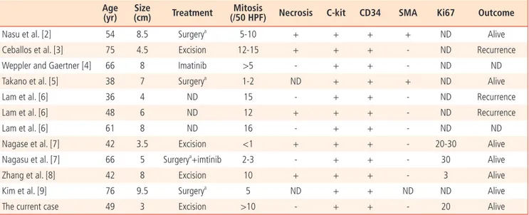
위장관기질종양은 주로 위장관에 발생하는 중간엽 종양으로 60-70%는 위, 20-30%는 소장, 그리고 10%는 식도와 대장에 발생한다[1]. 위 장관외에 발생하는 경우는 전체 위장관기질종양의 5-7%로 드물고 대 부분 장간막, 그물막, 후복막강에 발생한다.질에 발생한 경우는 매우 드 물며 전세계적으로 질-직장 사이벽에 발생한 것을 포함하여 8개의 논 문에 11예가 보고되어 있으며 국내 논문에는 보고된 바가 없다(Table 1). 대부분 질의 섬유종이나 평활근종으로 오인하기 쉬워 감별을 요하는 질환으로 문헌 고찰과 함께 이에 보고하는 바이다.
증 례
환 자: 김 O 자 나 이: 49세
주 소: 질내에 종괴 촉진 산과력: 2-0-1-1
월경력: 초경은 15세에 있었고 48세에 폐경되었다.
과거력: 특이사항 없었다.
가족력: 할머니는 위암이었고 아버지 간경화이었다.
현병력: 내원 3주전부터 잔변감이 있었으며 내진 중 질 후벽쪽으로 이 물감 만져져서 1차 병원 경유하여 수술위해 내원하였다.
이학적 소견: 혈압, 맥박, 체온은 정상이었으며 외부 생식기는 특이 소 견 없었다. 내진 시 질 입구 가까운 부위의 우측 후벽 쪽에 약 3.0 × 2.5 cm의 단단하고 비교적 경계가 잘 지워지는 종괴가 촉지되었다.
검사소견: 혈액 검사에서 혈색소 14.0 g/dL으로 정상이었으며, 백혈구 4,310/mm3, 혈소판 244,000/mm3이었다. 요검사, 간기능검사, 혈액응
CASE REPORT
Korean J Obstet Gynecol 2011;54(10):639-642 http://dx.doi.org/10.5468/KJOG.2011.54.10.639 pISSN 2233-5188 · eISSN 2233-5196
고검사, 심전도 및 단순 흉부 촬영 등은 모두 특이 소견을 보이지 않았 다. 종양표지자검사에서 carcino-embryonic antigen와 CA 19-9는 정 상 소견이었다. 자궁경부세포검사도 정상이었다.
질식초음파 소견: 질의 외후벽에 약 3.0
×
2.5 cm의 둥근 종괴가 관찰되 었고 혈류량은 증가되어 있었다. 자궁의 크기는 7.2×
4.6×
3.6 cm이었 으며 약 1.2×
0.6 cm의 자궁근종이 발견되었다. 양쪽 부속기의 이상 소 견은 없었다(Fig. 1)수술 소견: 종괴는 주변 조직의 침윤은 없었으며 잘 분리되었고 절제술 을 시행하였다.
병리조직학적 소견:
1. 육안소견: 절제한 종괴는 약 3.0 × 2.5 cm이었고 단단하였다. 단면 은 균일하였고 출혈이나 괴사는 관찰되지 않았다. 경계는 비교적 잘 지워졌다.
2. 현미경 소견: 길쭉한 모양의 방추형세포로 이루어져 있었으며 부
Th is is an Open Access article distributed under the terms of the Creative Commons Attribution Non-Commercial License (http://creativecommons.org/licenses/
by-nc/3.0/) which permits unrestricted non-commercial use, distribution, and reproduction in any medium, provided the original work is properly cited.
Copyright © 2011. Korean Society of Obstetrics and Gynecology
WWW.KJOG.ORG 640
KJOG Vol. 54, No. 10, 2011
분적으로 소용돌이 치는 양상이었다. 세포밀도는 높았고 괴사나 출혈은 관찰되지 않았다. 유사 분열수는 50군데의 고배율 시야당(50 HPFs) 10 개 이상이었다. 면역조직화학염색에서 종양세포는 CD34과 c-kit에 양 성이었으며, alpha-smooth muscle actin과 desmin에 음성이었고 Ki- 67 지수는 5%이었다(Fig. 2).
임상적 경과: 수술 후 3개월과 6개월 후에 찍은 골반
computed tomography 방사선검사에서 특이 소견은 관찰되지 않았다. 현재 1년 6
개월 추적 관찰 중이며 재발이나 다른 종괴는 발견되지 않았다.고 찰
위장관기질종양은 중간엽에서 발생하는 종양으로 Cajal 기질세포 혹
은 그 전구세포로부터 기원하는 것으로 예측되고 있다. 발생기전은 약 85%에서 KIT 유전자의 돌연변이이며, c-kit는 줄기세포인자(stem cell factor)의 수용체로서 tyrosine kinase의 기능을 가지고 있고 돌연변이 에 의해 활성화되면 신호전달계가 시작되어 세포의 증식이 일어난다.
KIT 유전자의 돌연변이 중 exon 11의 돌연변이는 70%, exon 9는 15%, exon 13과 exon 17은 각각 5% 미만에서 발견된다[10].
임상양상은 위장관의 경우 주로 남자에서 발생되며 50세 이상에서 호 발한다. 위에서 60-70%, 소장에서 20-30%, 그리고 식도와 대장에서 10%가 발생하므로 위장관외에 발생하는 경우는 드물다[1].
위장관외에
발생하는 경우 중 특히 질에서 관찰된 예는 매우 드물게 발견되며, 현재 까지 찾을 수 있었던 보고된 예는 총 11예 이었으며 1예가 외음질 중격 에 있었고 나머지는 모두 질의 후벽쪽인 직장질중격에 위치하였다. 종 괴로 인한 통증은 아무도 호소하지 않았으며 직장으로의 침윤은 관찰되 지 않았다.진단은 조직학적 소견과 면역조직화학검사 결과에 의하여 확진할 수 있다. 조직학적으로 위장관기질종양은 두 가지 종류가 있으며 길쭉한 모양의 방추형세포와 통통한 세포로 이루어진 상피형 세포로 나누어 지 며 두 가지의 세포가 혼합되어 관찰되는 경우도 있다. 이 종양 세포는 c-kit, CD34, PKC-theca, DOG-1이 발현되는 특징이 있어서 진단에 유 용하게 이용된다. 위장관기질종양 종양의 악성도는 저위험군과 고위험 군으로 나눌 수 있는데 유사분열상의 수와 종괴의 크기에 따라 구분된 다. 즉 유사분열수가 50개 고배율 시야당(50 HPFs) 5개 이상의 경우, 종 괴의 크기가 5 cm 이상인 경우에 악성의 경과를 보일 가능성이 높다.
본 예는 크기는 3 cm로 작고 현미경 소견에서 괴사는 관찰되지 않으나 세포밀도가 높고 유사분열이 50군데의 고배율 시야당(high power field) 10개 이상 관찰되므로 악성으로 진단할 수 있다[11].
통상적으로 질의 기질에 종양이 발생할 경우 위장관기질종양 보다는
Table 1. Review of clinicopathologic characteristics for gastrointestinal tumor in vagina and rectovaginal septum including current case
Age (yr)
Size
(cm) Treatment Mitosis
(/50 HPF) Necrosis C-kit CD34 SMA Ki67 Outcome
Nasu et al. [2] 54 8.5 Surgery
a5-10 + + + + ND Alive
Ceballos et al. [3] 75 4.5 Excision 12-15 + + + - ND Recurrence
Weppler and Gaertner [4] 66 8 Imatinib >5 - + + - ND ND
Takano et al. [5] 38 7 Surgery
a1-2 ND + + + ND Alive
Lam et al. [6] 36 4 ND 15 - + + - ND Recurrence
Lam et al. [6] 48 6 ND 12 + + + - ND Recurrence
Lam et al. [6] 61 8 ND 16 - + + - ND ND
Nagase et al. [7] 42 3.5 Excision <1 + + + - 20-30 Alive
Nagasu et al. [7] 66 5 Surgery
a+imtinib 2-3 - + + - 30 Alive
Zhang et al. [8] 42 8 Excision 10 + + + - 3 Alive
Kim et al. [9] 76 9.5 Surgery
a5 ND + + ND ND Alive
The current case 49 3 Excision >10 - + + - 20 Alive
ND, not described.
a
Surgery: resection of rectum with total hysterectomy and excision of posterior vagina.
Fig. 1. Transvaginal ultrasonography shows a 3.0 × 2.5 cm sized solid
mass in the posterolateral vagina with increased vascularity.
WWW.KJOG.ORG 641 Seung Yeon Ha, et al. Gastrointestinal stromal tumor in vagina: A case report
평활근종, 평활근육종, 슈반세포종, 섬유종 등을 먼저 생각하게 된다. 감 별진단은 c-kit과 CD34 면역조직화학염색이 도움이 된다. 평활근종과 평활근육종은 호산성의 세포질을 가진 방추형세포의 증식은 유사하나 c-kit과 CD34에 음성이며 smooth muscle actin과 desmin 에 양성이므 로 감별할 수 있다. 평활근육종의 경우는 크기가 5 cm 이상이 많으므 로 크기가 작을 때는 하나의 감별 점이 될 수 있을 것이라 생각된다. 슈 반세포종은 s-100 단백에 양성이며 역시 c-kit과 CD34에 음성이다. 그 러나 소장의 위장관기질종양 중의 2%, 위의 5%는 c-kit 음성으로 보고 되고 있다. 또 c-kit이 전체의 종양에 고르게 양성 반응을 보이지 않으므 로 작은 생검일 경우 음성으로 진단 될 수 있으므로 주의 해야 한다[12].
위의 사실을 고려하여 볼 때 질 생검의 경우는 진단에 제한 점이 있을 수 있다. 확진을 위해서는 종괴 절제가 필요할 것으로 생각된다. 조직학 적 소견상 위장관기질종양이 의심 되지만 c-kit에 음성이면, PDGFRA 돌연변이 분자검사를 하면 진단하는데 도움이 된다.
위장관기질종양의 예후에 대한 연구는 아직 정립되어 있지 않다[13].
소장에 발생한 위장관기질은 위에서 발생한 것 보다 예후가 더 좋지 않 다. 위장관기질종양은 재발률이 높기 때문에 보조적인 치료가 더 필요 하나 아직 문헌상 위장관기질외종양에 대한 항암치료와 방사선 치료의 유용성에 대한 문헌은 잘 되어있지 않다[8].하지만 재발된 위장관기질 종양에서는 발생기전에 언급하였듯이 티로신키나제 활성을 특이적으로 억제하는 imatinib으로 치료제가 개발됨으로써 환자의 5년 생존율이 높 아지고 있으며 심지어 전이를 한 경우에도 효과가 좋은 것으로 되어 있 다[13].약제에 대한 부분 반응률과 질병 안정화율은 각각 45%와 32%
이다[14].하지만 이차적인 KIT 유전자의 돌연변이가 생기면 imatinib 치 료에 저항을 보이므로 참고하여야 할 것으로 생각된다[10].
References
1. Miettinen M, Sarlomo-Rikala M, Lasota J. Gastrointestinal stromal tumors: recent advances in understanding of their biology. Hum Pathol 1999;30:1213-20.
2. Nasu K, Ueda T, Kai S, Anai H, Kimura Y, Yokoyama S, et al.
Gastrointestinal stromal tumor arising in the rectovaginal sep- tum. Int J Gynecol Cancer 2004;14:373-7.
3. Ceballos KM, Francis JA, Mazurka JL. Gastrointestinal stromal tumor presenting as a recurrent vaginal mass. Arch Pathol Lab Med 2004;128:1442-4.
4. Weppler EH, Gaertner EM. Malignant extragastrointestinal stromal tumor presenting as a vaginal mass: report of an unusual case with literature review. Int J Gynecol Cancer 2005;15:1169-72.
5. Takano M, Saito K, Kita T, Furuya K, Aida S, Kikuchi Y. Preop- erative needle biopsy and immunohistochemical analysis for gastrointestinal stromal tumor of the rectum mimicking vagi- nal leiomyoma. Int J Gynecol Cancer 2006;16:927-30.
6. Lam MM, Corless CL, Goldblum JR, Heinrich MC, Downs-Kelly E, Rubin BP. Extragastrointestinal stromal tumors presenting as vulvovaginal/rectovaginal septal masses: a diagnostic pit- fall. Int J Gynecol Pathol 2006;25:288-92.
7. Nagase S, Mikami Y, Moriya T, Niikura H, Yoshinaga K, Takano T, et al. Vaginal tumors with histologic and immunocytochemi- cal feature of gastrointestinal stromal tumor: two cases and review of the literature. Int J Gynecol Cancer 2007;17:928-33.
8. Zhang W, Peng Z, Xu L. Extragastrointestinal stromal tumor arising in the rectovaginal septum: report of an unusual case with literature review. Gynecol Oncol 2009;113:399-401.
9. Kim YJ, Jeong YY, Kim SM. Extragastrointestinal stromal tumor arising from the vagina: MR findings. Eur Radiol 2006;16:1860-1.
10. Korean Society of Pathologists. Textbook of pathology. 7th ed.
Seoul: Komoonsa; 2010.
11. Dow N, Giblen G, Sobin LH, Miettinen M. Gastrointestinal stromal tumors: differential diagnosis. Semin Diagn Pathol 2006;23:111-9.
12. Medeiros F, Corless CL, Duensing A, Hornick JL, Oliveira AM, Fig. 2. There are bundles of hypercellular spindle cells with frequent mitosis (A: H&E, ×200). The tumor cells are positive for c-kit (B: c-kit, ×200) and CD34 (C: CD34, × 200).
A B C
WWW.KJOG.ORG 642
KJOG Vol. 54, No. 10, 2011
Heinrich MC, et al. KIT-negative gastrointestinal stromal tu- mors: proof of concept and therapeutic implications. Am J Surg Pathol 2004;28:889-94.
13. Chou SQ, Tse KS, Wong WK, Chan SC. Vulval gastrointestinal stromal tumors with bone metastasis. J Hong Kong Col Radiol
2010;13:88-90.
14. Verweij J, Casali PG, Zalcberg J, LeCesne A, Reichardt P, Blay JY, et al. Progression-free survival in gastrointestinal stromal tumours with high-dose imatinib: randomised trial. Lancet 2004;364:1127-34.
자궁 질에 발생한 위장관기질종양 1예
가천의대 길병원 1병리과, 2산부인과 하승연1, 이기범2, 이순표2
위장관기질종양은 주로 위장관 벽에 발생하는 종양으로 위장관외에 발생한 예는 위장관기질종양의 약 5%에 불과하며 특히 질에 보고 된 예는 매우 희귀하다. 49세 여자환자가 수 주간 촉지된 질종양을 주소로 내원하였다. 종괴는 질 외후벽에 위치하고 있었으며 크기는 3.0 × 3.0 cm이었다. 조직학적으로 고밀도의 방추형세포로 구성되어 있었으며 유사 분열수는 50군데의 고배율(10 HPF) 시야당 10개 이 상이었다. 이 종양세포는 c-kit (CD117)과 CD34에 양성이었다. 치료는 수술이 제일 좋은 방법으로 되어 있으며 장기간 추척 관찰을 요한 다. 저자들은 드물지만 질의 기질 종양 중 감별을 요하는 위장관기질종양을 경험하여 이를 문헌 고찰과 함께 보고하는 바이다.
중심단어: 위장관기질종양, 질