Received: February 18, 2019, Revised: February 25, 2019, Accepted: February 27, 2019 eISSN 2233-7679
†Correspondence to: Soon-Jeong Jeong, https://orcid.org/0000-0002-8959-4663
Department of Dental Hygiene, College of Health Science, Youngsan University, 288 Junam-ro, Yangsan 50510, Korea Tel: +82-55-380-9453, Fax: +82-55-380-9305, E-mail: [email protected]
Copyright © The Korean Society of Dental Hygiene Science.
Biological Effects of Light-Emitting Diodes Curing Unit on MDPC-23 Cells and Lipopolysaccharide
Stimulated MDPC-23 Cells
Moon-Jin Jeong
1and Soon-Jeong Jeong
2,†1
Department of Oral Histology and Developmental Biology, School of Dentistry, Chosun University, Gwangju 61452,
2
Department of Dental Hygiene, College of Health Science, Youngsan University, Yangsan 50510, Korea
Background: Light-emitting diodes curing unit (LCU), which emit blue light, is used for polymerization of composite resins in many dentistry.
Although the use of LCU for light-cured composite resin polymerization is considered safe, it is still controversial whether it can directly or indirectly have harmful biological influences on oral tissues. The aim of this study was to elucidate the biological effects of LCU in wavelengths ranging from 440 to 490 nm, on the cell viability and secretion of inflammatory cytokines in MDPC-23 odontoblastic cells and inflammatory-induced MDPC-23 cells by lipopolysaccharide (LPS).
Methods: The MTT assay and observation using microscope were performed on MDPC-23 cells to investigate the cell viability and cytotoxic effects
on LCU irradiation.
Results:
MDPC-23 cells and LPS stimulated MDPC-23 cells were found to have no effects on cell viability and cell morphology in the LCU irradiation.
Nitric oxide (NO) and prostaglandin E
2which are the pro-inflammatory mediators, and interleukin-1 and tumor necrosis factor- (TNF-) which are the proinflammatory cytokines were significantly increased in MCPD-23 cells after LCU irradiation as time increased in comparison with the control. LCU irradiation has the potential to induce inflammation or biological damages in normal dental tissues, including MDPC-23 cells.
Conclusion:
Therefore, it is necessary to limit the use of LCU except for the appropriate dose and irradiation time. In addition, LCU irradiation of inflammatory-induced MDPC-23 cells by LPS was reduced the secretion of NO compared to the LPS alone treatment group and was significantly reduced the secretion of TNF- in all the time groups. Therefore, LCU application in LPS stimulated MDPC-23 odontoblastic cells has a photodynamic therapy like effect as well as inflammation relief.
Key Words: Inflammation, Light-emitting diodes curing unit, Lipopolysaccharide, MDPC-23 cell, Photodynamic therapy
Introduction
The tooth-colored composite resin used for restoration has been widely used since its introduction in the dental field in the 1970s due to the increasing problems of dental amalgam and demand for esthetic restoration of patients
1). In addition, the use of tooth-colored composite resin in posterior restorations as well as anterior restorations has increased and it is also used for the bonding of stainless- steel brackets and ceramic brackets in orthodontic treatment
2,3). In aesthetic dental restorations, the tooth-
colored composite resin is polymerized by the energy of
the laser produced by light curing units
4). Activated tooth-
colored composite resins that do not receive sufficient
energy by the light curing units break down along the
restored margins, weaken the bond strength with the tooth,
and lack hardness
5). Therefore, the light curing units have
been developed with the rapid increase of the aesthetic
dental restorative technique using the light-cured composite
resins
6). There are three types of light curing units that are
mainly used in dentistry for light-cured composite resins
polymerization, and these three types of LCU have
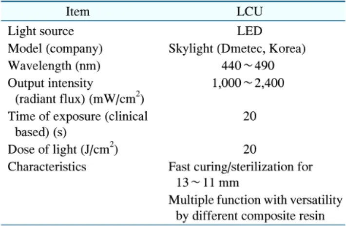
Table 1. Information and Characteristics of LED Curing Unit (LCU)
Item LCU
Light source LED
Model (company) Skylight (Dmetec, Korea)
Wavelength (nm) 440∼490
Output intensity (radiant flux) (mW/cm
2)
1,000∼2,400
Time of exposure (clinical based) (s)
20
Dose of light (J/cm
2) 20
Characteristics Fast curing/sterilization for 13∼11 mm
Multiple function with versatility by different composite resin LED: light-emitting diodes.
different light sources, namely, halogen curing units (HCU), light-emitting-diode curing units (LCU) and plasma arc curing units (PCU)
4). HCU has disadvantages in that only a small amount of the total energy is used for the polymerization of the light-cured composite resins and most of the energy generates heat, resulting in poor energy efficiency and long timings for polymerization
4). LCU and PCU have been developed to complement the shortcomings of HCU and to reduce the clinical chair time used for light curing time
5,6). PCU has a very high energy intensity of more than 2,000 mW/cm
2, which enables polymerization of the light-cured composite resins in a short time, but the rigidity of the composite resin during polymerization is significantly low, shrinkage is large, and it is very expensive
7). Light-emitting diodes (LEDs), introduced in 1990s, consist of a combination of two semiconductors and are an effective method of converting electrical energy into light or laser
6). The LCU, that uses LEDs as light source, generates up to 1,100 mW/cm
2energy at a wavelength of 402∼600 nm and has a semi-permanent, low price, and provide a strong vibration and shock resistance
6). Therefore, LCU, which is seen as blue light, is used for polymerization of composite resins in many dentistry
8). Although the use of light curing unit for polymerization of composite resin is considered safe, its safety is controversial whether it can directly or indirectly have harmful biological influences on oral tissues, including dental and non-dental tissues
9,10). Studies on the biological effects of light irradiation of LCUs using oral cell-derived cell lines are not sufficient
6). MDPC-23 cells are odontoblastic cells derived from dental papilla of fetal mouse molars. Odontoblast is a representative dental tissue cell that produces most of the extracellular matrix of dentin and is involved in mineralization of dentin
11). Odontoblast is directly influenced by LCU irradiation during resin polymerization in dental treatment when exposed to dental pulp and deep cavities
4).
We applied a LCU of blue light with distinct wavelengths ranging from 440 to 490 nm, which is widely used for polymerization of light-cured composite resins in dental clinics, for the most commonly used 20 seconds in other to evaluate biological effects on the cell viability and secretion of inflammatory cytokines in MDPC-23 odontoblastic
cells and inflammatory-induced MDPC-23 cells by lipopolysaccharide (LPS).
Materials and Methods
1. Cell culture and LCU irradiation
MDPC-23 cells were obtained from Dr. CT Hanks of University of Michigan (Ann Arbor, MI, USA). Cells were cultured in Dulbecco’s modified Eagle’s medium (DMEM; Gibco-BRL, New York, NY, USA) supplemented with a 1% antibiotic-antimycotic solution (Gibco-BRL) and 10% fetal bovine serum (Gibco-BRL). Cells were maintained in a humidified CO
2incubator at 37
oC.
LCU is a Dmetec Skylight product (Dmetec, Bucheon, Korea) with a wavelength of 440∼490 nm, which is widely used in dental clinics (Table 1). Cells were irradiated with LCU under the same conditions as when polymerizing a resin using LCU in dental clinic and the LCU held 2 cm away from the cells during exposure. The cells were divided into four groups as follows: the control, LPS alone treatment (100 ng/ml), LCU alone irradiation, and co- treatment group of LPS and LCU.
2. MTT assay
The cell viability and cytotoxic effects were assessed
using a 3-(4,5-dimethylthiazol-2-yl)-2,5-diphenyltetrazo-
lium bromide (MTT) assay. The MDPC-23 cells were
seeded in 48-well plates and treated with 100 ng/ml LPS,
irradiated with LCU for 20 seconds, or co-treated with
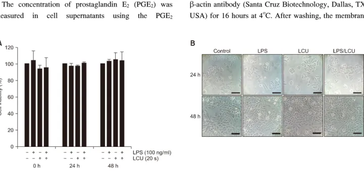
Fig. 1. Effects of light-emitting diodes curing unit (LCU) irradiation on cell viability and morphology of MDPC-23 cells and lip- opolysaccharide (LPS) stimulated MDPC-23 cells. The cells were irradiated for 20 seconds using LCU with wavelengths of 440 to 490 nm and LPS was treated with 100 ng/ml. (A) The cell viability on LCU irradiation was assessed with MTT assay. The results obtained from three independent experiments were expressed as a ratio of values in the surviving cell to the values in the control group (no treatment group of LPS and LCU), and the values were expressed as mean±standard deviation. (B) Morphological changes of MDPC-23 cells by the LPS alone treatment, the LCU alone irradiation and co-treatment group of LPS and LCU were observed by inverted phase contrast microscope. Scale bars=60 m.
LPS and LCU and then incubated for 0, 24, and 48 hours.
3. Microscopic observation
The culture medium was removed from the cells that were then washed three times with PBS and then fixed using a solution containing 3.7% formaldehyde (Sigma Chemical, St. Louis, MO, USA). The images of cells were obtained using an inverted phase contrast microscope (Carl Zeiss, Oberkochen, Germany).
4. NO production
Nitric oxide (NO) production was measured in cell supernatants of LPS alone treated, LCU alone irradiated, and LPS and LCU co-treated cell using the NO assay kit (R&D systems, Minneapolis, MN, USA). A 2-fold diluted culture supernatant was transferred to 96-well plate, and an equal amount of Griess reagent was added. The plate was then incubated for 10 minutes, and the absorbance was measured at 540 nm on a microplate reader (BioTek Instruments, Winooski, VT, USA). The amount of NO was calculated using sodium nitrite standard curve.
5. PGE
2ELISA
The concentration of prostaglandin E
2(PGE
2) was measured in cell supernatants using the PGE
2enzyme-linked immunosorbent assay (ELISA) kit (R&D systems). The absorbance was recorded at 490 nm using a microplate reader (BioTek Instruments). The PGE
2con- centration in cell supernatants was determined using a PGE
2standard curve.
6. Western blotting
MDPC-23 cells were lysed using an NP-40 containing lysis buffer (150 mM NaCl, 1% NP-40, 50 mM Tris-HCl [pH 7.4], 2 mM Na
3VO
4, 2 mM Na
4P
2O
7, 50 mM NaF, 2 mM EDTA [pH 7.4], 0.1 g/ml leupeptin, and 1 g/ml aprotinin). Following, the total protein concentration of the samples was estimated using protein estimation kit (Bio-Rad, Hercules, CA, USA) and 30 g of the protein samples was subjected electrophoresis in 10% SDS- polyacrylamide gel. After electrophoresis, the protein was transferred to a polyvinylidene difluoride (PVDF) membrane (Merck Millipore, Darmstadt, Germany). The membrane was blocked with 5% bovine serum albumin (Bioshop, Burlington, Canada) for 1 hour. The membranes were blotted with 1:1,000 anti-rabbit interleukin-1 (IL-1) and tumor necrosis factor- (TNF-) antibodies (Abcam, Cambridge, United Kingdom), and 1:2,500 anti-mouse
-actin antibody (Santa Cruz Biotechnology, Dallas, TX,
USA) for 16 hours at 4
oC. After washing, the membrane
Fig. 2. Effects of light-emitting diodes curing unit (LCU) irradiation on nitric oxide and prostaglandin E2 (PGE2) production of MDPC-23 cells and lipopolysaccharide (LPS) stimulated MDPC-23 cells. MDPC-23 cells were irradiated for 20 seconds using LCU and LPS was treated with 100 ng/ml. The values of nitric oxide (A) and PGE2 (B) were measured in the supernatants of the culture medium using the Griess reaction and a commercial enzyme-linked immunosorbent assay kit. Values are represented the mean±standard deviation of results obtained in three independent experiments. *p<0.05 and **p<0.01 compared with the control group.
was probed with 1:5,000 HRP-conjugated goat anti-rabbit or goat anti-mouse secondary antibody (Santa Cruz Bio- technology) for 1 hour. The signals were developed on X-ray film (Fuji Film, Tokyo, Japan) after detection using an ECL solution (Merck Millipore). The intensities of bands corresponding to respective proteins were measured using Science Lab Image Gauge (Fuji Film).
7. Statistical analysis
All experiments were carried out in triplicate. All the data is reported as the mean and standard deviation determined using Microsoft Excel 2010 software (Microsoft, Redmond, WA, USA). The significant differences (p<0.05, p<0.01) were determined using a Student’s t-test.
Results
1. Effect of LCU irradiation on the cell viability and morphology of MDPC-23 cells and LPS stimulated MDPC-23 cells
The cells were irradiated for 20 seconds using LCU within 440 to 490 nm wavelength range, which is widely used for composite resin polymerization in actual clinical practice. Cells were also induced inflammation with 100 ng/ml of LPS, and each treatment group was incubated for 0, 24, and 48 hours. The MTT assay was performed to
investigate the cell viability and cytotoxicity of LCU irradiation on MDPC-23 cells. MDPC-23 cells were found to have no significant effects on the cytotoxicity and cell viability in all the three group, i.e., LCU alone irradiation, LPS alone treatment and the co-treatment group of LCU and LPS (Fig. 1A). The morphological changes were observed under microscope, and the elongation of the protrusions was seen in the LPS alone treated and in the co-treatment group of LPS and LCU, however, no morphological change was observed in the LCU irradiation alone group (Fig. 1B).
2. Effects of LCU irradiation on NO and PGE
2production of MDPC-23 cells and LPS stimulated MDPC-23 cells
The production of NO by MDPC-23 cells was
significantly increased in comparison to the control,
depending on LPS treatment, regardless of whether or not
the LCU irradiation, but there was no change in LCU
irradiation alone group (p<0.05; Fig. 2A). On the other
hand, co-treatment group of LCU and LPS at 24 hours
showed a decrease in the amount of NO compared to the
24 hours LPS alone treatment group but the differences
were not found to be significant (Fig. 2A). The production
of PGE
2in MDPC-23 cells was also significantly
increased following LPS treatment, regardless of the
presence or absence of LCU irradiation (p<0.05, p<
Fig. 3. Total protein expression of interleukin-1 (IL-1) and tumor necrosis factor- (TNF-) by the light-emitting diodes curing unit (LCU) irradiation in MDPC-23 cells and lipopolysaccharide (LPS) stimulated MDPC-23 cells. The cells were irradiated with LCU for 20 seconds and LPS (100 ng/ml) was treated. (A) Total protein was extracted from the MDPC-23 cells and was subjected to western blot analysis using indicated antibodies. Actin was used as internal control for the western blot assays. The values of IL-1 (B) and TNF- (C) in the culture supernatants were measured by commercial enzyme-linked immunosorbent assay kits. Value are represented the mean±standard deviation of results from three independent experiments. *p<0.05 and **p<0.01 compared with the control group. #p<0.05 and ##p<0.01 compared with the LPS groups.