Inhibition of ERK1/2 by silymarin in mouse mesangial cells Cha Kyung Youn1
8
0
0
전체 글
(2) 118 production of oxygen radicals [4]. Further, IFN-γ, TNF-α, and IL1β synergistically increase iNOS expression and NO generation [10]. Silymarin, a standardized extract isolated from the fruit and seeds of milk thistle, Silybum marianum, is known to protect against hepatotoxicity caused by a variety of agents [11-14]. Protective effects of silymarin against nephrotoxic drugs including chloroform and ferric nitrilotriacetate have been reported [15]. Silymarin also prevented ischemia/reperfusion-induced renal injury and morphology changes in Sprague-Dawley rats, and has exhibited anticancer activities against renal cell carcinoma [1618]. Possible mechanisms for the anticancer effects of silymarin include inhibition of cell proliferation, enhancement of apoptosis, decrease of angiogenesis, and blockage of cell cycle regulators. Silymarin attenuated diabetic nephropathy in streptozotocininduced diabetic rats [19] and led to recovery of the endocrine function of damaged pancreatic tissue in alloxan-induced diabetic rats [20]. Silymarin treatment increased catalase and glutathione peroxidase activities and reduced lipid peroxidation in the renal tissue [19] and increased the expression of both Pdx1 and insulin genes, while increasing β -cell proliferation, in the pancreatic tissue [21]. Although its mechanisms of action are largely unknown, silymarin does exert a direct antioxidant activity by scavenging free radicals and modulating antioxidant and inflammatory enzymes [22,23]. In the present study, we investigated the synergistic effects of cytokines on NO production and the effects of silymarin on the regulation of iNOS and p44/42 in pro-inflammatory cytokine-stimulated mesangial cells.. METHODS Materials Mesangial cells purchased from ATCC (Manassas, VA) were grown in Dulbecco’s Modified Eagle’s Medium supple mented with 10% fetal bovine serum, 2 mM L-glutamine, 100 U/ml penicillin, 100 μg/ml streptomycin, and 50 μM 2-mercaptoethanol. For each experiment, cells (5×10⁵ cells/ml) were plated in 100-mm dishes. Silymarin was purchased from Sigma (St. Louis, MO) and CalBiochem (San Diego, CA). The anti-iNOS antibody and antibodies against phospho-p44/42, p44/42, phospho-p38, and p38 were purchased from Upstate Biotechnology (Lake Placid, NY) and Cell Signaling Technology, Inc. (Beverly, MA), respectively.. Youn CK et al. in culture supernatants was measured as an indicator of NO production in the medium as previously described [24,25].. Reverse transcriptase-polymerase chain reaction (RTPCR) Total RNA was isolated using TRI Reagent (Molecular Research Center, Cincinnati, OH, USA). Forward and reverse primer sequences were as follows: iNOS: 5'-CTG CAG CAC TTG GAT CAG GAA CCT G-3', 5'-GGG AGT AGC CTG TGT GCA CCT GGA A-3', respectively; and β-actin: 5'-TGG AAT CCT GTG GCA TCC ATG AAA C-3', 5'-TAA AAC GCA GCT CAG TAA CAG TCC G-3', respectively. Equal amounts of RNA were reverse-transcribed into cDNA with oligo(dT)15 primers. PCR was performed using cDNA and each of the primers. PCR reaction conditions were as follows: 94oC for 5 min, 30 cycles at 94oC for 1 min, 55oC for 1.5 min, and 94oC for 1 min, followed by an additional extension step at 72oC for 5 min. PCR products were separated using 8% SDS-polyacrylamide gels, followed by staining with ethidium bromide. The iNOS and β-actin primers produced amplified products of 311 and 349 bp, respectively.. Western immunoblot analysis Cell lysates were separated by 10% SDS-polyacrylamide gels and then electro-transferred to nitrocellulose membranes (Amersham International, Buckinghamshire, UK). The membranes were incubated for 1 h at room temperature in Tris-buffered saline (TBS) pH 7.6, containing 0.05% Tween-20 and 3% bovine serum albumin, followed by incubation with iNOS, phosphorylated p44/42, and phospho-p38antibodies. Immunoreactive bands were detected by incubation with conjugates of anti-rabbit IgG with horseradish peroxidase and enhanced chemiluminescence reagent (Amersham).. Statistical analysis Data were expressed as the mean±SD for each treatment group in a given experiment. When significant differences were noted, treatment groups were compared to the respective vehicle controls using a Student’s two-tailed t-test.. RESULTS. Nitrite determination. Synergistic induction of nitrite production by cytokines in mesangial cells. Mesangial cells were treated with the indicated concentrations of silymarin in the presence of cytokine mixture (CM 1×: TNF-α, 20 ng/ml; IFN- γ, 20 ng/ml; IL-1β, 5 U/ml) for 24 h. Culture supernatants were collected, and the accumulation of nitrate. Cytokines, including IL-1β, IFN-γ, and TNF-α, are known to induce or potentiate iNOS expression and NO production [26,27]. To investigate the effects of cytokines on NO production in mouse mesangial cells, we prepared a cytokine mixture (CM 1×: TNF-α,. Korean J Physiol Pharmacol 2017;21(1):117-124. https://doi.org/10.4196/kjpp.2017.21.1.117.
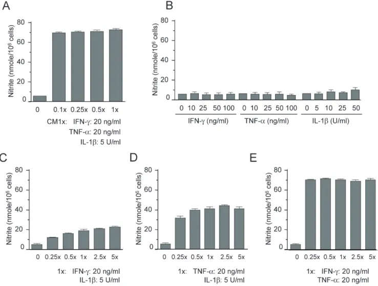
(3) 119. Silymarin inhibits mesangial cell function. Fig. 1. Synergistic induction of nitrite production by cytokines in mesangial cells. (A) Mesangial cells were treated with the indicated concentrations of cytokine mixture (CM: TNF-α, IFN-γ, and IL-1β) for 24 h. (B) Mesangial cells were treated with each cytokine for 24 h. (C) Cells were co-treated with IFN-γ and IL-1β for 24 h. (D) Cells were co-treated with TNF-α and IL-1β for 24 h. (E) Cells were co-treated with TNF-α and IFN-γ for 24 h. Supernatants were subsequently isolated and analyzed for nitrite. Each column shows the mean±SD of triplicate measurements. *p<0.05 compared with the control group, as determined by Student’s two-tailed t-test.. 20 ng/ml; IFN-γ, 20 ng/ml; IL-1β, 5 U/ml), and treated mesangial cells with various concentrations of CM (0.1×, 0.25×, 0.5×, and 1×) for 24 h. Supernatants were then prepared and analyzed for NO production by measuring nitrite, a stable end-product of NO. Treatment with CM increased the production of nitrite ≥ 10-fold over basal levels in mesangial cells (Fig. 1A). However, treatment with each cytokine alone could not increase NO production, even when applied at a dose 5–10 fold higher than that present in 1× CM (Fig. 1B). When we co-treated mesangial cells with two cytokines together, we noted a synergistic induction of NO production (Fig. 1C, 1D, and 1E). Co-treatment with TNF- α and IFN-γ showed a strong synergistic effect on the induction of NO production (Fig. 1E), and the level of NO induced was similar to that induced after three-cytokine co-treatment. These results demonstrate that TNF-α, IFN-γ, and IL-1β synergistically interact to stimulate NO production in mouse mesangial cells, and that the NO induction stimulated by TNF-α and IFN-γ cowww.kjpp.net. treatment is as strong as the induction by treatment with CM containing the three cytokines.. Inhibition of iNOS expression by silymarin in cytokine-stimulated mesangial cells We investigated the effects of silymarin on NO production and iNOS expression in cytokine-stimulated mouse mesangial cells. Mesangial cells were treated with silymarin in the presence of CM for 24 h, and nitrite generation was analyzed. CM-induced nitrite generation was inhibited by silymarin in a dose-dependent manner (Fig. 2A). To test the cytotoxic effect of silymarin, we performed an MTT assay for evaluating tetrazolium dye reduction activity by mitochondrial succinate dehydrogenase. The viability of all the silymarin-treated cells exceeded 90%, except for those treated with the highest dose of 100 μg/ml, which showed slight cytotoxicity (Fig. 2B). Korean J Physiol Pharmacol 2017;21(1):117-124.
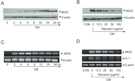
(4) 120. Youn CK et al. Fig. 2. Inhibition of nitrite production by silymarin in CM-stimulated mesangial cells. Mesangial cells were treated with the indicated concentrations of silymarin in the presence of cytokine-mixture (CM) for 24 h. (A) Supernatants were subsequently isolated and analyzed for nitrite. (B) Cells were analyzed for viability by MTT assay. Each column shows the mean±SD of triplicate measurements. *p<0.05 compared with the control group, as determined by Student’s two-tailed t-test.. Fig. 3. Inhibition of iNOS gene expression by silymarin in CM-stimulated mesangial cells. (A and B) Mesangial cells were treated with CM for the indicated duration. (A) Cell lysates were prepared and the expression of iNOS were analyzed by Western blot using an antibody specific for murine iNOS. (B) Total RNA was isolated and mRNA expression levels of iNOS and β-actin were analyzed by RT-PCR. (C and D) Mesangial cells were treated with the indicated concentrations of silymarin in the presence of CM for 16 h (C) or 6 h (D). Cell lysates (C) or total RNA (D) were prepared and analyzed by Western blot or RT-PCR, respectively.. We further analyzed the effects of silymarin on iNOS expression by Western blot and RT-PCR analyses. Western blot analysis showed that the expression of iNOS protein was detected 4 h after CM treatment, peaked at 16 h, and was maintained until 24 h (Fig. 3A). RT-PCR analysis showed that iNOS mRNA expression was detected 2 h after CM treatment, peaked at 6 h, and was maintained at a slightly lower level until 24 h (Fig. 3B). To analyze the effect of silymarin on iNOS expression, we treated mouse mesangial cells with silymarin and CM for 16 h and 6 h for Western blot and RT-PCR analyses, respectively. Korean J Physiol Pharmacol 2017;21(1):117-124. Silymarin inhibited the CM-induced iNOS protein expression in a dose-dependent manner (Fig. 3C). The β-actin loading control was constitutively expressed and was not affected by silymarin treatment. Silymarin inhibited iNOS mRNA expression, although the sensitivity of iNOS mRNA to inhibition was relatively lower than that of iNOS protein (Fig. 3D). These results showed that silymarin decreased the expression of iNOS, which is involved in chronic kidney disease.. https://doi.org/10.4196/kjpp.2017.21.1.117.
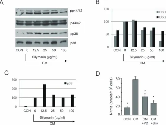
(5) 121. Silymarin inhibits mesangial cell function. Fig. 4. Inhibition of p44/42 phosphorylation by silymarin in CM-stimulated mesangial cells. (A) Mesangial cells were pretreated with the indicated concentrations of silymarin for 1 h and then incubated in the presence of CM for 20 min. The phosphorylation of p44/42 and p38 was analyzed by Western blot. The relative band densities of phosphorylated p44/42 (B) and p38 (C) were analyzed using Image J software. (D) Cells were treated with PD98059 (50 μM) or silymarin (50 μg/ml) for 48 h in the presence of CM. The supernatants were subsequently isolated and analyzed for nitrite. Each column shows the mean±SD of triplicate measurements. *p<0.05 compared with the control group, as determined by Student’s twotailed t-test.. Inhibition of p44/42 (ERK1/2) phosphorylation by silymarin in CM-stimulated mesangial cells Because p44/42 kinase is important for NO generation in CM-stimulated mesangial cells and is a possible target of silymarin, we further determined the role of p44/42 in NO inhibition by silymarin. Mesangial cells were pretreated with silymarin for 1 h and then incubated for 20 min in the presence of CM. MAPK phosphorylation was analyzed by Western blot. The phosphorylation of p44/42 was strongly increased by CM treatment, while silymarin pretreatment decreased the phosphorylation of p44/42 in a dose-dependent manner (Fig. 4A). We also analyzed the effect of silymarin on the phosphorylation of p38, another important MAPK in the production of NO. CM significantly induced the phosphorylation of p38, and silymarin inhibited this phosphorylation (Fig. 4A). We analyzed relative band densities of phosphorylated p44/42 (Fig. 4B) and p38 (Fig. 4C) using Image J software. These results demonstrate that silymarin inhibits p44/42 and p38, which are important factors in signal transduction pathways that regulates iNOS expression in CM-stimulated mouse mesangial cells. We tested the role of ERK1/2 in iNOS expression using PD98059, a specific inhibitor of mitogen activated protein kinase/extracellular signal-regulated www.kjpp.net. kinase 1 (MEK-1), which is responsible for ERK1/2 activation. PD98059 inhibited CM-induced production of nitrite (Fig. 4D).. DISCUSSION In the present study, we reported that silymarin, isolated from milk thistle (Silybum marianum), is a potent anti-inflammatory agent in mouse mesangial cells. Silymarin inhibited cytokineinduced production of NO, an important mediator of inflammatory responses. The inhibition of NO generation is related to the attenuation of iNOS gene expression. The protective role of silymarin against cytokines, including TNF-α and IL1β, in human mesangial cells is demonstrated [28]. Silymarin inhibits nuclear factor kappa-light-chain-enhancer of activated B cells (NF-κB) activation and monocyte chemoattractant protein-1 (MCP-1) expression through the inhibition of increase in intracellular calcium levels [28]. Inhibition of iNOS gene expression by silymarin has been reported in various cells including macrophages, pancreatic beta cells, fibroblasts, and mesenchymal stem cells. Because NO production by mesangial cells, macrophages, and pancreatic beta cells plays critical roles in the pathogenesis of diabetes and diabetic nephropathy, the Korean J Physiol Pharmacol 2017;21(1):117-124.
(6) 122 protective role of silymarin against these conditions is likely due, at least in part, to an anti-inflammatory effect. In addition, silymarin protects pancreatic beta cells from pro-inflammatorycytokine-induced NO production and cell death [29,30]. Proinflammatory cytokines are known to induce the expression of iNOS mRNA and production of NO, resulting in death of beta cells [26,27]. A relationship between diabetic nephropathy and NO production has been suggested to exist [31]. During the development of diabetic nephritis, immune cells, including macrophages, invade and initiate local inflammatory responses. Cytokines produced by infiltrated cells induce the expression of iNOS gene and the production of NO [32,33], which then enhances glomerular mesangial cell relaxation and hyperfiltration [34]. Hyperfiltration exacerbates glomerular damage and causes fibrotic changes that lead to renal dysfunction. In the present study, we showed that IFN- γ and TNF- α synergistically induce mouse mesangial cells to express iNOS. Co-treatment with low doses of IFN-γ (5 ng/ml) and TNF-α (5 ng/ml) synergistically induced NO production, whereas treatment with each cytokine alone did not increase production of NO at doses up to 100 ng/ml (Fig. 1B). Because similar induction of NO production was observed following co-treatment of mesangial cells with IFN-γ and TNF-α as with the three-cytokine mixture (IFN-γ, TNF-α, and IL-1β), the contribution of IL-1β towards NO induction was concluded to be minimal. However, cotreatment with IL-1β together with either IFN-γ or TNF-α clearly showed the synergistic induction of NO (Fig. 1C, 1D). Synergistic induction of NO by IFN-γ and TNF-α is further supported by the results of a previous study [10]. Although the mechanism for the synergistic induction of NO by cytokines is not known, signal transduction pathways involving their membrane receptors might play a role in regulating NO production. Cytokine binding to their receptors initiate signal transduction events leading to the activation of transcription factors such as NF-κ B, signal transducers and activators of transcription family of transcription factors (STAT)1, and interferon regulatory factor (IRF)-1. Activated transcription factors cooperate to produce the maximal induction of iNOS gene expression (Fig. 5). TNF-α binds to TNF receptors (TNFR1 and TNFR2) that are preferentially expressed in the glomeruli in the normal kidney [35]. The role of TNF-α-TNFR1 signal transduction was shown using TNFR-deficient mice [36]. Chemokine secretion was absent and glomerular leukocyte infiltration was abrogated in TNFstimulated TNFR1-deficient glomeruli. Signal transduction through TNFR1 activates NF-κB, an important transcription factor of inflammation-regulating genes [37]. Following its activation, NF-κB binds to the promoters of many inflammatory genes, including iNOS [38]. The involvement of IFN- γ in glomerulonephritis was shown by animal studies using IFN-γreceptor-deficient mice and soluble IFN-γ receptor treatment [39,40]. Signal transduction through the IFN-γ receptor induces Korean J Physiol Pharmacol 2017;21(1):117-124. Youn CK et al. Fig. 5. Diagram showing cytokine-induced signal transduction pathways and possible targets of silymarin. TNF-α and IFN-γ binding to their receptors activate signal transduction pathways, including MAPKs, STAT1, NF-κB, and IRF-1. The iNOS gene promoter contains binding sites for NF-κB, STAT1, and IRF-1. Some possible targets of silymarin including ERK1/2 are shown.. STAT1 and IRF-1, which then bind to the gamma-interferon activated site (GAS) motif and IFN-stimulated response elements (ISRE) of the iNOS promoter, respectively [10]. Use of a reporter gene assay demonstrated that the binding sites of IRF-1, STAT1, and NF-κB were essential for the synergistic response. Ectopic expression experiments further showed that these transcription factors synergistically induce NO accumulation [10]. We demonstrated that silymarin inhibits the p44/42 pathway in cytokine-stimulated mesangial cells. Silibinin, a major component of silymarin, has been shown to inhibit TPA- or TNF-α-induced MMP-9 expression through inhibition of the MAPK pathway [41-43]. ERK activity is required for iNOS gene expression in insulin-producing INS-1E cells and beta cells [31,44]. In summary, these experiments demonstrated that silymarin inhibits CM-induced iNOS gene expression in mouse mesangial cells. Based on our findings, the most likely mechanism that can account for this biological effect involves inhibition of the ERK1/2 kinase pathway. Owing to the critical roles that NO and ERK1/2 play in mediating inflammatory responses in glomerular mesangial cells, inhibition of these activities by silymarin is potentially a useful strategy for protecting against chronic kidney disease.. ACKNOWLEDGEMENTS The present study was supported by grants from the Clinical Medicine Research Institute at Chosun University Hospital, 2013.. https://doi.org/10.4196/kjpp.2017.21.1.117.
(7) Silymarin inhibits mesangial cell function. CONFLICTS OF INTEREST The authors declare no conflicts of interest.. REFERENCES 1. Vianna HR, Soares CM, Tavares MS, Teixeira MM, Silva AC. Inflammation in chronic kidney disease: the role of cytokines. J Bras Nefrol. 2011;33:351-364. 2. Betjes MG. Immune cell dysfunction and inflammation in endstage renal disease. Nat Rev Nephrol. 2013;9:255-265. 3. Radeke HH, Resch K. The inf lammatory function of renal glomerular mesangial cells and their interaction with the cellular immune system. Clin Investig. 1992;70:825-842. 4. Klahr S. Oxygen radicals and renal diseases. Miner Electrolyte Metab. 1997;23:140-143. 5. Marx M, Sterzel RB, Sorokin L. Renal matrix and adhesion in injury and inflammation. Curr Opin Nephrol Hypertens. 1993;2:527-535. 6. Zhao J, Jiang T, Li H, Zhang Y, Zhang N. Aldose reductase regulates TNF- α-induced inducible nitric oxide synthase expression in human mesangial cells. Mol Biol Rep. 2012;39:1815-1822. 7. Wang W, Zolty E, Falk S, Summer S, Zhou Z, Gengaro P, Faubel S, Alp N, Channon K, Schrier R. Endotoxemia-related acute kidney injury in transgenic mice with endothelial overexpression of GTP cyclohydrolase-1. Am J Physiol Renal Physiol. 2008;294:F571-576. 8. Schwartz D, Mendonca M, Schwartz I, Xia Y, Satriano J, Wilson CB, Blantz RC. Inhibition of constitutive nitric oxide synthase (NOS) by nitric oxide generated by inducible NOS after lipopolysaccharide administration provokes renal dysfunction in rats. J Clin Invest. 1997;100:439-448. 9. Szabó C, Southan GJ, Thiemermann C. Beneficial effects and improved survival in rodent models of septic shock with S-methylisothiourea sulfate, a potent and selective inhibitor of inducible nitric oxide synthase. Proc Natl Acad Sci U S A. 1994;91:12472-12476. 10. Paludan SR, Malmgaard L, Ellermann-Eriksen S, Boscá L, Mogensen SC. Interferon (IFN)-gamma and Herpes simplex virus/ tumor necrosis factor-alpha synergistically induce nitric oxide synthase 2 in macrophages through cooperative action of nuclear factor-kappa B and IFN regulatory factor-1. Eur Cytokine Netw. 2001;12:297-308. 11. Valenzuela A, Garrido A. Biochemical bases of the pharmacological action of the flavonoid silymarin and of its structural isomer silibinin. Biol Res. 1994;27:105-112. 12. Zhang W, Hong R, Tian T. Silymarin's protective effects and possible mechanisms on alcoholic fatty liver for rats. Biomol Ther (Seoul). 2013;21:264-269. 13. Bektur NE, Sahin E, Baycu C, Unver G. Protective effects of silymarin against acetaminophen-induced hepatotoxicity and nephrotoxicity in mice. Toxicol Ind Health. 2016;32:589-600. 14. Mereish KA, Bunner DL, Ragland DR, Creasia DA. Protection against microcystin-LR-induced hepatotoxicity by Silymarin: biochemistry, histopathology, and lethality. Pharm Res. 1991;8:273277. 15. Dashti-Khavidaki S, Shahbazi F, Khalili H, Lessan-Pezeshki M. www.kjpp.net. 123 Potential renoprotective effects of silymarin against nephrotoxic drugs: a review of literature. J Pharm Pharm Sci. 2012;15:112-123. 16. Senturk H, Kabay S, Bayramoglu G, Ozden H, Yaylak F, Yucel M, Olgun EG, Kutlu A. Silymarin attenuates the renal ischemia/ reperfusion injury-induced morphological changes in the rat kidney. World J Urol. 2008;26:401-407. 17. Cheung CW, Gibbons N, Johnson DW, Nicol DL. Silibinin--a promising new treatment for cancer. Anticancer Agents Med Chem. 2010;10:186-195. 18. Li L, Gao Y, Zhang L, Zeng J, He D, Sun Y. Silibinin inhibits cell growth and induces apoptosis by caspase activation, downregulating survivin and blocking EGFR-ERK activation in renal cell carcinoma. Cancer Lett. 2008;272:61-69. 19. Vessal G, Akmali M, Najafi P, Moein MR, Sagheb MM. Silymarin and milk thistle extract may prevent the progression of diabetic nephropathy in streptozotocin-induced diabetic rats. Ren Fail. 2010;32:733-739. 20. Soto C, Mena R, Luna J, Cerbón M, Larrieta E, Vital P, Uría E, Sánchez M, Recoba R, Barrón H, Favari L, Lara A. Silymarin induces recovery of pancreatic function after alloxan damage in rats. Life Sci. 2004;75:2167-2180. 21. Soto C, Raya L, Juárez J, Pérez J, González I. Effect of silymarin in Pdx-1 expression and the proliferation of pancreatic β-cells in a pancreatectomy model. Phytomedicine. 2014;21:233-239. 22. Lettéron P, Labbe G, Degott C, Berson A, Fromenty B, Delaforge M, Larrey D, Pessayre D. Mechanism for the protective effects of silymarin against carbon tetrachloride-induced lipid peroxidation and hepatotoxicity in mice. Evidence that silymarin acts both as an inhibitor of metabolic activation and as a chain-breaking antioxidant. Biochem Pharmacol. 1990;39:2027-2034. 23. Zhao J, Sharma Y, Agarwal R. Significant inhibition by the flavonoid antioxidant silymarin against 12-O-tetradecanoylphorbol 13-acetate-caused modulation of antioxidant and inflammatory enzymes, and cyclooxygenase 2 and interleukin-1alpha expression in SENCAR mouse epidermis: implications in the prevention of stage I tumor promotion. Mol Carcinog. 1999;26:321-333. 24. Green LC, Wagner DA, Glogowski J, Skipper PL, Wishnok JS, Tannenbaum SR. Analysis of nitrate, nitrite, and [15N]nitrate in biological fluids. Anal Biochem. 1982;126:131-138. 25. Huong PT, Lee MY, Lee KY, Chang IY, Lee SK, Yoon SP, Lee DC, Jeon YJ. Synergistic induction of iNOS by IFN-γ and glycoprotein isolated from dioscorea batatas. Korean J Physiol Pharmacol. 2012;16:431-436. 26. Cetkovic-Cvrlje M, Eizirik DL. TNF-alpha and IFN-gamma potentiate the deleterious effects of IL-1 beta on mouse pancreatic islets mainly via generation of nitric oxide. Cytokine. 1994;6:399406. 27. Darville MI, Eizirik DL. Regulation by cytokines of the inducible nitric oxide synthase promoter in insulin-producing cells. Diabetologia. 1998;41:1101-1108. 28. Chang JW, Kim CS, Kim SB, Park SK, Park JS, Lee SK. Pro inflammatory cytokine-induced NF-kappaB activation in human mesangial cells is mediated through intracellular calcium but not ROS: effects of silymarin. Nephron Exp Nephrol. 2006;103:e156165. 29. Kim MJ, Yoo YC, Kim HJ, Shin SK, Sohn EJ, Min AY, Sung NY, Kim MR. Aged black garlic exerts anti-inflammatory effects by Korean J Physiol Pharmacol 2017;21(1):117-124.
(8) 124 decreasing NO and proinflammatory cytokine production with less cytoxicity in LPS-stimulated RAW 264.7 macrophages and LPSinduced septicemia mice. J Med Food. 2014;17:1057-1063. 30. Matsuda T, Ferreri K, Todorov I, Kuroda Y, Smith CV, Kandeel F, Mullen Y. Silymarin protects pancreatic beta-cells against cytokinemediated toxicity: implication of c-Jun NH2-terminal kinase and janus kinase/signal transducer and activator of transcription pathways. Endocrinology. 2005;146:175-185. 31. Youn CK, Park SJ, Li MH, Lee MY, Lee KY, Cha MJ, Kim OH, You HJ, Chang IY, Yoon SP, Jeon YJ. Radicicol inhibits iNOS expression in cytokine-stimulated pancreatic beta cells. Korean J Physiol Pharmacol. 2013;17:315-320. 32. Ikeda M, Ikeda U, Ohkawa F, Shimada K, Kano S. Nitric oxide synthesis in rat mesangial cells induced by cytokines. Cytokine. 1994;6:602-607. 33. Song MY, Kim KA, Lee SY, Kim EK, Lv N, Lee JH, Park JW, Ryu DG, Kwon KB, Park BH. Radix asari extract protects pancreatic beta cells against cytokine-induced toxicity: implication of the NFkappaB-iNOS signaling cascade. Int J Mol Med. 2007;20:769-775. 34. Stocka nd JD, Sa nsom SC . Glomer u la r mesa ng ia l cel ls: electrophysiology and regulation of contraction. Physiol Rev. 1998;78:723-744. 35. Vielhauer V, Mayadas TN. Functions of TNF and its receptors in renal disease: distinct roles in inflammatory tissue injury and immune regulation. Semin Nephrol. 2007;27:286-308. 36. Taubitz A, Schwarz M, Eltrich N, Lindenmeyer MT, Vielhauer V. Distinct contributions of TNF receptor 1 and 2 to TNF-induced glomerular inflammation in mice. PLoS One. 2013;8:e68167. 37. Micheau O, Tschopp J. Induction of TNF receptor I-mediated apoptosis via two sequential signaling complexes. Cell. 2003;114:181190.. Korean J Physiol Pharmacol 2017;21(1):117-124. Youn CK et al 38. Jia J, Liu Y, Zhang X, Liu X, Qi J. Regulation of iNOS expression by NF-κB in human lens epithelial cells treated with high levels of glucose. Invest Ophthalmol Vis Sci. 2013;54:5070-5077. 39. Schwarting A, Wada T, Kinoshita K, Tesch G, Kelley VR. IFNgamma receptor signaling is essential for the initiation, acceleration, and destruction of autoimmune kidney disease in MRL-Fas(lpr) mice. J Immunol. 1998;161:494-503. 40. Ozmen L, Roman D, Fountoulakis M, Schmid G, Ryffel B, Garotta G. Experimental therapy of systemic lupus erythematosus: the treatment of NZB/W mice with mouse soluble interferon-gamma receptor inhibits the onset of glomerulonephritis. Eur J Immunol. 1995;25:6-12. 41. Oh SJ, Jung SP, Han J, Kim S, Kim JS, Nam SJ, Lee JE, Kim JH. Silibinin inhibits TPA-induced cell migration and MMP9 expression in thyroid and breast cancer cells. Oncol Rep. 2013;29:1343-1348. 42. Kim S, Choi JH, Lim HI, Lee SK, Kim WW, Kim JS, Kim JH, Choe JH, Yang JH, Nam SJ, Lee JE. Silibinin prevents TPA-induced MMP9 expression and VEGF secretion by inactivation of the Raf/MEK/ ERK pathway in MCF-7 human breast cancer cells. Phytomedicine. 2009;16:573-580. 43. Kim S, Choi MG, Lee HS, Lee SK, Kim SH, Kim WW, Hur SM, Kim JH, Choe JH, Nam SJ, Yang JH, Kim S, Lee JE, Kim JS. Silibinin suppresses TNF-alpha-induced MMP-9 expression in gastric cancer cells through inhibition of the MAPK pathway. Molecules. 2009;14:4300-4311. 44. Larsen L, Størling J, Darville M, Eizirik DL, Bonny C, Billestrup N, Mandrup-Poulsen T. Extracellular signal-regulated kinase is essential for interleukin-1-induced and nuclear factor kappaBmediated gene expression in insulin-producing INS-1E cells. Diabetologia. 2005;48:2582-2590.. https://doi.org/10.4196/kjpp.2017.21.1.117.
(9)
수치

관련 문서
The matrix A show the cost per computer (in thousands of dollars) and B the production figures for the year 2005 (in multiples of 1000 units).. Find a matrix C that
Pro-allergic cytokines were important mediators of allergic inflammation, cell recruitment and allergenic response decided to further investigate the
This study aimed to evaluate the site and extent of injury, injury mechanism, player position, and the reinjury incidence in the hamstring by using magnetic
The purpose of this study is to present the background, necessity and purpose of the study in Chapter 1, and the second part of the theoretical study is related to the
tricuspidata on the production of proinflammatory cytokines in TNFα+IFNγ-stimulated HaCaT cells ...15 Fig.5: The cell viability of sub-fractions from 70% EtOH
In conclusion, the data acquired in this study demonstrate that pinosylvin can inhibit LPS-induced expression of pro-inflammatory mediators, and the
Key word: Cinnamomum camphora, pro-inflammatory cytokines, iNOS, COX-2.. Systematic purification using solvent partitioning from Cinnamomum camphora .... Sequences
iMSC‑EVs decreased itching, which was supported by reduced inflammatory cell infiltration and mast cells in AD mouse skin; reduced IgE receptor expression and
