Lab Anim Res 2017: 33(4), 283-290 https://doi.org/10.5625/lar.2017.33.4.283
ISSN 2233-7660 (Online)
Dendropanax morbifera Léveille extract ameliorates D -galactose- induced memory deficits by decreasing inflammatory
responses in the hippocampus
Kwon Young Lee
1, Hyo Young Jung
2, Dae Young Yoo
2, Woosuk Kim
2, Jong Whi Kim
2, Hyun Jung Kwon
3, Dae Won Kim
3, Yeo Sung Yoon
2, In Koo Hwang
2, Jung Hoon Choi
1,*
1
Department of Anatomy, College of Veterinary Medicine and Institute of Veterinary Science, Kangwon National University, Chuncheon 24341, South Korea
2
Department of Anatomy and Cell Biology, College of Veterinary Medicine, and Research Institute for Veterinary Science, Seoul National University, Seoul 08826, South Korea
3
Department of Biochemistry and Molecular Biology, Research Institute of Oral Sciences, College of Dentistry, Gangneung- Wonju National University, Gangneung 25457, South Korea
In the present study, we examined the effects of Dendropanax morbifera Léveille leaf extract (DML) on
D- galactose-induced morphological changes in microglia and cytokines, including pro-inflammatory cytokines (interleukin [IL]-1β, IL-6, and tumor necrosis factor [TNF]-α) and anti-inflammatory cytokines (IL-4 and IL-10) in the hippocampus. Administration of DML to
D-galactose-treated mice significantly improved
D-galactose-induced reduction in escape latency, swimming speed, and spatial preference for the target quadrant. In addition, administration of DML to
D-galactose-treated mice significantly ameliorated the microglial activation and increases of IL-1β, IL-6, and TNF-α levels in the hippocampus.
Administration of
D-galactose significantly reduced IL-4 levels in the hippocampus, while administration of DML to
D-galactose-treated mice significantly increased IL-4 level. However, we did not observe any significant changes in IL-10 levels in hippocampal homogenates. These results suggest that DML reduces
D
-galactose-induced mouse senescence by reducing pro-inflammatory cytokines such as IL-1 β, IL-6, and TNF-α, as well as increasing anti-inflammatory cytokine IL-4.
Keywords: Dendropanax morbifera extract,
D-galactose, hippocampus, inflammation, memory
Received 23 May 2017; Revised version received 14 September 2017; Accepted 19 December 2017
Nervous tissue is more susceptible to oxidative damage than other tissue, because it has a high content of unsaturated fatty acids. Moreover, the brain has high metabolic activity and relatively low antioxidant defense [1]. In aging progresses, oxidative damage accumulates and the brain undergoes morphologic and functional changes [2]. Finally oxidative damage causes chronic inflammation in the aged brain. Neuroinflammation induces the activation of microglia in the brain and active microglia secrete pro-inflammatory cytokines (M1 microglia) or have important roles in brain repair
and plasticity (M2 microglia) [3]. Activated microglia release neurotoxic substances and pro-inflammatory cytokines such as interleukin (IL)-1 β, IL-6, and tumor necrosis factor- α (TNF-α) [4-6]. In contrast, anti- inflammatory cytokines such as IL-4 and IL-10 may decrease neuroinflammation by regulating the production of IL-1 β or IL-6 [7].
There are several animal models for the study of aging. However, the D -galactose model is the most convenient and can be compared to natural aging studies. Under normal condition, diet-fed D -galactose
*Corresponding author: Professor Jung Hoon Choi, DVM, PhD, Department of Anatomy, College of Veterinary Medicine and Institute of Veterinary Science, Kangwon National University, Chuncheon 24341, South Korea
Tel:+82-33-250-8682; Fax: +82-33-244-2367; E-mail: [email protected]
This is an Open Access article distributed under the terms of the Creative Commons Attribution Non-Commercial License (http://creativecommons.org/licenses/
by-nc/3.0) which permits unrestricted non-commercial use, distribution, and reproduction in any medium, provided the original work is properly cited.
can be metabolized by two enzymes in the body: D - galactokinase and galactose-1-phosphate uridyl transferase.
However, repeated treatment of D -galactose can be converted into hydrogen peroxide in the presence of galactose oxidase, leading to the formation of a superoxide anion [8]. Administration of D -galactose shows aging phenotypes such as impairment of spatial learning and memory, and neurodegeneration and neuroinflammation [8].
Dendropanax morbifera Léveille (D. morbifera) is an endemic species distributed in the southwest costal region and Jeju island of South Korea. Several lines of evidence demonstrate that D. morbifera leaf extract (DML) has antioxidant [9-11] and anti-inflammatory [12,13] effects. In particular, in our previous study, we demonstrated that D. morbifera stem extract ameliorates streptozotocin-induced memory impairment, microglial activation, and release of TNF- α and IL-1β in the hippocampus [13].
However, there are few reports on the effects of DML on age-related changes of hippocampal function. Therefore, in the present study, we evaluated the effects of DML on hippocampal function using a Morris water maze task, and microglial activation and subsequent cytokine release using immunohistochemistry and enzyme-linked immunosorbent assay (ELISA), respectively, in D - galactose treatment-induced aged mouse hippocampus.
Materials and Methods
Experimental animals
Male C57BL/6 mice were purchased from Orient Bio Inc. (Seongnam, South Korea). Rats were housed in a conventional animal facility at 23
oC with 60% humidity, a 12-h/12-h light/dark cycle, and free access to food and tap water. The handling and care of the animals conformed to the guidelines established to comply with current international laws and policies (NIH Guide for the Care and Use of Laboratory Animals, NIH Publication No.
85-23, 1985, revised 1996). Ethical and experimental protocol approvals were obtained from the Institutional Animal Care and Use Committee (IACUC) of Kangwon National University (KW-161014-1). All of the experiments were conducted with an effort to minimize the number of animals used and the suffering caused by the procedures employed.
Preparation of DML
Fresh D. morbifera was supplied by HBJ Biofarm (Jeju, South Korea). The plant was identified by two practitioners of traditional Asian medicine. Leaves from the plant samples (100 g) were chopped, blended, soaked in 2 L of 80% ethanol, and then refluxed three times at 20
oC for 2 h. Insoluble materials were removed by centrifugation at 10,000×g for 30 min, and the resulting supernatant was concentrated and freeze-dried to yield a powder. Before each experiment, dried extracts were dissolved in distilled and deionized water.
Administration of D -galactose and (DML)
Animals were divided into 3 groups (n=10 in each group): vehicle-treated, D -galactose-treated, and D -galactose with DML treated group. D -Galactose (100 mg/kg) and DML (100 mg/kg) were subcutaneously and orally administered to 7-week-old mice once a day for 10 weeks.
Water maze performance
During the 10th week after D -galactose administration, spatial memory was assessed using a Morris water maze as described previously [14]. Morris water maze tests were performed in order to ensure objectivity in blind conditions. Three days after the training, the time individual mice spent to find the submerged platform (within 2 min) (escape latency) and the swimming distance were monitored by a digital camera and a computer system for 4 consecutive days during 4 trials per day. The administration of D -galactose and DML was continued during the water maze performance. For each trial, a mouse was placed in the water facing the wall at one of four starting positions and released. The swimming speed and the time required for the mouse to find the hidden platform were recorded via visual tracking system. The probe test was done on day 5; the platform was removed and the time that the mouse spent swimming in the target quadrant and in the three non- target quadrants (right, left, and opposite quadrants) was measured in the training and opposite quadrants for 60 s.
In addition, the number of times the mouse crossed the platform site was recorded.
Tissue processing
For immunohistochemical analysis, vehicle-treated, D -
galactose-treated, and D -galactose with DML treated mice (n=5 per group) were anesthetized with 1 g/kg urethane (Sigma-Aldrich, St. Louis, MO, USA) and perfused with 0.1 M phosphate-buffered saline (PBS, pH 7.4) followed by 4% paraformaldehyde in 0.1 M phosphate-buffer, pH 7.4. Brains were removed and postfixed in the same fixative for 24 h. Subsequently, the brain was dehydrated with graded concentrations of alcohol before being embedded in paraffin. Paraffin- embedded tissues were sectioned into 3- μm coronal sections using a microtome (Leica Microsystems GmbH, Wetzlar, Germany) and were mounted onto silane-coated slides (Muto Pure Chemicals Co., Ltd, Tokyo, Japan).
Immunohistochemistry for Iba-1
To ensure that the immunohistochemical data was comparable among groups, sections were carefully processed under the same conditions. Sections were hydrated and treated with 0.3% hydrogen peroxide (H
2O
2) in phosphate-buffered saline (PBS) for 30 min.
For antigen retrieval, the sections were placed in 400-mL jars filled with citrate buffer (pH 6.0) and heated in a 2100-retriever (Prestige Medical, Lancashire, UK). After antigen retrieval, slides were allowed to cool at room temperature and were washed in PBS. After washing, sections were incubated in 10% normal goat serum in PBS for 30 min. Sections were then incubated with rabbit anti-Iba-1 antibody (1:500, Wako, Osaka, Japan) for 48 h at 4
oC. Sections were subsequently exposed to biotinylated goat anti-rabbit IgG, or anti-mouse IgG (diluted 1:200, Vector Laboratories, Inc., Burlingame, CA, USA), and streptavidin peroxidase complex (diluted 1:200, Vector Laboratories). Finally, sections were stained with 3,3-diaminobenzidine tetrahydrochloride (Sigma, St. Louis, MO, USA) in 0.1 M Tris-HCl buffer (pH 7.4).
Analysis of the regions of interest in the hippocampal CA1 region was performed using an image analysis system. Images were calibrated into an array of 512×
512 pixels corresponding to a tissue area of 140 μm×
140 μm (40× primary magnification). Pixel resolution was 256 gray levels. The intensity of Iba-1 immuno- reactivity was evaluated by relative optical density (ROD), which was obtained after transformation of the mean gray level using the formula: ROD=log(256/mean gray level). The ROD of the background was determined in unlabeled portions (white matter region) and this value was subtracted to normalize the corrected optical
densities using ImageJ 1.50 software (National Institutes of Health, Bethesda, MD, USA). The ratio of the ROD was calculated as percentage relative to control (vehicle- treated group).
ELISA for cytokines
To measure changes in TNF- α, IL-1β, IL-4, IL-6, and IL-10 levels in the hippocampus, vehicle-treated, D - galactose-treated, and D -galactose with DML treated mice (n=5) were sacrificed and used for ELISA. After sacrificing the mice and removing the hippocampus, hippocampal tissues were homogenized in ice-cold 50 mM sodium phosphate buffer (pH 7.4) containing 0.1 mM ethylenediaminetetraacetic acid (EDTA) by using a glass-teflon homogenizer (Heidolph Silent Crusher M, Germany). The supernatant was separated by centrifugation at 20,000×g for 20min at 4
oC. TNF- α, IL-1 β, IL-4, IL-6, and IL-10 were measured in the supernatant of homogenized hippocampal tissue by using ELISA kits (EMD Millipore, Billerica, MA, USA). The procedures were carried out according to the manufacturer’s instructions. TNF- α, IL-1β, IL-4, IL-6, and IL-10 levels were determined from a standard curve and were expressed in pg/mg protein.
Statistical analysis
Data are expressed as means for each experiment. The differences among the means were statistically analyzed by one- or two-way analysis of variance (ANOVA) with repeated measures and Bonferroni’s post hoc test using GraphPad Prism 5.01 software (GraphPad Software, Inc., La Jolla, CA). Threshold for statistical significance was set to P<0.05.
Results
Effects of D -galactose and/or DML on spatial memory in mice
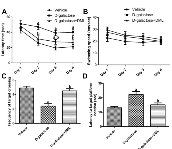
Mean escape latency in the D -galactose-treated group
was longer than in the vehicle-treated group on all days
of experiments. However, differences were only significant
on days 2, 3, and 4. Administration of DML to the D -
galactose-treated group resulted in shorter mean escape
latencies compared to escape latencies in the D -galactose-
treated group. Differences were statistically significant
on days 2 and 4. There was no significant difference in
escape latency between the vehicle-treated group and D -
galactose-treated group with DML (Figure 1A).
Mean swimming speed tended to be decreased after successive trials. In addition, mean swimming speed was slower in the D -galactose-treated group than in the vehicle-treated group. Administration of DML to the D - galactose-treated group increased the swimming speed in all trials although this tendency was not statistical significant (Figure 1B).
Significantly fewer platform crossings in the probe trial were observed in the D -galactose-treated group compared to the vehicle-treated group. Administration of DML to the D -galactose-treated group significantly increased the frequency of crossing over the platform site relative to that in the D -galactose-treated group and similar to that in the vehicle-treated group (Figure 1C).
In the probe trial for the escape latency task, mice form the D -galactose-treated group took significantly longer to find the target platform location than mice from the vehicle-treated group. Administration of DML to the D - galactose-treated group significantly reduced the time mice required to find the target platform (Figure 1D).
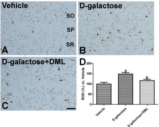
Effects of D -galactose and/or DML on the morphology of Iba-1-immunoreactive microglia
In the vehicle-treated group, Iba-1-immunoreactive microglia were present throughout hippocampus. These microglia showed small cytoplasms and long processes (Figure 2A). In the D -galactose-treated group, the distribution pattern was similar to that in the vehicle- treated group. However, Iba-1 immunoreactive microglia showed hypertrophy of the cytoplasm with highly ramified processes (Figure 2B). In this group, Iba-1 immunoreactivity was significantly increased by 148.6%
relative to the vehicle-treated group (Figure 2D). In the
D -galactose-treated group with DML, only a few Iba-1 immunoreactive microglia had hypertrophied cytoplasm, while most cells had small cytoplasms (Figure 2C). In addition, Iba-1 immunoreactivity was significantly decreased in this group compared to that in the D -galactose-treated group (Figure 2D).
Figure 1. Escape latency training trials (A), average speed (cm/sec) (B), frequency of target crossing (C) and time spent in correct quadrant (D) of vehicle-treated,
D-galactose-treated, and
D-galactose-treated group with Dendropanax morbifera Léveille leaf extract (
D-galactose+DML) in the Morris water maze task (n=10;
aindicates a significant difference from the vehicle-treated group;
b