J. Exp. Biomed. Sci. 2011, 17(3): 225~230
Anti-inflammatory Effect of Indirubin-3'-Monoxime-5-Sulphonic Acid on Lipopolysaccharide-stimulated Murine Macrophage
Gang Baek Park, Hyun Jin Kim, Hye Seon Heo, Geun-Mook Park, Kyung Woo Park and Jin-Kyung Kim†
Department of Biomedical Science, Catholic University of Daegu, Gyeongsan-Si 700-702, Korea
Indirubin is the active ingredient of Danggui Longhui Wan, a mixture of plants that is used in traditional Chinese medicine to treat chronic diseases. In this study we investigated the anti-inflammatory effects of an indirubin derivative, indirubin-3'-monoxime-5-sulphonic acid (I3M-5S, C16H11N3O5S). We found that I3M-5S inhibits the production of various inflammatory mediators such as nitric oxide (NO) and prostaglandin E2 (PGE2) as well as inflammatory cytokines, tumor necrosis factor-α and interleukin-6 in lipopolysaccharide (LPS) stimulated murine macrophage, RAW264.7 cells.
In addition, the expression of inducible nitric oxide synthase and cyclooxygenase-2, which are essential enzymes to produce NO and PGE2, respectively, was blocked by I3M-5S treatment in LPS-stimulated RAW264.7 cells. Present data suggest that I3M-5S exhibits potent anti-inflammatory activity in cultured macrophages and merit further study as potential therapeutic agents for inflammatory disorders.
Key Words: Inflammation, Indirubin-5-sulphonic acid, Macrophage, Lipopolysaccaride, RAW264.7 cell
서 론
염증이란 인체에 유해한 자극에 노출되었을 때 세포 손상을 일으키는 원인을 제거하거나 희석하기 위한 생체 방어로서 , 조직변질, 순환장애, 조직증식의 세 가지를 병 발하는 복잡한 현상이다 (Tizard, 1986). 생체에서 염증반 응이 유도되면 충혈, 부종, 발열, 통증 등의 증상이 나타 난다 (Tizard, 1986). 이 과정에서 호중구를 포함한 다양 한 면역세포, 특히 대식세포가 활성화되면서 산화질소 (Nitric oxide, NO)나 프로스타글란딘 (prostaglandin, PG) E
2등의 염증매개물질들과 tumor necrosis factor-α (TNF-α), interleukin-6 (IL-6), IL-1β 등의 염증성 사이토카인들을 분 비하게 된다 (Sharma et al., 2007; Goldberg, 2009; Ricciotti and FitzGerald, 2011).
NO는 미생물의 침입 혹은 사이토카인의 자극으로 인
해 대식세포를 포함한 다양한 세포가 활성화 되어 생성 되는 물질로, NO synthase (NOS)에 의해 L-arginine으로부 터 생성된다 (McCartney-Francis et al., 1993; Weisz et al., 1996; Sharma et al., 2007). NOS는 neuronal NOS (nNOS), inducible NOS (iNOS), endothelial NOS (eNOS)의 3종류가 존재한다 (Sharma et al., 2007). 이중 iNOS는 염증반응을 조절하는데 중요한 역할을 담당하고 있으며, iNOS에 의 해 증가된 NO는 패혈성 쇼크, 조직손상, 류마티스 관절 염 (rheumatoid arthritis) 등과 같은 질병을 유발하는 중요 원인물질의 하나이다 (McCartney-Francis et al., 1993; Weisz et al., 1996; Sharma et al., 2007). PG는 생체 내에 존재하는 다양한 세포로부터 cyclooxygenase (COX)라는 효소에 의 해 생성된다 (Ricciotti and FitzGerald, 2011). COX는 두 종 류가 존재하는데, COX-1은 대부분의 조직에서 PG 생성 에 관여한다. 반면, COX-2는 성장인자, 세포증식인자, 사 이토카인 등과 같은 요인에 의해 발현이 증가되어 다량 의 PG를 생성함으로써, 염증관련 질병을 유발하는 것으 로 알려져 있다 (Botting, 2006; Ricciotti and FitzGerald, 2011).
내독소로 잘 알려진 lipopolysaccharide (LPS)는 그람 음성균의 세포 외막에 존재하며, 대식세포는 toll-like receptor (TLR)-4를 통해 LPS를 인지하여 다양한 염증매개
*접수일: 2011년 8월 3일 / 수정일: 2011년 9월 18일 채택일: 2011년 9월 20일
*Gang Baek Park, Hyun Jin Kim, Hye Seon Heo, contributed equally to this work.
†Correspondence: Jin-Kyung Kim, Department of Biomedical Science, Catholic University of Daugu, Gyeongsan-Si 712-702, Korea.
Tel: 053-850-3774, Fax: 053-850-3774 e-mail: [email protected]
물질들의 생성을 촉진하는 것으로 알려져 있다 (Butchar et al., 2006). 이와 같은 염증매개물질들이 생성되기 위 해서는 nuclear factor-κB (NF-κB)의 활성화가 필수적이다 (Siebenlist et al., 1994). NF-κB는 면역과 염증반응에 관여 하는 유전자의 enhancer 부위에 결합하여 그 유전자의 발 현을 조절하는 인자이다 (Siebenlist et al., 1994). 자극되지 않은 세포에서 NF-κB는 inhibitor κB (IκB)라 불려지는 억제 단백질에 결합되어 세포질 내에 존재한다 (Baldwin, 1996). 세포가 자극되면 IκB는 수 분 내에 인산화 과정을 거치게 되고 이어서 proteosomes에 의해 급격히 분해된 다 (Palombella et al., 1994; Baldwin, 1996). IκB에서 분리된 NF-κB는 핵 내로 이동해 표적유전자의 promotor 부위의 특정염기서열에 결합하여 iNOS, COX-2, TNF-α, IL-6 등 다양한 염증매개물질의 전사를 촉진하는 것으로 알려져 있다 (Palombella et al., 1994; Baldwin, 1996).
본 연구팀은 쪽풀 (Persicaria tinctoria)의 생리활성성 분의 하나인 indirubin 및 그 유도체가 갖는 다양한 생리 활성에 대한 연구를 진행하고 있다. 쪽풀은 마디풀과 (polygonaceae)에 속하는 1년 생초로, 그 구성 단백질인 indican은 가수분해 시 산화반응을 거쳐 색소성분인 indigo와 indirubin으로 변형된다 (Christie, 2007). Indigo와 indirubin은 예로부터 천연염료로 사용되어 왔으며 특히, indirubin은 해독, 강장, 해열작용 등 다양한 약리작용이 있어 민간에서는 약용으로도 이용되어 왔다 (Xiao et al., 2002). 최근에는 indirubin이 백혈병 및 치매의 치료효과 를 나타내는 주성분으로 보고되면서 이의 유도체의 합성 에 관한 연구가 활발히 진행되고 있다 (Eisenbrand et al., 2004). 그러나 indirubin은 용해도가 매우 낮아 체내에서 흡수가 어렵기 때문에 각종 치료제 개발을 위해서는 제 한이 따랐다. 본 연구에서는 indirubin에 술폰기 (-SO
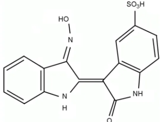
3H) 를 결합시켜 그 용해도를 개선시킨 indirubin의 유도체인 indirubin-3'-monoxime-5-sulphonic acid (3-[3-(Hydroxyimino)- 1,3-dihydro-2H-indol-2-ylidene]-2-oxo-2,3-dihydro-1H-indole -5-sulfonic acid, I3M-5S, Fig. 1)의 항염증효과를 살펴보고 자 하였다.
재료 및 방법
시약 및 재료
생쥐의 대식세포주인 RAW264.7 세포는 한국세포주은 행 (Seoul, Korea)에서 구입을 하여 사용하였으며, I3M-5S 는 Merck (Gibbstown, NJ, USA)에서 구입하여 사용하였다.
LPS와 β-actin 항체는 Sigma-Aldrich (St. Louis, MO, USA) 에서 구입을 하였다. Dulbecco's modified eagle's medium (DMEM), fetal bovine serum (FBS), penicillin 및 strepto- mycin은 Hyclone (Logan, UT, USA)에서 구입하였다.
CellTiter 96 AQueous One Solution과 Griess reagent system 은 Promega (Madison, WI, USA)에서 구입하였고, PGE
2enzyme-linked immunosorbent assay (ELISA) kit는 R&D (Minneapolis, MI, USA)에서 구입하였으며, TNF-α와 IL- 6 ELISA kit는 eBioscience (San Diego, CA, USA)에서 구 입하여 사용하였다. iNOS, COX-2 항체는 Cell signaling (Danvers, MA, USA)에서 구입하였다. Pro-prep
TM는 iNtRON Biotechnology (Sungnam, Korea)에서 구입하였다. BCA protein assay kit은 Thermo (Waltham, MA. USA)에서 구 입하였다 . PVDF Western blotting membranes은 Roche (Basel, Switzerland)에서 구입하였다. UVP는 GelDoc-It
TMTS Imaging System (Cambridge UK, USA)의 제품을 이용하 였다.
세포 배양 및 세포증식 측정
세포 배양액은 DMEM 배양액에 10% FBS와 100 μg/
ml penicillin을 첨가한 배양액을 사용하였다. I3M-5S의 독 성을 측정하기 위해 RAW264.7 세포를 96 well plate에 well 당 2×10
4cells/well이 되도록 분주한 후 12시간 배 양하였다 . 세포를 배양중인 세포 배양액에 I3M-5S을 각 각 0, 1.5625, 3.125, 6.25, 12.5, 25, 50, 100 μM 농도로 처리 하여 24시간 배양한 후, CellTiter 96AQueous One solution assay를 이용하여 세포증식에 미치는 I3M-5S의 영향을 조사하였다.
Fig. 1. The structure of indirubin-3'-monoxime-5-sulphonic acid
(I3M-5S).NO, PGE
2의 측정
RAW264.7 세포를 48 well plate에 2.5×10
5cells/well이 되도록 분주한 후 12시간 배양하였다. 다양한 농도의 I3M-5S를 LPS (100 ng/ml)와 함께 처리하여 24시간 배 양한 후, 세포 배양액을 얻어 배양액 중에 함유된 NO 의 양을 Griess Reagent System을 이용하여 측정하였고, PGE
2는 PGE
2ELISA kit을 이용하여 측정하였다.
IL-6 및 TNF-α의 측정
RAW264.7 세포를 48 well plate에 2.5×10
5cells/well이 되도록 분주한 후 12시간 배양하였다. 다양한 농도의 I3M-5S를 LPS (100 ng/ml)와 함께 처리하여 24시간 배양 한 후, 세포 배양액을 얻어 배양액 중에 함유된 TNF-α와 IL-6의 양을 ELISA kit을 이용하여 측정하였다.
Western blot assay
RAW264.7 세포에 Pro-prep
TMlysis 용액을 넣은 후 얼음 에서 40분간 처리하고 원심분리 (12,000 rpm, 5분, 4℃)하 여 단백질이 함유된 상층액만을 회수하였다. 정제한 단 백질은 BCA protein assay kit을 이용하여 정량하였다. 정 량한 단백질은 SDS-polyacrylamide gel에서 분리시킨 후 Eletroblot system (Bio Rad)을 사용하여 250 mA로 1시간 동안 PVDF membrane에 옮겼다. 항체의 비특이적 결합을 억제시키기 위해 membrane을 PBS-T (PBS, Tween-20)로 세척하고 , 3% skim milk로 1시간 실온에서 blocking한 후, iNOS와 COX-2의 항체를 이용하여 4℃에서 12시간 반 응시켰다 . 세척 후, horseradish peroxidase (HRP)가 붙어 있는 secondary antibody로 실온에서 1시간 반응 후 HRP chromogenic substrate를 이용하여 iNOS와 COX-2 발현 양을 관찰했다. 그 수치를 UVP로 확인하고 분석하였다.
자료의 통계처리
실험결과는 평균 ± 표준편차로 표시하였으며, 통계처 리는 GraphPad Prism 4.0 프로그램 (GraphPad software, San Diego, CA, USA)을 이용하여, One-way analysis variance (ANOVA)를 따랐다. P value가 0.05 이하일 때만 통계적 유의성이 있는 것으로 판단하였다.
결과 및 고찰
대식세포는 내재면역반응에 핵심적인 역할을 담당하고
있는 면역세포로 내독소, 세포증식인자, 바이러스 등에 의해서 활성화 된다 (Laskin and Pendino, 1995). 활성화된 대식세포는 탐식작용을 하고 면역반응을 조절하는 다양 한 물질을 분비한다 (Laskin and Pendino, 1995). 이들 물 질 중에는 NO와 같은 산소대사산물, PG와 같은 지질대 사물 및 단백질류 (사이토카인 및 케모카인) 등이 있으 며, 대식세포는 이들을 생산 · 분비하여 다양한 생체기능 을 수행하게 된다. 따라서 본 연구에서는 생쥐의 대식세 포주인 RAW264.7 세포를 이용하여 I3M-5S의 항염증작용 을 관찰하였다.
먼저 , I3M-5S가 생쥐의 대식세포 RAW264.7 세포의 생 존에 영향을 끼치는지 알아보기 위해 CellTiter 96AQueous One solution assay를 이용하여 세포증식에 미치는 I3M-5S 의 영향을 조사하였다. 그 결과 실험에서 사용한 최고농 도인 100 μM의 농도에서도 세포의 증식에 영향을 끼치 지 않음이 밝혀졌다 (Fig. 2). 이 결과는 본 실험에서 사 용한 I3M-5S가 생쥐의 대식세포에 대해 독성을 나타내 지 않음을 시사한다. 따라서 이하의 실험에서는 I3M-5S 의 최고농도를 100 μM로 하여 실험을 진행하였다.
내독소로 잘 알려진 LPS는 그람음성균의 세포 외막 에 존재하며, 대식세포 또는 단핵구에 존재하는 염증매 개물질의 생성을 증강시키는 것으로 알려져 있다 (Laskin and Pendino 1995; Baldwin, 1996; Butchar et al., 2006). LPS 는 대식세포에 존재하는 TLR4를 활성화시켜 세포 내 정 보전달 흐름 (intracellular signaling cascades)의 활성이 유 Fig. 2. Effects of I3M-5S on murine macrophage viability.
RAW264.7 cells were treated with indicated concentrations of I3M-5S for 24 hr, and proliferation was determined as described in Materials and Methods.
도되고 결과적으로 다양하면서도 독특한 염증 및 면역 반응 관련 유전자들의 전사가 이루어진다 (Laskin and Pendino 1995; Baldwin, 1996; Butchar et al., 2006; Ricciotti and FitzGerald, 2011). 따라서 본 연구에서는 LPS로 RAW264.7 세포를 자극하여 염증반응을 유도하였다.
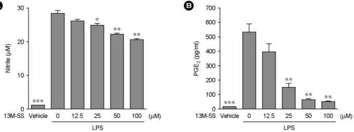
LPS에 의해 활성화된 RAW264.7 세포의 배양액 중에 생성된 NO의 양에 미치는 I3M-5S의 영향을 관찰하고자 RAW264.7 세포에 LPS와 I3M-5S를 동시처리 후, 세포 배양액 중으로 분비된 NO와 PGE
2의 농도를 측정하였다.
I3M-5S의 농도를 12.5 μM로 처리한 그룹에서는 LPS를 단독으로 처리한 그룹과의 NO 생성량의 유의적인 차이 는 관찰할 수 없었으나, I3M-5S의 농도를 25 μM 이상으 로 처리한 그룹에서는 LPS 자극에 의한 NO의 생성을 유 의적으로 억제함을 관찰할 수 있었다 (Fig. 3A).
또한 , I3M-5S를 처리한 그룹에서의 PGE
2생성량을 관 찰한 결과, I3M-5S의 농도를 25 μM 이상으로 처리한 그 룹에서의 PGE
2생성량은 LPS 자극에 의한 PGE
2의 생성 을 유의적으로 억제하였다 (Fig. 3B). NO및 PGE
2가 염증 반응을 매개하는 핵심적인 물질임을 고려할 때, I3M-5S 는 NO 및 PGE
2의 생성을 억제하여 항염증작용을 나타 내는 것으로 사료된다.
NO와 PGE
2의 억제와 관련된 I3M-5S의 작용 기전을 확인하기 위해, LPS에 의해 유도되는 염증반응에 핵심적 으로 작용하는 주요 효소, iNOS 및 COX-2의 발현량에 대한 I3M-5S의 효과를 관찰하였다. RAW264.7 세포에 다 양한 농도의 I3M-5S와 LPS를 처리하여 iNOS와 COX-2 의 발현량을 Western blot 방법을 이용하여 관찰한 결과,
RAW264.7 세포에 LPS를 단독처리 했을 때 iNOS와 COX-2 단백질의 발현이 유도되었고, I3M-5S의 처리가 iNOS와 COX-2의 단백질 발현을 농도의존적으로 억제함 을 관찰할 수 있었다 (Fig. 4). 이와 같은 결과는 I3M-5S 의 처리에 의한 iNOS와 COX-2의 발현량의 감소가 NO 와 PGE
2생성을 억제시킴을 시사하고 있다.
LPS에 의해 자극된 대식세포는 염증반응을 조절하 는 다수의 사이토카인을 생성 · 분비한다. 그 중 TNF-α 는 T 세포와 대식세포를 활성화 시켜 다른 염증성 사 이토카인들의 생성을 유도하여 염증반응을 촉진한다 (Parameswaran and Patial, 2010). IL-6는 LPS의 자극에 의해 대식세포에서 분비되는 중요한 염증성 사이토카인이다 (Bryniarski et al., 2003). 따라서, I3M-5S가 염증성 사이토카
A B
Fig. 3. Effects of I3M-5S on LPS-induced NO and PGE
2production. RAW264.7 cells were treated with indicated concentrations of I3M-5S in the presence of 100 ng/ml of LPS or with LPS alone for 24 hr, and NO (A) and PGE2 (B) release were determined. The results are reported as mean ± SE of three independent experiments in triplicate. Statistical significance is based on the difference when compared with LPS-stimulated cells (*P<0.05, **P<0.01, ***P<0.001).Fig. 4. Effects of I3M-5S on COX-2 and iNOS expression.
RAW264.7 cells were treated the same as in Fig. 3. Thirty μg of protein obtained from each cell lysate was resolved on 10% SDS- PAGE for iNOS and COX-2 determination. β-actin expression is shown as a loading control.
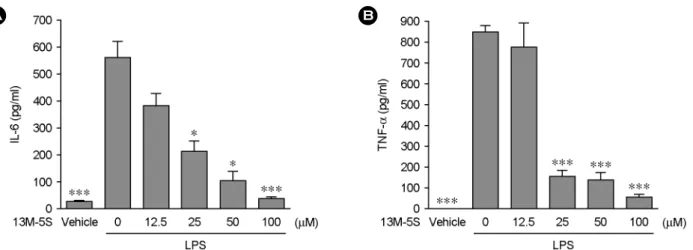
인의 생성에 미치는 영향을 관찰하고자, RAW264.7 세포 에 LPS와 함께 다양한 농도의 I3M-5S를 처리하여 세포 배양액 중으로 분비된 IL-6 및 TNF-α의 양을 측정하였다.
LPS의 처리에 의해 유도된 IL-6와 TNF-α의 농도는 각 각 561.8±59.2 pg/ml과 851.4±36.7 pg/ml이였으나 50 μM 의 I3M-5S를 LPS와 함께 처리한 결과, IL-6는 92.4±40.9 pg/ml로 TNF-α는 135.1±33.18 pg/ml로 그 분비량이 유의 적으로 감소한 것을 확인할 수 있었다 (Fig. 5).
최근 다양한 식물자원으로부터 분리된 생리활성성분을 중심으로 기능성식품 및 의약품의 개발에 대한 연구가 활발히 이루어지고 있다. 식물자원 중 쪽풀은 그 색소로 인해 오래 전부터 염료로 사용되어 왔다. 또한, 쪽풀은 해독, 강장, 해열작용이 있어 민간에서 약용으로 이용되 어 왔고, 특히, 쪽풀의 성분인 indirubin은 백혈병을 치 료하는데 효과가 있다고 보고되었다 (Xiao et al., 2002;
Eisenbrand et al., 2004). 이후 indirubin-3'-oxime을 포함한 다양한 indirubin 유도체가 합성되고 있다 (Eisenbrand et al., 2004). Indirubin-3'-oxime은 glycogen synthase kinase (GSK)-3β와 cyclin-dependent kinases의 강력한 저해제로 항종양효과를 나타내는 것으로 보고되고 있다 (Eisenbrand et al., 2004). 또한 본 연구팀은 indirubin의 또 다른 유도 체인 indirubin-3'-monoxime이 혈관신생을 억제한다는 것 을 밝혔다 (Kim et al., 2011). 이와 같이 다양한 indirubin 유도체가 상이한 생리활성을 보이고 있어 이에 대한 자 세한 연구가 앞으로도 진행되어야 할 것으로 사료된다.
본 연구에서는 indirubin 유도체 중의 하나인 I3M-5S가 대식세포에서 LPS로 유도되는 NO, PGE
2, 염증성 사이토
카인들의 생성을 억제하여 항염증효과를 나타내는 것을 밝혔다 . 본 연구결과를 바탕으로 차후 I3M-5S의 항염증 작용의 기전 및 in vivo에서의 I3M-5S의 항염증작용에 대 한 검증이 필요할 것으로 사료된다.
감사의 글
이 연구는 학부교육선진화 선도대학 지원 사업에 의해 지원되었음에 이에 감사 드립니다.
REFERENCES
Baldwin AS Jr. The NF-κB and IκB proteins: new discoveries and insights. Annu Rev Immunol. 1996. 14: 649-683.
Botting RM. Inhibitors of cyclooxygenases: mechanisms, selectivity and uses. J Physiol Pharmacol. 2006. 5: S113-S124.
Bryniarski K, Maresz K, Szczepanik M, Ptak M, Ptak W.
Modulation of macrophage activity by proteolytic enzymes.
Differential regulation of IL-6 and reactive oxygen inter- mediates (ROIs) synthesis as a possible homeostatic mechanism in the control of inflammation. Inflammation 2003. 27: 333-340.
Butchar JP, Parsa KV, Marsh CB, Tridandapani S. Negative regulators of toll-like receptor 4-mediated macrophage inflam- matory response. Curr Pharm Des. 2006. 12: 4143-4153.
Christie RM. Why is indigo blue? Biotech Histochem. 2007. 82:
51-56.
Eisenbrand G, Hippe F, Jakobs S, Muehlbeyer S. Molecular mechanisms of indirubin and its derivatives: novel anticancer
A B
Fig. 5. Effects of I3M-5S on LPS-induced IL-6 and TNF-α production. RAW264.7 cells were treated with indicated concentrations of
I3M-5S in the presence of 100 ng/ml of LPS or with LPS alone for 24 hr, and IL-6 (A) and TNF-α (B) release were determined. The results are reported as mean ± SE of three independent experiments in triplicate. Statistical significance is based on the difference when compared with LPS-stimulated cells (*P<0.05, ***P<0.001).molecules with their origin in traditional Chinese phyto- medicine. J Cancer Res Clin Oncol. 2004. 130: 627-635.
Goldberg RB. Cytokine and cytokine-like inflammation markers, endothelial dysfunction, and imbalanced coagulation in development of diabetes and its complications. J Clin Endocrinol Metab. 2009. 94: 3171-3182.
Kim JK, Shin EK, Kang YH, Park JH. Indirubin-3'-monoxime, a derivative of a chinese antileukemia medicine, inhibits angio- genesis. J Cell Biochem. 2011. 112:1384-1391.
Laskin DL, Pendino KJ. Macrophages and inflammatory mediators in tissue injury. Annu Rev Pharmacol Toxicol. 1995. 35: 655 -677.
McCartney-Francis N, Allen JB, Mizel DE, Albina JE, Xie QW, Nathan CF, Wahl SM. Suppression of arthritis by an inhibitor of nitic oxide synthase. J Exp Med. 1993. 178: 749-754.
Palombella VJ, Rando OJ, Goldberg AL, Maniatis T. The ubiquitin-proteasome pathway is required for processing the NF-κB1 precursor protein and the activation of NF-κB. Cell 1994. 78: 773-785.
Parameswaran N, Patial S. Tumor necrosis factor-α signaling in macrophages. Crit Rev Eukaryot Gene Expr. 2010. 20: 87-103.
Ricciotti E, FitzGerald GA. Prostaglandins and inflammation.
Arterioscler Thromb Vasc Biol. 2011. 31: 986-1000.
Sharma JN, Al-Omran A, Parvathy SS. Role of nitric oxide in inflammatory diseases. Inflammopharmacology 2007. 15: 252 -259.
Siebenlist U, Franzoso G, Brown K. Structure, regulation and function of NF-κB. Annu Rev Cell Biol. 1994. 10: 405-455.
Tizard IR. Immunology: an introduction inflammation. 2nd ed.
1986. pp. 423-441. Saunders College Publishing.
Weisz A, Cicatiello L, Esumi H. Regulation of the mouse inducible- type nitric oxide synthase gene promoter by interferon- gamma, bacterical lipopolysaccharideand NG-monomethyl- L-arginene. Biochem J. 1996. 316: 209-215.
Xiao Z, Hao Y, Liu B, Qian L. Indirubin and meisoindigo in the treatment of chronic myelogenous leukemia in China. Leuk Lymphoma 2002. 43: 1763-1768.