Received: June 2, 2014 / Accepted: June 2, 2014 Address for correspondence: Ohyun Kwon, MD
Department of Neurology, Eulji Hospital, Eulji University College of Medicine, 14 Hangeulbisuk-gil, Nowon-gu, Seoul 139-872, Korea Tel: +82-2-970-8312, Fax: +82-2-974-7785, E-mail:[email protected], [email protected]
신경근육이음부의 해부 및 생리
을지대학교 의과대학 을지병원 신경과학교실
권 오 현
Anatomy and Physiology of Neuromuscular Junction
Ohyun Kwon
Department of Neurology, Eulji Hospital, College of Medicine, Eulji University, Seoul, Korea
KEYWORDS Acetylcholine, Motor endplate, Neuromuscular junction, Nicotinic acetylcholine
receptor
Neuromuscular junction (NMJ) has a sophisticated structure to transfer neural signal securely to muscle fiber. Synaptogenesis of NMJ is developmentally well preserved along invertebrate and vertebrate, and the morphology of NMJ itself corresponds with its physiological role. In this review, Subcellular specialization of the NMJ, pre- and post‐synaptic compartment and synaptic cleft, and their physiology will be described briefly.
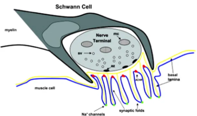
Figure 1. Organization of the NMJ. A terminal Schwann cell caps rather than wraps the motor nerve; terminal branches of the motor axon, which accumulate mitochondria (mc) and synaptic vesicles (sv); and an AChR‐rich postsynaptic endplate. In the presynaptic terminal, active zones (az) form along the junctional surface, and secondary synaptic clefts creates folds in the postsynaptic mem- brane with AChR concentrated at the crests of secondary folds, and voltage‐gated sodium channels concentrated in the troughs of the folds. Synaptic basal lamina contains components, which or- ganize synaptic specializations in all three cells.
신경근육이음부(neuromuscular junction, NMJ)는 생체 신 호가 전달 물질을 매개로 한 세포에서 접한 다른 세포로 전달되는 시냅스(synapse)의 일종으로 신경과 신경 사이 의 전달과 달리, 운동신경과 대상인 근육세포로 이루어진 다. NMJ의 주름 영역은 전체 근육섬유 표면의 0.1%에 지 나지 않는다.1 시냅스전(presynaptic) 부위는 운동신경의 말단부(terminal)로 NMJ의 신경전달물질인 아세틸콜린 (acetylcholine, Ach)의 시냅스소포(synaptic vesicle)가 밀집 해 있는 시냅스전 활성부위(presynaptic active zone)이며, 시냅스후(postsynaptic) 부위는 아세틸콜린수용체(acetylcholine receptor, AchR)가 밀집해 있는 시냅스주름(synaptic fold)으 로 구성된다. 세포외 공간인 시냅스틈새(synaptic cleft)는 acetylcholine esterase (AchE)의 작용으로 시냅스소포에서 분비된 Ach이 AchR에 결합하고 분해되는 과정에 관여한 다(Fig. 1).
Figure 2. The agrin–MuSK–Lrp4 and neuregulin–ErbB pathways.
The agrin–MuSK–Lrp4 and neuregulin–ErbB pathways induce NMJ assembly positive signals. A. Agrin is released by the motor axon terminal and induces AChR clustering, phosphorylation, and stabilization at the postsynaptic membrane. Lrp4 associates with MuSK in the absence of agrin. Agrin binds to the preformed MuSK–Lrp4 complex by interacting with Lrp4 and promotes MuSK transphosphorylation and activation. Once phosphory- lated, MuSK recruits the adapter protein Dok‐7 which binds Crk and CrkL and stimulates further MuSK phosphorylation and kin- ase activity. This induces phosphorylation and stabilization of nascent AChR clusters. Rapsyn is a coeffector in AChR assem- bly which anchorates AChRs at the muscle membrane. B.
Neuregulin (NGR‐1) is released by the nerve and induces AChR transcription in synaptic nuclei. NRG‐1 acts by binding tyrosine kinases receptors ErbBs. ErbB phosphorylation induced by NRG stimulates ERK and JNK kinase activity which phosphor- ylates GABP‐α and GABP‐β transcription factors. GABP‐α heter- odimerizes with GABP‐β and binds DNA at the N‐box thereby enhancing transcription of AChR genes.
신경근육이음부의 발생과 구조
개체 발생 과정 중에 아직 NMJ의 형성되기 이전의 무 신경근육섬유(aneural muscle fiber)에는 AchR 군집(AchR cluster)이 섬유 전반에 걸쳐 발현한다. 발생이 진행하면서 운동신경 말단부가 근육섬유에 도달하여 신경지배를 하면 신경과 접하는 부위에만 AchR 군집이 더욱 발달하고, 다 른 부위에 발현되었던 AChR는 사라진다. 그리고 지배하 는 운동신경은 하나만 남게 되어 운동신경과 근육섬유는 1대 1 대응하여 시냅스를 이룬다. 운동신경과 근육섬유는 다양한 인자를 분비하며 이를 통해 주변의 분화 발달 과 정에 관여한다. 근육섬유는 neurotrophin 3 등을, 운동신경 은 neuroregulin 1을 발현하여 NMJ의 Schwann cell의 생존 과 발달에 영향을 미친다. 반대로 Schwann cell도 다양한 신호물질을 분비하는데 그 중 하나인 transforming growth factor β (TGF β)는 NMJ의 발달을 촉진한다.
흥미롭게도 Ach에 의해 유발된 근육섬유의 전기적 활동 은 AchR의 군집, 발현 그리고 안정화를 방해한다. 이에 대 항하여 운동신경은 당단백질인 agrin을 생성하여 시냅스바 닥판(synaptic basal lamina)으로 분비하며 agrin은 AchR 군 집 등 시냅스후 분화를 촉진한다.
AchR와 함께 분포하는 muscle-specific kinase (MuSK)는 agrin의 NMJ에서의 역활을 매개한다. Agrin과 MuSK는 직 접 결합하지 않으며 Agrin이 lipoprotein-related protein 4 (Lrp4)에 직접 결합하고 Lrp4가 MuSK의 dimerization을 촉 진하여 MuSK의 활성화가 진행한다(Fig. 2). MuSK의 활성 화하는 downstream-of-tyrosine-kinase-7 (Dok7), AchR의 군 집화를 유도하는 rapsyn 등 다양한 adaptor 단백질들이 NMJ의 발생과 향상성에 관여하는데 MuSK의 활성화, MuSK의 세포내섭취(endocytosis)와 분해, AchR의 군집화, AchR 군집의 안정화, nuclear anchoring, scaffolding 역활 등 에 관여 한다.2
시냅스후 구조의 retrograde signal이 시냅스전 발달도 영 향을 미치는데 nerve growth factor (NGF), TGF β, fibroblast growth factor (FGF), glial cell line-derived neurotrophic factor (GDNF), β-catenin, β1 integrin, neurotrophin-3가 대표적인 물질들이다.
NMJ에만 AchR가 발현하는 기전으로 신경 지배를 받으 면 운동 섬유 표면에 퍼져 있던 AchR 들이 시냅스 부위로 이동을 하여 시냅스아래 세포골격(cytoskeleton)에 의해 고 정되고, 신경에서 유래한 인자들에 의해 시냅스 바로 아래 에 있는 세포핵에서 AchR의 전사(transcription)가 활성화되 고, Ach에 의한 근육섬유의 전기적 활동은 시냅스와 떨어
진 세포핵에서의 AchR 발현을 억제한다.3
NMJ에 접한 말단부 Schwann세포는 신호전달, 신경말단 성장과 유지, 축삭돋음(axonal sprouting) 그리고 신경의 재 생 등 NMJ의 기능과 형성에 매우 중요한 역활을 수행한다.
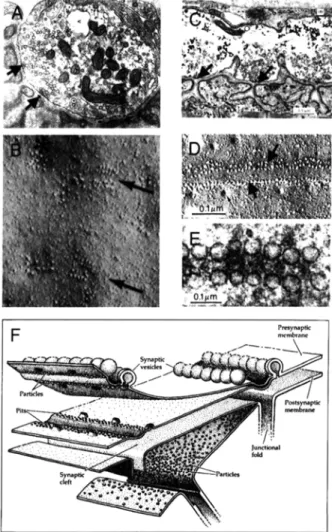
Figure 3. Active zones in the presynaptic membrane at mamma- lian (left column) and frog (right column and lower diagram) NMJs. In the upper electron micrographs (A, C), transverse sec- tions through active zones (indicated by arrows) are shown; these are often much more distinct in frogs than in mammals. In both groups, the active zones lie immediately opposite the openings of the synaptic folds. In freeze‐fracture preparations, the active zones both mammals (B) and frogs (D) are seen to be made up of two parallel rows of intramembranous particles (arrows). In frogs, these rows are up to 1 mm long and run perpendicular to the long axis of the motor axon terminal. In mammals the rows are much shorter, and apparently randomly oriented in the plane of the membrane. In views on the cytoplasmic side of the active zones, synaptic vesicles can be seen aligned with the much smaller intra- membranous particles (E). The diagram in (F) depicts the 3‐di- mensional arrangement of intramembranous particles.
시냅스틈새는 약 50~100 nm의 공간으로 바닥판(basal lamina)로 구성되어 있고 AchE의 anchor 등의 기능을 한다.
AchR는 α- 그리고 β-dystroglycans을 통해 세포막에 부착되 는데 rapsyn이 β-dystroglycan을 AchR에 부착시키는 역활 을 하며 β-dystroglycan은 시냅스후주름의 둔덕(crest)에 AchR와 함께 위치하는 utrophin과 결합한다.
신경근육접합부의 생리
Safety factor는 종말판전위(end plate potential, EPP)의 크 기와 세포막전위와 역치전위(threshold potential)의 차 사이 의 비율을 말한다. EPP는 시냅스소포 하나에 의한 미세종 말판전위(miniature end plate potential, MEPP) 크기와 quan- tal content, 운동신경 말단부에 도달한 하나의 활동전위에 의해 분비되는 시냅스소포의 갯수에 의해 결정된다. 포유류 에서는 한번의 활동전위에 20~300개의 시냅스소포가 분비 된다. 결국 safety factor는 quantal release, Ach 분해 효소인 아세틸콜린에스테라제(acetylcholine esterase, AchE), AchR의 전도 특성, AchR의 밀도, 시냅스후 sodium channel 활동 밀 도 그리고 시냅스주름의 형태 등에 의해 결정된다.4,5
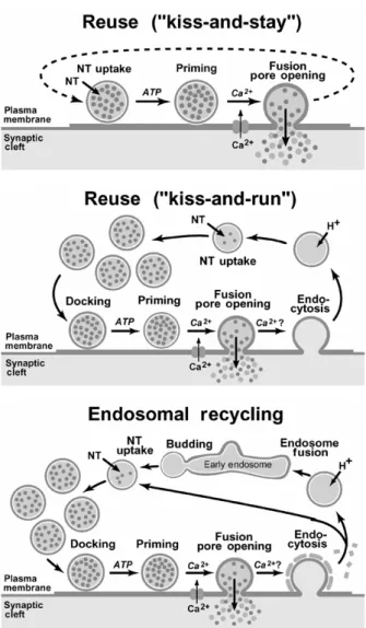
시냅스전 운동신경 말단부가 활동전위로 탈분극이 되면 P/Q type voltage-gated Ca2+ channels에 의해 Ca2+가 유입되 고 AchR가 많은 시냅스후주름에 정확히 대응하여 시냅스 소포는 위치하는데 이 영역을 active zone이라 한다. active zone에는 소포 내용물을 분비하는 칼슘통로가 두 줄로 나열 해 있다6(Fig. 3). 신경 말단부에 calcium의 유입되면 다양한 단백질들의 상호작용으로 시냅스소포가 active zone의 세포 막과 docking, priming, fusion, pore opening의 과정을 거쳐 Ach를 세포외 배출한다. 세포막과 만난 소포는 다시 3가지 기전(“kiss and stay”, “kiss and run”, endosomal recycling)으 로 세포내로 유입되며 재순환된다6(Fig. 4).
Nicotinic AchR(nAchR)는 cys-loop ligand-gated ion chan- nels (LGICs) 군에 포함되며, 척추동물에서는 17개의 nAChR subunits (α1– α 10, β1– β 4, γ, δ, and ε)로 다양한 조합로의pentameric assembliesfh NMJ 뿐만 아니라, 신경계 및 비신경계 세포에서 발현한다. 근육의 nAChR는 태아기 에는 α12βγδ로 구성되고, 성인에서는 γ subunit가 ε subunit 로 대치된다(α12β1εδ)7(Fig. 5). β1 subunit는 AchR의 군집에 관여하는 것으로 알려져 있다. 발생 과정에서 근육섬유가 신경지배를 받게 되면 근육세포는 γ subunit 대신 ε sub- unit를 발현한다.3,8 성인형 AchR는 태아형에 비해 채널의 개방 시간이 짧고, calcium에 비투과성인데 발생학적으로 태아형 AChR는 신경의 근육지배를 유도하는데 관여하지
만, 이후의 발달 과정에서는 성인형 AchR가 NMJ의 안정 및 성숙에 기여하고, 과도한 calcium의 유입에 의한 나쁜 영향을 최소화하는 것으로 알려져 있다.3
AchR는 allosteric protein으로서 전류의 흐름이 없고 ago-
Figure 4. Synaptic vesicle recycling pathways. Three pathways are proposed: a pathway in which vesicles endocytose by clo- sure of the fusion pore and are refilled with neurotransmitters while remaining docked to the active zone (kiss‐and‐stay); a lo- cal recycling pathway that is clathrin independent but results in mixing vesicles with the reserve pool after endocytosis (kiss‐and
‐run); and a pathway whereby vesicles undergo clathrin‐medi- ated endocytosis and recycle either directly or via endosomes.
Figure 5. Structure of acetylcholine receptor at the NMJ. A. The threading pattern of receptor subunits through the membrane. B.
A schematic representation of the quaternary structure, showing the arrangement of the subunits in the muscle‐type receptor, the location of the two ACh‐binding sites (between an ‐ and a ‐sub- unit, and an ‐ and a ‐subunit), and the axial cation‐conducting channel. C. A cross‐section through the 4.6‐Å structure of the re- ceptor determined by electron microscopy of tubular crystals of Torpedo membrane embedded in ice. Dashed line indicates pro- posed path to binding site.
nist에 낮은 affinity를 보이는 폐쇄 및 안정 상태, agonist에 높은 affinity를 보이며 양이온의 전류 흐름이 있는 개방 상 태 그리고 높은 gonist affinity를 보이나 전류의 흐름이 없 는 탈감작 상태(desensitized state)를 가질 수 있다. Ach을 비롯한 agonist의 결합으로 닫힌 상태에서 개방 상태로 바 뀔 수 있으나 지속적인 agonist의 노출은 탈감작으로 진행 하여 양이온의 흐름이 더 이상 없게 된다. AchR는 Na+와 K+에 대해 비선택적으로 투과성을 보이나 electrochemical gradient에 의해 보다 세포내 K+가 세포 밖으로 유출되는
것보다 많은 Na+가 유입되어 탈분극되고, 세포막 전위가 역치 전위에 도달하면 주변의 voltage-gated Na channel이 개방되어 근유섬유의 활동전위가 발생한다9(Fig. 6). 종말 판 주변에서 발생한 활동전위는 근육섬유의 t-tubule sys- tem의 dihydropyridine receptors인 L-type voltage-gated Ca2+
channels에 의해 Ca2+이 유입되고, 이는 또한 근육세포의 sarcoplasmic reticulum (SR) 막에 존재하는 ryanodine re- ceptor인 Ca2+ channel을 통해 SR의 Ca2+가 세포질내로 유 입하고 actin myofilament의 troponin C와 결합하여, ex- citation-contraction coupling을 통해 최종적으로 근육 수축 이 발생한다.
NMJ의 신호 전달은 대단히 안정적인 과정으로 극심한 피로 상태에서도 수의적 운동에서 신경근육전달의 실패는 생기지 않는다. 이는 장기간의 강한 수준의 근육 수축을 위해 지속적이고 반복적인 운동신경의 활동전위에 의해 NMJ의 가용한 신경전달물질이 감소하여 quantal content가 감소함에도 불구하고 정상적인 신경근육전달은 정상적으 로 유지된다.
A C
B
Figure 6. Reaction sequence and timing of synaptic transmission. The principal reactions with the associated time constants are shown on the left, and traces from the corresponding re- actions in the calyx of Held synapses are illustrated on the right. The time calibration bar at the bottom applies to all traces.
Figure 7. The quantal content depends on the frequency of activation. (A) Isolated human intercostal nerve‐muscle preparations were stimulated at different frequencies. The muscle fibers were pretreated with glycerol to block action potentials. During stimulation, there is an initial rapid decline of quantal content. This is followed by a slower decline which is not seen in this figure (from Kamenskaya et al., 1975). (B) Quantal release is better maintained during repetitive activity at NMJs in slow than in fast rat muscles. The quantal content was determined in isolated nerve‐muscle preparations from fast (EDL) and slow (Soleus) rat muscles.
The nerves were stimulated at 20 Hz for 10 min.
한 개의 소포는 5,000~10,000개의 Ach 분자를 포함한다.
신경의 활동전위가 없는 상태에서도 자발적인 quantum의 분 비가 1초에 수 회에서 1분에 수 회 빈도로 발생하는데, 이 는 NMJ에서 약 1mV의 작은 탈분극들(miniature end plate potentials, MEPPs)로 반영된다. 신경의 활동전위가 도달하 면 active zone에서 종에 따라 20~200개의 quanta가 수 천분 의 1초동안 분비된다.5
생리적 근육 수축은 운동신경의 지속 반복 자극으로 유 지된다. 반복 자극이 유지됨에 따라 각 신경자극에 따른 quantal content는 변하는데, 20Hz 이상의 지속적인 신경 자 극은 신경 말단부의 Ca2+의 축적을 유도하여 소포 분비에 우호적인 환경을 조성한다. 이에 반해 소포의 분비가 지속 함에 따라 active zone에 부착된(“docked”) 즉시 사용할 수 있는(“readily available”) 소포의 갯수가 줄어서, 자극이 반 복될수록 보통 10~20번의 자극만에 분비되는 소포 갯수도
급격히 줄어들어 최초의 quantal content의 절반 수준으로 감소하기도 한다(Fig. 7). 시간이 좀 더 지나면 신경 활동 에 따른 second messenger cascade가 관여해서 synapsin에 부착되어 있는 더 많은 수의 “mobilization” 소포를 동원하 여 생리적 활동에 대응하도록 quantal content가 upregulate 된다. 이 외에도 수 초에서 수 분이내로 Ach외 다른 수용 체들이 신경 말단부의 활성을 조절하는데, 시냅스전 AchR, ADP/ATP에 반응하는 purinergic receptors, 그리고 adrenoreceptor들이 관여하지만 그들의 생리적 기능은 분명 하게 밝혀지지 않았다.
분비된 대부분의 Ach은 시냅스틈을 지나 AchR에 반응 한다. 그러나 AchR에서 해리되면 곧 바로 AchE에 의해 분 해되는데 이 과정을 통해 분비된 Ach의 활동을 공간적으 로나 시간적으로 국소화할 수 있으며, 이는 높은 빈도의 근육 수축을 가능하게 한다. Nodes of Ranvier나 axon hil-
A B
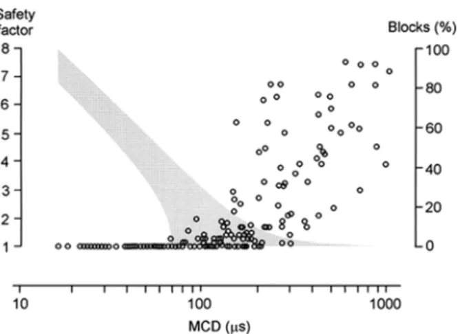
Figure 8. Relationship between the jitter and the safety factor.
Relationship between the jitter and the safety factor (shaded area) estimated on the basis of jitter studies of 170 myasthenic NMJs at 10‐Hz stimulation (circles). If the upper values of the safety factor in human skeletal muscle are between 8 and 10, the lowest jitter value in this material (15 μs) corresponds to safety factor values of 6 to 8. A safety factor of close to 1 (a safe- ty margin close to zero) is assumed to be indicated by inter- mittent blocking. In this sample of myasthenic NMJs, the lowest MCD at which blocking is observed is about 60 μs, and the high- est MCD at which no blocking is seen is about 200 μs. Nonlinear interpolation is used to delineate the approximate range of safe- ty factor values for a given jitter value.
lock과 같은 예로 시냅스후주름의 깊은 골에는 기타 부위 에 비해 10배 정도의 높은 밀도로 VGSCs이 존재하여, 역 치를 낮추는데 기여한다. 뿐만 아니라 촘촘한 시냅스후주 름은 전류에 대해 높은 저항을 가지는데, 이를 통해 AchR 에 의한 작은 전류에도 충분한 탈분극을 유도하여 VGSCs 의 활성화를 보다 더 용이롭게 한다.
단일섬유근전도검사를 통해 중증근무력증 환자에서의
safety factor와 mean consecutive differences (MCDs), block- ing의 관계를 비교한 연구에서, safety factor가 감소할수록 MCDs가 차츰 증가하고, MCDs가 60msec에 이르면, 간헐 적으로 blocking이 확인되는데, 이 상태의 safety factor는 1 에 근접한 상태이다10(Fig. 8).
REFERENCES
1. Wu H, Xiong WC, Mei L. To build a synapse: signaling pathways in neuromuscular junction assembly. Development 2010;137:
1017-1033.
2. Ferraro E, Molinari F, Berghella L. Molecular control of neuromuscular junction development. Journal of cachexia, sarcopenia and muscle 2012;3:13-23.
3. Kalamida D, Poulas K, Avramopoulou V, et al. Muscle and neuronal nicotinic acetylcholine receptors. Structure, function and pathogenicity.
The FEBS journal 2007;274:3799-3845.
4. Hughes BW, Kusner LL, Kaminski HJ. Molecular architecture of the neuromuscular junction. Muscle & nerve 2006;33:445-461.
5. Wood SJ, Slater CR. Safety factor at the neuromuscular junction.
Progress in neurobiology 2001;64:393-429.
6. Sudhof TC. The synaptic vesicle cycle. Annual review of neuroscience 2004;27:509-547.
7. Zouridakis M, Zisimopoulou P, Poulas K, Tzartos SJ. Recent advances in understanding the structure of nicotinic acetylcholine receptors. IUBMB life 2009;61:407-423.
8. Karlin A. Emerging structure of the nicotinic acetylcholine receptors. Nature reviews Neuroscience 2002;3:102-114.
9. Goodman BE. Channels active in the excitability of nerves and skeletal muscles across the neuromuscular junction: basic function and pathophysiology. Advances in physiologyeducation 2008;32:127-135.
10. Trontelj JV, Mihelin M, Khuraibet A. Safety margin at single neuromuscular junctions. Muscle & nerve Supplement 2002;
11:S21-27.