pISSN 1598-9992 eISSN 2233-6869
CASE REPORT
다발성 전이를 동반한 후복막강 악성 혈관주위상피모양세포종양 1예
오혜원
1,3, 김태효
1,3, 차라리
1,3, 김나영
1,3, 김현진
1,3, 정운태
1,3, 이옥재
1,3, 이정희
2,3경상대학교 의학전문대학원 내과학교실1, 병리학교실2, 건강과학원3
A Case of Malignant Perivascular Epithelioid Cell Tumor of the Retroperitoneum with Multiple Metastases
Hye Won Oh1,3, Tae Hyo Kim1,3, Ra Ri Cha1,3, Na Young Kim1,3, Hyun Jin Kim1,3, Woon Tae Jung1,3, Ok Jae Lee1,3 and Jeong Hee Lee2,3 Departments of Internal Medicine1 and Pathology2, Institute of Health3, Gyeongsang National University School of Medicine, Jinju, Korea
Perivascular epithelioid cell tumors (PEComas) are unusual mesenchymal neoplasms composed of histologically and im- munohistochemically distinct perivascular epithelioid cells (PECs). Although PEComas have the potential to behave in a malignant fashion, malignant PEComas arising from the retroperitoneum are extremely rare. A 68-year-old woman presented with a painful palpable mass in her left upper abdomen. Computed tomography of the abdomen showed a 9 cm sized heterogeneous mass in left para-aortic space and multiple hypervascular nodules in the liver. 18F-fludeoxyglucose-PET/CT showed multifocal hyper- metabolic lesions in retroperitoneum, liver, and skeletal bones. Percutaneous needle biopsies were done on the retroperitoneal and hepatic mass. Both specimens were positive for human melanoma black-45 (HMB-45) on histological and im- munohistochemical staining which was compatible with PEComas. Herein, we report a rare case of retroperitoneal PEComa with multiple metastases involving liver and bone at initial diagnosis that exhibited aggressive behavior and resulted in a devastating prognosis. (Korean J Gastroenterol 2014;64:302-306)
Key Words: Perivascular epithelioid cell tumor; Metastasis; Retroperitoneal; Liver; Bone
Received February 7, 2014. Revised March 17, 2014. Accepted March 18, 2014.
CC This is an open access article distributed under the terms of the Creative Commons Attribution Non-Commercial License (http://creativecommons.org/licenses/
by-nc/3.0) which permits unrestricted non-commercial use, distribution, and reproduction in any medium, provided the original work is properly cited.
교신저자: 김태효, 660-702, 진주시 강남로 79, 경상대학교병원 내과
Correspondence to: Tae Hyo Kim, Department of Internal Medicine, Gyeongsang National University Hospital, 79 Gangnam-ro, Jinju 660-702, Korea. Tel: +82-55-750- 8726, Fax: +82-55-755-9078, E-mail: [email protected]
Financial support: None. Conflict of interest: None.
서 론
혈관주위상피모양세포종양(perivascular epithelioid cell tumor, PEComa)은 중간엽에서 발생하는 종양으로 평활근 및 멜라닌 세포의 분화를 동시에 보이는 종양이며, 병리조직 학적으로 혈관주위상피모양세포가 존재하고 면역조직화학염 색에서 근육 멜라닌 세포 표지자에 양성인 것이 특징이다.1-3 혈관주위상피모양세포종양은 신장, 폐, 췌장, 자궁, 방광, 전 립선, 간, 장간막, 위장관, 후복막강 등 대부분의 장기에서 발 생할 수 있는데, 특히 후복막강에 발생한 악성 혈관주위상피모 양세포종양은 매우 드물어 국내에서는 아직 보고된 바 없다.4,5
저자들은 후복막강 종괴로 내원한 68세 여자에서 진단 당 시 이미 간과 뼈에 다발성 전이를 동반한 후복막강 악성 혈관 주위상피모양세포종양 1예를 국내에서 처음 경험하였기에 문 헌고찰과 함께 보고하는 바이다.
증 례
68세 여자가 내원 2개월 전부터 좌상복부에 동통을 동반한 큰 종괴가 있어 내원하였다. 과거력에서 9년 전 유두상 갑상 선암으로 갑상선 전절제술을 시행받았으며 가족력과 사회력 에서는 특이사항은 없었다.
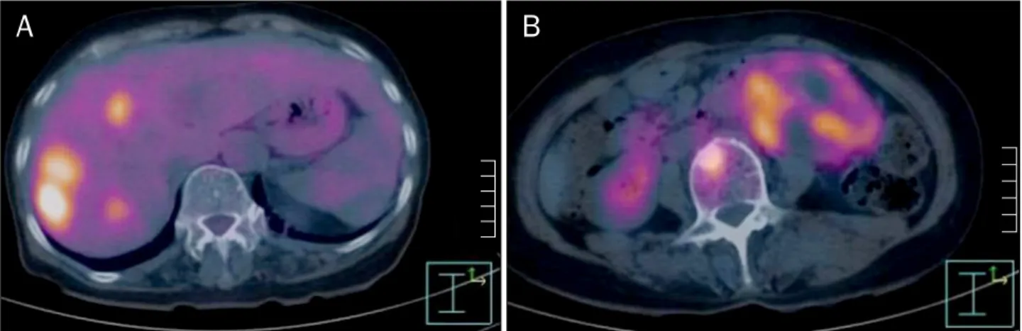
Fig. 2. PET/CT scan shows 18F-fludeoxyglucose hot uptakes in the liver (A) and left para-aortic space mass (B).
Fig. 1. Abdominal enhanced computer tomography shows multiple hypervascular nodules in the liver (A) and 9 cm sized heterogenous enhancing mass in left para-aortic space (B).
내원 당시 만성 병색이었으나 의식은 명료하였고, 신체활 력징후는 혈압은 120/90 mmHg, 맥박수는 분당 68회, 호흡 수는 분당 14회, 체온은 36.7oC였다. 신체검사에서 좌측 상복 부에 손바닥 크기의 고정되고 단단한 종괴가 촉지되었으며, 압통은 있었으나 반발통은 없었다. 검사실 소견에서 백혈구 9,400/mm3, 혈색소 12.5 g/dL, 헤마토크리트 37%, 혈소판 281,000/mm3였다. 생화학검사에서 혈액요소질소 18.2 mg/dL, 크레아티닌 0.63 mg/dL, 총 단백질 6.8 g/dL, 알부민 4.2 g/dL, 총 빌리루빈 1.27 mg/dL, AST/ALT 20/18 IU/L, LDH 424 U/L, HBsAg 음성, anti-HBs 양성, anti-HCV 음성, anti-HIV 음성이었고, 혈청내 종양표지자 검사에서 AFP 1.71 ng/mL, CEA 1.19 ng/mL, CA 19-9 2.95 U/mL, CA 125 13.77 U/mL였다. 복부 초음파와 전산화단층촬영에서 좌측 복부 대 동맥과 요근, 신장, 요관, 신동맥과 신정맥에 인접하여 비균질 한 조영증강을 보이는 약 9 cm 종괴가 관찰되었다. 또한 간양 엽에 높은 조영증강을 보이는 다발성 종괴가 관찰되었다(Fig. 1).
18F-fludeoxyglucose (18F-FDG)-PET/CT 전신 스캔에서는 좌
측 복부 대동맥 주변과 간 뿐만 아니라 5번 흉추의 왼쪽 횡돌 기, 2번 및 3번 요추 체부, 4번 요추 왼쪽 추궁판, 왼쪽 장골에 도 18F-FDG 섭취 증가 소견이 관찰되었다(Fig. 2).
간 및 뼈에 다발성 전이를 동반한 악성 후복막강 종양 또는 유두상 갑상선암의 재발로 인한 후복막강, 간 및 뼈전이를 의 심하고 입원 2일째 초음파 유도하에 후복막강과 간 종괴에 합병증 없이 경피적 조직생검을 시행하였다. 후복막강과 간 종괴의 조직병리 검사에서 동일하게 과립형 호산구세포질로 구성된 다양한 모양의 상피모양세포들이 둥지를 형성하고 있 었고, 유사분열지수는 2/10 HPF였으며 중등도의 핵 이형성 이 관찰되었다(Fig. 3A). 그리고 면역조직화학염색에서 cyto- keratin 음성, vimentin 음성, CD56 음성, S-100 protein 음 성, human melanoma black-45 (HMB-45) 양성, CD117 (c-KIT) 음성, inhibin 음성, D2-40 음성, smooth muscle ac- tin 음성을 보여 혈관주위상피모양세포종양으로 진단되었다 (Fig. 3B-D). 진단 후 환자에게 항암치료를 권유하였으나 거 절하고 지내다 2개월 후 사망하였다.
Fig. 3. (A) The tumor shows nested epithelioid tumor cells with round nucleus and granular eosinophilic or clear cytoplasm (H&E, ×200). The tumor cells are positive for HMB-45 (B, ×200), but negative for smooth muscle actin (C, ×200) and S-100 (D, ×200).
고 찰
1992년 Bonetti 등1이 혈관주위상피모양세포종양이라는 용 어를 처음으로 사용한 이후 지금은 발생 위치에 관계없이 혈 관근육지방종(angiomyolipoma), 림프관근종증(lymphangio- leiomyomatosis), 폐의 clear cell “sugar” tumor를 포함하는 중간엽 종양의 한 범주에 속하는 종양이다.2,6 혈관주위상피모 양세포종양은 공통적으로 투명세포질 또는 호산구세포질을 가진 혈관 주위에 분포한 다양한 모양의 상피모양세포로 이루 어져 있고 세포 내부에 과립물질이 존재하며 면역조직화학염 색에서 smooth muscle actin 및 HMB-45 (gp100 protein) 같은 melanocytic marker에 양성을 보인다.1,7 HMB-45와 Melan-A는 혈관주위상피모양세포의 가장 민감한 melano- cytic marker이다.
이처럼 혈관주위상피모양세포종양은 병리조직 및 면역조 직화학염색으로 진단하는 드문 종양으로서 자연사나 예후에 대해서 알려진 바가 별로 없으나 대부분 양성종양으로서 완전
한 수술적 절제가 이루어진 후에는 재발을 하지 않는 것으로 알려져 있다. 하지만 국소적인 침습적 재발 또는 전이를 보이 는 악성종양도 드물게 발견된다. Folpe 등3,4이 제안한 악성을 시사하는 소견으로는 종양의 크기(직경 >5 cm), 침윤성 성장 경향, 높은 세포밀도 및 높은 핵분화도, 높은 유사분열 정도 (고배율상 >1/50), 조직학적 괴사, 혈관침범으로 이중 2개 이 상이 해당되면 악성으로 분류한다(Table 1). 현재까지 후복막 강에서 발견된 악성 혈관주위상피모양세포종양은 매우 드물며 특히 국내에서는 아직 보고된 바가 없다. Table 2에서는 현재 까지 국외문헌에 보고된 후복막강에 발생한 악성 혈관주위상피 모양세포종양의 8개 증례들을 정리하여 특성을 살펴보았다.8-15 Table 2의 8예들은 대부분 여성이고, 흔히 우연히 발견되 는 양성 혈관주위상피모양세포종양과 달리 복통, 요통, 복부 압박감, 촉지되는 종괴 등의 증상이 대부분 있었다. 일차치료 로 모든 예에서 수술적 절제를 시행하였고, 1예11를 제외한 모든 예에서 재발하였다. 재발하지 않은 1예도 추적기간이 짧 아 이후 재발하였을 가능성도 배제하기 어렵다. 또한, 2예8,12
Table 1. Proposed Classification of Perivascular Epithelioid Cell Tumors by Folpe et al.3,4
Risk features Risk category
Size >5 cm
Infiltrative growth pattern High nuclear grade and cellularity Mitotic rate >1/50 HPF Necrosis
Vascular invasion
Benign; <2 high risk features and size <5 cm
Uncertain malignant potential; size >5 cm with no other high risk features or nuclear pleomorphism/multinucleated giant cells only
Malignant; 2 or more high risk features
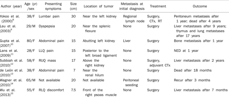
Table 2. Summary of Previously Reported Cases of Malignant Retroperitoneal Perivascular Epithelioid Cell Tumors
Author (year) Age (yr) /sex
Presenting symptoms
Size
(cm) Location of tumor Metastasis at
initial diagnosis Treatment Outcome Yokoo et al.
(2000)8
38/F Lumbar pain 30 Near the left kidney Regional lymph node
Surgery, CTx, RT
Peritoneum metastasis after 1 year; dead after 4 years Lau et al.
(2003)9
29/M Dyspepsia 20 Near the splenic flexure
None Surgery Liver metastases after 9 years;
thymus and lung metastases after 17 years
Gupta et al.
(2007)10
80/F Abdominal pain 15 Abutting left kidney Liver Surgery Bone metastasis after 1 year
Lans et al.
(2009)11
28/F LLQ pain 15 Posterior to the left broad ligament
None Surgery NED at 1 year
Subbiah et al.
(2010)12
58/F RUQ mass 17 Above the
right kidney
None Surgery, adjuvant CTx
Liver metastasis after 2 years
de León et al.
(2010)13
38/F Abdominal pain 7 Near the renal hilum
None Surgery Dead after 18 months
Wagner et al.
(2010)14
65/M Not available 20 Not available Peritoneal seeding
Surgery Recur after 3 months
Wu et al.
(2013)15
55/F RLQ discomfort 7.5 Front of the right psoas muscle
None Surgery Liver metastasis after 7 months
CTx, chemotherapy; RT, radiation therapy; LLQ, left lower quadrant; RUQ, right upper quadrant; RLQ, right lower quadrant; NED, no evidence of disease.
에서 수술 후 화학요법을 추가로 시행하였지만 결국 1-2년 내에 재발하였다. 일반적으로 악성 혈관주위상피모양세포종 양에서 평균 약 71%에서 주위 장기로의 전이, 침범 등의 공격 적인 성향을 보인다고 보고된 바 있으나, 후복막강의 악성 혈 관주위상피모양세포종양은 상대적으로는 드물게 발생하지만 더 높은 침습성과 불량한 예후를 가짐을 확인하였다.8-15 이번 증례도 이미 진단 당시 후복막강으로부터 간과 뼈로 다발성 전이를 하는 매우 공격적인 성향의 악성 혈관주위상피모양세 포종양으로 수술에 의한 종괴의 완전 제거가 불가능하였으며 항암치료 없이 단지 보존적인 치료 중 조기 사망하여 아주 나쁜 예후를 보였다.
악성 혈관주위상피모양세포종양의 치료로 광범위한 수술 적 절제 외에 mammalian target of rapamycin complex 1 (mTORC1) 억제제인 sirolimus로 치료한 사례 및 sirolimus 에 반응이 없는 환자에서 everolimus 또는 doxorubicin과 ifosfamide로 항암치료를 시도한 사례가 보고되고 있으나, 혈 관주위상피모양세포종양이 일반적으로 드물어 아직 수술적 치료 외 항암치료 또는 방사선치료의 효능을 평가하기 매우
어려우므로 현재까지 치료기준이 확립되어 있지 않다.14,16-18 따라서 혈관주위상피모양세포종양의 발생과 관련된 분자생물 학적 기전과 유전적 요인에 대한 추후 연구를 토대로 종양의 악성도 평가 기준 및 수술적 절제와 더불어 항암 또는 방사선 치료의 치료기준의 새로운 정립이 꼭 필요하다.
결론으로 혈관주위상피모양세포종양은 중간엽에서 기원하 는 드문 종양으로, 후 복막강에서 특히 악성으로 발생하는 경 우는 매우 드물고 예후가 매우 나쁘다는 것을 국외 보고와 이번 증례로 확인할 수 있었다. 따라서 후복막강에 발생한 혈 관주위상피모양세포종양에 대해 수술이 가능하여 근치적 절제 술을 하더라도 악성으로 판정된 경우에는 재발과 전이가 매우 흔하여 보다 적극적인 추적관찰 및 치료가 필요할 것으로 생각 된다. 향후 악성 혈관주위상피모양세포종양에 대한 보다 많은 경험보고와 발병원인 및 치료에 대한 연구가 더 필요할 것이다.
REFERENCES
1. Bonetti F, Pea M, Martignoni G, Zamboni G. PEC and sugar. Am J Surg Pathol 1992;16:307-308.
2. Pea M, Martignoni G, Zamboni G, Bonetti F. Perivascular epi- thelioid cell. Am J Surg Pathol 1996;20:1149-1153.
3. Folpe AL, Kwiatkowski DJ. Perivascular epithelioid cell neo- plasms: pathology and pathogenesis. Hum Pathol 2010;41:1-15.
4. Folpe AL, Mentzel T, Lehr HA, Fisher C, Balzer BL, Weiss SW.
Perivascular epithelioid cell neoplasms of soft tissue and gyne- cologic origin: a clinicopathologic study of 26 cases and review of the literature. Am J Surg Pathol 2005;29:1558-1575.
5. Hornick JL, Fletcher CD. Sclerosing PEComa: clinicopathologic analysis of a distinctive variant with a predilection for the retroperitoneum. Am J Surg Pathol 2008;32:493-501.
6. Ruco LP, Pilozzi E, Wedard BM, et al. Epithelioid lymphangioleio- myomatosis-like tumour of the uterus in a patient without tuber- ous sclerosis: a lesion mimicking epithelioid leiomyosarcoma.
Histopathology 1998;33:91-93.
7. Hornick JL, Fletcher CD. PEComa: what do we know so far?
Histopathology 2006;48:75-82.
8. Yokoo H, Isoda K, Nakazato Y, et al. Retroperitoneal epithelioid angiomyolipoma leading to fatal outcome. Pathol Int 2000;
50:649-654.
9. Lau SK, Marchevsky AM, McKenna RJ Jr, Luthringer DJ.
Malignant monotypic epithelioid angiomyolipoma of the retroperitoneum. Int J Surg Pathol 2003;11:223-228.
10. Gupta C, Malani AK, Gupta V, Singh J, Ammar H. Metastatic retro- peritoneal epithelioid angiomyolipoma. J Clin Pathol 2007;60:
428-431.
11. Lans TE, van Ramshorst GH, Hermans JJ, den Bakker MA, Tran TC, Kazemier G. Perivascular epithelioid cell tumor of the retro-
peritoneum in a young woman resulting in an abdominal chyloma. J Gastrointest Surg 2009;13:389-392.
12. Subbiah V, Trent JC, Kurzrock R. Resistance to mammalian tar- get of rapamycin inhibitor therapy in perivascular epithelioid cell tumors. J Clin Oncol 2010;28:e415.
13. de León DC, Pérez-Montiel D, Bandera A, Villegas C, Gonzalez- Conde E, Vilchis JC. Perivascular epithelioid cell tumor of ab- dominal origin. Ann Diagn Pathol 2010;14:173-177.
14. Wagner AJ, Malinowska-Kolodziej I, Morgan JA, et al. Clinical ac- tivity of mTOR inhibition with sirolimus in malignant perivascular epithelioid cell tumors: targeting the pathogenic activation of mTORC1 in tumors. J Clin Oncol 2010;28:835-840.
15. Wu JH, Zhou JL, Cui Y, Jing QP, Shang L, Zhang JZ. Malignant peri- vascular epithelioid cell tumor of the retroperitoneum. Int J Clin Exp Pathol 2013;6:2251-2256.
16. Scheppach W, Reissmann N, Sprinz T, Schippers E, Schoettker B, Mueller JG. PEComa of the colon resistant to sirolimus but re- sponsive to doxorubicin/ifosfamide. World J Gastroenterol 2013;19:1657-1660.
17. Koenig AM, Quaas A, Ries T, et al. Perivascular epitheloid cell tu- mour (PEComa) of the retroperitoneum - a rare tumor with un- certain malignant behaviour: a case report. J Med Case Rep 2009;3:62.
18. Gennatas C, Michalaki V, Kairi PV, Kondi-Paphiti A, Voros D.
Successful treatment with the mTOR inhibitor everolimus in a patient with perivascular epithelioid cell tumor. World J Surg Oncol 2012;10:181.