www.krspine.org
Acute Spontaneous Cervical Spinal Epidural Hematoma with Spontaneous Resolution
-A Case Report-
Young-Do Koh, M.D.,Seung Hwan Kook, M.D.
J Korean Soc Spine Surg 2011 Sep;18(3):163-168.
Originally published online September 30, 2011;
http://dx.doi.org/10.4184/jkss.2011.18.3.163
Korean Society of Spine Surgery
Department of Orthopedic Surgery, Inha University School of Medicine
#7-206, 3rd ST. Sinheung-Dong, Jung-Gu, Incheon, 400-711, Korea Tel: 82-32-890-3044 Fax: 82-32-890-3467
©Copyright 2011 Korean Society of Spine Surgery pISSN 2093-4378 eISSN 2093-4386
The online version of this article, along with updated information and services, is located on the World Wide Web at:
http://www.krspine.org/DOIx.php?id=10.4184/jkss.2011.18.3.163
This is an Open Access article distributed under the terms of the Creative Commons Attribution Non-Commercial License (http://
creativecommons.org/licenses/by-nc/3.0) which permits unrestricted non-commercial use, distribution, and reproduction in any medium, provided the original work is properly cited.
Journal of Korean Society of
Spine Surgery
Received: December 31, 2010 Revised: May 31, 2011 Accepted: June 10, 2011
Published Online: September 30, 2011 Corresponding author: Young-Do Koh, M.D
Department of Orthopedic Surgery, Ewha Womans University Mokdong Hospital, 911-1 Mokdong, Yangcheon-ku, Seoul, Korea
TEL: 82-2-2650-5114, FAX: 82-2-2655-0984 E-mail: [email protected]
“This is an Open Access article distributed under the terms of the Creative Commons Attribution Non-Commercial License (http://
creativecommons.org/licenses/by-nc/3.0/) which permits unrestricted non-commercial use, distribution, and reproduction in any medium, provided the original work is properly cited.”
Acute Spontaneous Cervical Spinal Epidural Hematoma with Spontaneous Resolution
-A Case Report-
Young-Do Koh, M.D.,Seung Hwan Kook, M.D.
Department of Orthopedic Surgery, Ewha Womans University Mokdong Hospital, Seoul, Korea
Study Design: This is a case report.
Objectives: We wanted to report on the spontaneous dissolution of acute spontaneous epidural hematoma of the cervical spine and the effectiveness of conservative treatments, in the cases where the symptoms related to spontaneous epidural hematoma improve by themselves in a short period.
Summary of Literature Review: Cases of acute spontaneous epidural hematoma of the cervical spine are rarely reported; surgical decompression procedures have been performed in most of the cases as treatment. However, there are some reported cases of hematoma dissolving spontaneously after a certain period of conservative treatment.
Material and Methods: A 29 year-old female, who had no history of recent trauma, appealed neck pain with radiating pain in her upper right extremity due to acute epidural hematoma of the cervical spine which was diagnosed with MRI. The neck pain and radiating pain showed early recovery and gradual improvement during a period of the following 7 days after occurrence. MRI studies were done after 3 days, 7days, and 1 month from the day of occurrence of the symptoms.
Results: In comparing MRI studies there were significant decreases in the sizes of hematoma, which implied spontaneous dissolution.
Almost all the symptoms related to acute spontaneous epidural hematoma vanished after a 1-month period of conservative treatment.
Conclusions: Decompressive surgical procedure may not be necessary, if the symptoms related to spontaneous epidural hematoma improve by themselves in short period of conservative treatment.
Key Words: Cervical, Epidural hematoma, Spontaneous, Resolution
INTRODUCTION
Although medication such as aspirin, or blood vessel malformations, coagulopathy complications, etc. are being thought of as causes of epidural hematoma of the cervical spine, cases are reported as spontaneous epidural hematoma of which causes are not known.1) Spontaneous epidural hematoma had been reported seldomly after Jackson’s first report2) in 1869, however, with advances in the diagnostic technique including magnetic resonance imaging, it has resulted in an increasing number of cases being reported.3) The typical clinical symptoms, which vary according to the regions of epidural hematoma, can be neurological disorders such as motor and sensory disorders, urination, defecation, etc, and can rapidly progress into paralysis of the limbs. In cases of epidural hematoma, neck pain and radiating pains such as, arm pain and shoulder pain can occur,
Young-Do Koh et al Volume 18 • Number 3 • September 2011
www.krspine.org 164
and this can be suspected of being disc herniation in the early diagnosis. Surgical decompression of the hematoma has been performed as traditional a treatment of epidural hematoma, however, there are some reports of spontaneous resolving neurological abnormalities and dissolving of hematoma in short time of period.4-6) Epidural hematoma of the cervical spine cases have been, albeit rarely, reported overseas, however, no cases of epidural hematoma of the cervical spine have been reported in Korea.
CASE REPORT
A 29-year-old female patient without traumatic history, started experiencing acute neck pain and acute radiating pain, since 4 hours prior to her hospital visit, that started from her right periscapular area and ran through the inner arm to the 4th and 5th fingers of her hand; the pain was severe enough for her to visit our emergency department. At the time of her visit, her vital signs were stable: blood pressure 121/63 mmhg, pulse
Fig. 1. MR images on the 3rd hospital day, demonstrating an epidural lesion at the dorsal C6~T1 level as a intermediate to low intensity area on the (A) T2-weighted sagittal image (B) T2-weighted axial image and (C) intermediate to high intensity area on the T1-weighted image (D) T1-weighted axial image.
64 beats/min, body temperature of 36.7 C, and respiratory rate of 20 breaths/min were shown, and she had no history of coagulopathy, vascular deformity nor taking aspirins. There were no signs of cervical deformity nor tenderness, and there was a pattern of her radiating pain in her upper right arm improving at abduction of the shoulder and worsening when her neck was positioned at flexion state. Physical examination showed normal motor strength in her upper and lower limbs, and there were no sensory abnormalities. Her brachial biceps, brachioradialis muscles and triceps muscle’s deep tendon reflex showed no hyperactivity findings; there were no ankle clonus or Babinski signs. There were no abnormal signs sufficient to suspect cervical myelopathy, in terms of finger escape signs, Hoffmann’s signs,
and signs from the grip-and-release test. Cervical spine disc herniation was suspected and bed rest was recommended and symptomatic treatment for her pain and outpatient follow-ups were conducted. On the 3rd day of occurrence she revisited our emergency department. There were no notable change in her continuous radiating pain on her upper right arm with no other presentation of neurological symptoms. Based on the magnetic resonance image findings, the right posterior epidural space between the 6th cervical vertebra and the 1st thoracic vertebra showed lesions of irregularly lobulated configurations with T1WI displaying mid-to-high signal intensity and T2WI displaying mid-to-low signal intensity. Epidural hematoma with mass effect that compress the spinal cord to the left was diagnosed;
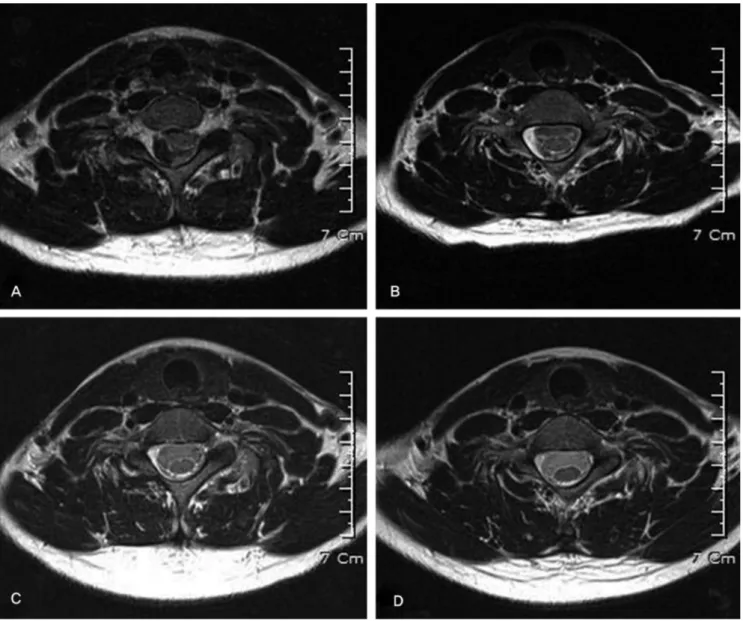
Fig. 2. Serial T2-weighted axial MR images, (A) Heterogeneous lobulated contour lesion compressed the spinal cord on the 3rd hospital day (B) On the 7th hospital day, a vary thin low intensity lesion was observed at the hematoma site (C) One month later, almost hematoma had disappeared (D) 3 months later, hematoma with complete resolution.
Young-Do Koh et al Volume 18 • Number 3 • September 2011
www.krspine.org 166
the size of the lesion was 6.9 x 11.6 x 5.0 mm (Fig. 1). Since there were no neurological symptoms, rather than an immediate surgical decompression procedure, bed rest was ordered and symptomatic treatment for the pain and any changes in symptoms were closely monitored. On the 5th day of symptom onset, no neurological abnormalities were observed and the radiating pain on her right upper arm showed improvement.
On the 7th day, the neck pain and the radiating pain almost
resolved and since the size of the hematoma showed significant decrease in the follow-up magnetic resonance imaging, the mass effect did not accompany; only remnant lesions of irregularly lobulated configurations with high signal intensity in T1WI and low signal intensity in T2WI were remaining in a size of 4.0 x 11.4 x 3.3 mm in the posterior epidural area of the 7th cervical vertebra. After one month, the results of follow-up observations showed that the pain was not felt in normal daily life and that
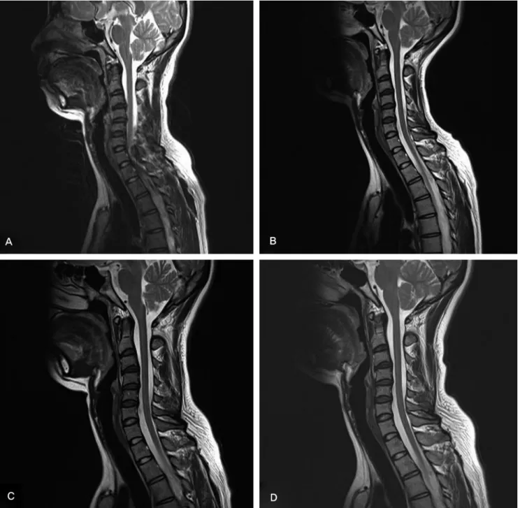
Fig. 3. Serial T2-weighted sagittal MR images, (A) Heterogeneous lobulated contour lesion compressed the spinal cord on the 3rd hospital day (B) On the 7th hospital day, a vary thin low intensity lesion was observed at the hematoma site (C) One month later, almost hematoma had disappeared (D) 3 months later, hematoma with complete resolution.
there was some radiating pain in the right shoulder area if the neck was hyperflexed. The follow-up magnetic resonance imaging showed low signal intensity in T1WI, and low signal intensity in T2WI, which indicated that fine lesions remained.
Three months after the onset of neck pain, the results of follow- up observations showed no pain and only a minor pain was felt during strenuous exercise around the shoulder area, and magnetic resonance imaging showed no remaining lesions. (Fig.
2,3).
DISCUSSION
Occurrences of acute epidural hematoma without a trauma or unusual medical history are extremely rare, hence, not many cases have been reported domestically or internationally.7) Up until the diagnostics techniques such as magnetic resonance imaging were introduced in 1987, the annual occurrence rate was 2.4 cases on average in terms of reported cases in literature, but it increased to 6.4 cases after 1987.3) The causes of epidural hematoma are usually related to previous anticoagulation treatments for coagulopathy, vascular malformations, aspirin usage, and tumors,1) however, since in many cases with unknown causes, it has been reported as spontaneous epidural hematoma. The locations of epidural hematoma are mainly in the thoracic region, and often located in the posterior of the dura.8) The clinical symptoms are acutely-progressing spinal cord compression and neurological abnormalities such as muscle strength or sensory function abnormalities, urination and defecation disorders, and paralysis of the limbs. In the case of epidural hematoma of the cervical spine, occipital and cervical pain, radiating pain to the upper extremities can result; in the case of thoracolumbar epidural hematoma, chest pain, back pain, and radiating pain to the lower extremities could result.
In terms of literature, it has been reported that the cases of pain and radiating pain as the onset symptom for disc herniation being the only clinical symptoms were 8.4%, and the cases of additional neurological symptoms accompanying, were 91.6%.5) Magnetic resonance imaging is more useful than myelography and computerized tomography in terms of identifying accurate location, size and properties.1) The magnetic resonance imaging of the hematoma shows the same extent of signal intensity as the spinal cord during the highly-acute stage in T1WI, and in T2WI,
higher or equal signal intensity as of that of the the spinal cord is revealed. In the case of acute hematoma within 24 hours, T1WI shows the same signal intensity as the spinal cord, and in T2WI although it may show higher signal intensity as a whole, it may also show low signal intensity partially. After a few days from onset, during the sub-acute stage, T1WI shows a high signal intensity, and T2WI shows a low signal intensity.7) In our study, after 3 days from onset, during the sub-acute phase, T1WI showed a mid-to-high signal intensity level, and T2WI showed a mid-to-low signal intensity level. These signal intensity levels indicated a highly-acute stage to sub-acute stage transition phase.
Generally, in most of the cases displaying paralysis of the limbs and rapidly progressing neurological symptoms, surgical decompression led to improved results.5) However, in some cases, rehabilitative treatments and conservative treatments resulted in improved conditions as well, in cases with rapidly progressing clinical symptoms; it was reported that spontaneous dissolutions of hematoma were verified with magnetic resonance imaging and computed tomography.4-7,9) However, there is a report of conservative treatment resulting in fast recovery without any sequelae, for a patient with neurologic symptom and severe clinical progression in which surgical decompression being impossible due to combined comorbidity.5) In our report, disc herniation was suspected in a rationale for rapid symptom appearance of cervical nerve compression without neurologic abnormality, which eventually led us to perform conservative management resulting in improvment within few days. On the 3rd day from onset MRI revealed hematoma, observation under admission was done showing no additional symptoms nor neurologic symptoms change. Therefore, bed rest and conservative treatments were performed instead of surgical decompression.
As for the principle of spontaneous dissolution, Inamasu et al.9) suggested leakage of hematoma into the intervertebral foramen;
Le Coz et al.10) stated that as the epidural hematoma enlarge in size, the dural sac decompresses itself by releasing dural filaments that cross the epidural space, and proposed that during hematoma formation, a temporary pressure to the anterior spinal artery possibly plays a role in acute symptom development and relief. Groen et al.5) reported in a retrospective study of surgical treatment and conservative treatment of spontaneous
Young-Do Koh et al Volume 18 • Number 3 • September 2011
www.krspine.org 168
epidural hematoma, that the hematoma in the cases that showed spontaneous dissolution was longer in length than the hematoma which surgical decompressive procedure was performed. The lengthening was thought to be due to hematoma expansion, and it was suggested as a clue to understanding spontaneous dissolution.
We believe that there may be a number of cases of spontaneous hematoma, of which rapid symptomatic relief leads to exempting the need to seek hospital care; we also believe that, even if hospital care was sought, there may a number of cases of which conservative treatment leads to symptomatic relief after diagnosed as a simple disc herniation. As in our study, when neurologic symptoms such as paralysis of the limbs are not present and the recovery is rapid, bed rest and conservative treatments may bring about sound clinical results due to spontaneous hematoma resolution.
REFERENCES
1. Al-Mutair A, Bender DA. Spinal Epidural Hematoma. J am Acad Orthop Surg. 2010;18:494-502.
2. Jackson R. Case of spinal apoplexy. Lancet. 1869;2:5-6.
3. Jamjoom ZA. Acute spontaneous spinal epidural hematoma: the influence of magnetic resonance imaging on
diagnosis and treatment. Surg Neurol. 1996;46:345-9.
4. Kato S, Seki H, Koshu K. Acute Cervical spinal Epidural Hematoma with Spontaneous Resolution --case report.
Neurol Med Chir (Tokyo). 1994;34:23-6.
5. Groen RJ. Non-operative treatment of spontaneous spinal epidural hematomas: a review of the literature and a comparison with operative cases. Acta Neurochir(Wien).
2004;146:103-10.
6. Wagner S, Forsting M, Hacke W. Spontaneous Resolution of a Large Spinal Epidural Hematoma. Neurosurgery.
1996;38:816-8.
7. Shim DM, Jeung UO, Kim CS. Conservative Treatment of Spontaneous Spinal Epidural Hematoma. J Korean Soc Spine Surg. 2006;13:319-22.
8. Ogawa T, Abe S, Nakahara S, Sekino H, Tani S.
Spontaneous extradural hematoma localized in the ventral side of the thoracic spinal cord --a case report. No Shinkei Geka. 1985;13:439-43.
9. Inamasu J, Hori S, Aoki K, Aikawa N, Maruiwa H, Toyama Y. Spontaneous spinal epidural hematoma. Am J Emerg Med. 2000;18:837-9.
10. Le Coz P, Helias A, Woimant F, Haguenau M. Transient neurological manifestations disclosing spontaneous acute cervical epidural hematoma.Rev Neurol (paris).
1997;153:325-30.
급성 경추 경막외 혈종의 자발적 용해 -1예 보고-
고영도 • 국성환
이화여자대학교 의과대학 정형외과학교실
연구 계획: 증례 보고
목적: 자발적 급성 경추 경막외 혈종의 자발적으로 용해된 예를 보고하고, 짧은 시간내에 증상호전이 있는 경우 보존적 치료가 유용함을 제시하고자 한 다.
선행문헌의 요약: 자발적 급성 경추 경막외 혈종은 드물게 보고되고 있으며, 대부분의 증례에서 수술적 감압술이 시행되어 왔다. 하지만 보존적 치료를 시행하였을 때 자발적으로 용해됐다는 증례가 드물게 보고되고 있다.
대상 및 방법: 29세 여자 환자는 특이 외상력 없이 경부 통증과 우측 상지 방사통이 유발되었으며, MRI상 급성 경추 경막의 혈종이 확인되었다. 증상은 빠른 회복을 보여 보존적 치료를 시행하였으며 발병 3일, 일주일, 한달, 세 달 후 MRI 추시관찰 하였다.
결과: 증상은 빠른 회복을 보이며 7일간 점진적으로 개선되었다. MRI 소견을 비교 결과 혈종의 크기는 점차 감소하여 자발적으로 용해되었음을 알 수 있었다. 증상 발생 한달 후 대부분의 임상적 이상소견은 사라졌다.
결론: 자발적으로 유발된 급성 경추 경막외 혈종이 빠르게 임상증상의 호전을 보일 경우 보존적 치료가 가능할 것으로 생각된다.
색인 단어 : 경추, 경막외 혈종, 자발적, 용해 약칭 제목 : 경추 혈종의 자발적 용해