Introduction
The mediastinum is a unique anatomic area containing several structures and pluripotent cells that allow for the development of a range of tumors. Uncommon neoplasms of the mediastinum account for less than 10% of all mediastinal masses and include primary thymic carcinomas, neuroendocrine carcinomas, germ-cell tumors, lymphomas, and neurogenic, endocrine, and mesenchymal tumors.1-2)
Primary mediastinal lymphomas are rare and the most common types are Hodgkin's lymphoma(HL), large B-cell lymphoma, and lymphoblastic lymphoma(LBL).1-3) Distinction of the specific histologic subtype is important as it influences treatment and prognosis.3-4) LBL is well known as a highly malignant tumor in children, but is relatively rare in adults.5) T-LBL is a rare subtype of adult non-Hodgkin lymphoma(NHL). Despite its frequently dramatic presentation and aggressive clinical course, few
description of LBL in adults can be found in the radiologic literature.5) And no description of primary mediastinal T-LBL can be found in the radiologic literature.
We report the radiologic findings in a 46-year-old woman pathologically proven primary mediastinal precursor T-cell lymphoblastic lymphoma.
Case Report
A 46-year-old woman was admitted to the hospital because of chest discomfort for 15 days. She did not complain of fever, night sweating, weight loss. Her peripheral WBC was 6800/uL with 26 percent normal lymphocytes. The laboratory test of LDH was 505 IU/L, She had no palpable peripheral lymphadenopathy and the remainder of physical examination was unremarkable.
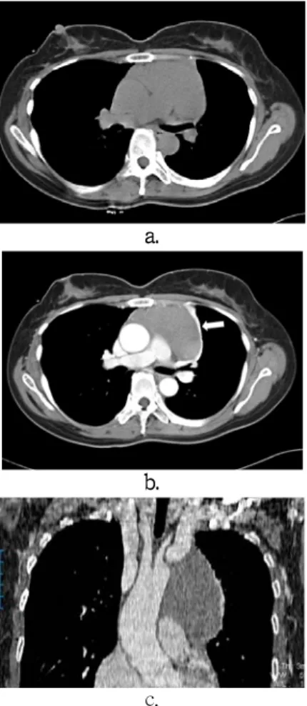
The initial chest radiograph reveal a large anterior mediastinal mass(Fig. 1). The precontrast chest computerized tomography(CT) demonstrated homogeneous attenuation of anterior mediastinal mass without destruction of sternum (Fig. 2a). On postcontrast CT, a mildly homogeneous enhanced left anterior mediastinal large mass
고신대학교 의과대학 학술지 제 권 제 호23 4 Kosin Medical Journal
Vol. 23. No. 4, pp. 216 219, 2008∼
성인에서 발생한 원발성 종격동 세포 림프아구성 림프종 증례보고 T :
이운하 정미희 정경순, , 고신대학교 의과대학 복음병원 영상의학과
Woon-Ha Lee, Mi-Hee Jung, Kyung-Soon Jeong
Department of Diagnostic Radiology, Kosin University College of Medicine, Busan, Korea
――― Abstract ――――――――――――――――――――――――――――――――――――――――
Lymphoblastic lymphoma is high-grade malignancy and rapidly fatal course without appropriate treatment. It is well known as a highly malignant tumor in children, but is rare in adults. Radiologic report of lymphoblastic lymphoma is rare. We report here on a case of a primary mediastinal precursor T-cell lymphoblastic lymphoma in a 46-year-old woman.
―――――――――――――――――――――――――――――――――――――――――――――――――
Key words : Mediastinum, Lymphoma
교신저자 Mi-Hee Jung:
ADD : 602-902, 34 Amnam dong, Seo-Gu, Busan Korea Department of Radiology, Gospel Hospital, Kosin University College of Medicine TEL : +82-51-990-6341, FAX : +82-51-255-2764 E-mail: [email protected]
성인에서 발생한 원발성 종격동 세포 림프아구성 림프종 증례보고T :
was noted (Fig. 2b and 2c). Surface lobulation, pericardial or pleural effusion, involving vascular structure were not found.
Systemic nodal involvement including cervical, axillary, paraaortic, mesenteric and inguinal area was not noted.
Fig. 1. Posteroanterior (a) and left lateral (b) chest radiograph sharply marginated spheric mass in the anterior mediastinum.
c.
Fig. 2. Unenhanced chest CT scan obtained with mediastinal window setting at the level of bronchus intermedius (a) shows sharply marginated homogeneous isodense anterior mediastinal mass. Enhanced chest CT scan obtained at the same level (b) shows slightly homogeneous enhancement of mass and compressive segmental atelectasis of left upper lobe (arrow).
Coronal multiplanar reformatted image (c) shows the mass that extend from left brachiocephalic vein to left heart without vascular encasement.
Patient underwent mediastinal mass excision because of needle biopsy was inconclusive. The specimen was rubbery mass with grayish tan and brownish surface. The photomicrograph shows delicate convolutions of the nuclear membrane of the tumor cells (Fig. 3a). The immunohistochemical staining was positive for CD45RO (Fig. 3b). These features were consistent with a diagnosis of T-cell lymphoblastic lymphoma.
Discussion
LBL is one of the most common mediastinal lymphomas in children and comprise about one-third of all NHL in this age group,2,5-8) but rare subtype of adult NHL, with an incidence of less than 2% (1.7% for T-LBL and <1% for B-LBL).9) T-LBL patients are younger and show a higher rate of mediastinal or bone marrow involvement and stage IV disease than B-LBL.9) Men are affected more often than women.
a.
b.
Fig. 3. Photomicrograph of mediastinal mass (a) shows delicate convolutions of the nuclear membrane of the tumor cells and tingible body macrophages (H-E, x400).
Immunohistochemical staining of tumor cells (b) are positive for CD45RO.
고신대학교 의과대학 학술지 제 권 호24 4 , 2008
The rapid course and aggressive clinical behavior observed in children area equally prevalent in adults.6)LBL usually present with masses or lymphadenopathy above the diaphragm, with rapidly disseminate throughout the body, including even the bone marrow and central nervous system.5,6) Only mediastinal involvement of LBL is rare.3,6) Our case showed only mediastinal mass without mediastinal lymphadenopathy and dissemination throughout the body, including bone marrow and central nervous system.
Tkihide et al.3) reported that the different subtypes of primary mediastinal lymph node often have characteristic manifestions that allow their distinction on CT. HL commonly presents as a mediastinal mass with surface lobulation and involves cervical, mediastinal, hilar and paraortic nodules. Mediastinal diffuse large B-cell lymphoma demonstrates mediastinal mass without surface lobulation, often associated with vascular involvement, and pleural or pericardial effusion. T-LBL is characterized by mass without surface lobulation involving vascular structures often associated with pleural or pericardial effusion, by systemic nodal involvement including cervical, axillary, paraaortic, mesenteric, and inguinal, and by hepatomegaly and splenomegaly.
Schwartz et al.6) reported six cases of mediastinal LBL.
These patients were 20 to 38 years of age. All six cases had large mediastinal mass, and 4 of them had symptoms of compression. Cardiac tamponade was a dramatic early occurrence in 3 cases. 4 of them had pleural effusion.
Schwartz et al.6)assumed that the rapid growth within the closed spaces of the mediastinum which characterizes this lesion may lead to pericardial effusion or obstruct the superior vena cava.
In our patient, CT revealed homogeneous enhanced anterior mediastinal mass without surface lobulation. This finding was compatible with previous report.3,6) But systemic lymph node involvement of cervical, axillary, paraaortic, mesenteric, and inguinal lymphadenopathy was not found. Pericardial effusion and pleural effusion was not noted.
Distinction of the specific histologic subtype is important as it influences treatment and prognosis.3,4) Because the specific diagnosis should be confirmed by
immunohistochemical analysis and hence requires large tissue samples, it is not always easy to make a confident diagnosis on biopsy specimens.3) Our patient underwent mediastinal mass excision because of needle biopsy was inconclusive.
In conclusion, primary mediastinal precursor T-cell lymphoblastic lymphoma is rare. Our case showed a homogeneous enhanced anterior mediastinal mass without pleural effusion, pericardinal effusion and vascular obstruction. Although these findings are not unique and differentiation of anterior mediastinal masses is difficult on the basis of imaging findings alone, recognition about radiologic findings of primary mediastinal T-cell lymphoblastic lymphoma could help differential diagnosis of anterior mediastinal mass.
국문요약
성인에서 발생한 원발성 종격동 세포T 림프아구성 림프종 증례보고:
림프아구성 림프종은 고등급 악성종양으로서 적절한 치료를 하지 않을시 빠르게 치명적인 경과로 진행한다.
원발성 종격동 림프종은 대부분 호지킨 림프종 큰, B세
포 림프종(large B cell lymphoma), 림프모구 림프종
이다 원발성 세포 림프아구
(lymphoblastic lymphoma) . T
성 림프종은 성인에서 드문 형태의 종격동 림프종이며
영상학적 보고는 드물다 이에 저자들은 성인에서 원발.
성으로 종격동에 발생한 세포 기원의 림프아구성 림프T
종 예를 경험하였기에 보고하고자 한다1 .
Index words: Mediastinum, T-cell lymphoblastic lymphoma
References
1) Strollo DC, Rosado-de-Christenson ML, Jett JR: Primary mediastinal tumors: Part II. Tumors of the middle and posterior mediastinum. Chest. 112:1344 1357, 1997–
2) Macchiarini P, Ostertag H: Uncommon primary mediastinal tumours. Lancet Oncol. 5:107-118,2004
3) Tateishi U, M ller NL, Johkoh T, Onishi Y, Arai Y, Satake M,ü
성인에서 발생한 원발성 종격동 세포 림프아구성 림프종 증례보고T :
Matsuno Y, Tobiani K: Primary mediastinal lymphoma:
characteristic feature of the various histological subtypes on CT. J Comput Assist Tomogr 28;782-789, 2004
4) The non-Hodgkin's lymphoma classification project: A clinical evaluation of the international lymphoma Study Group classification of non-Hodgkin's lymphoma. Blood 89:3909-3918, 1997
5) Streuli RA, Kaneko Y, Variakojis D, Kinnealey A, Golomb HM, Rowley JD: Lymphoblastic lymphoma in adults. Cancer 47:2510-2516, 1981
6) Schwartz EE, Conroy JF, Bonner H: Mediastinal involvement in adults with lymphoblastic lymphoma. Acta Radiol. 28:403–
407,1987
7) Reiser M, Josting A, Soltani M, Staib P, Salzberger B, Diehl V, Engert A: T-cell Non-Hodgkin's Lymphoma in adults:
Clinicopathological characteristics, response to treatment and prognostic factors: Leukemia and Lymphoma. 43:805-811, 2002
8) Hoelzer D, Gokbuget N, Digel W, Faak T, Kneba M, Reutzel R,Jarosinska JR, Zwolinski J, Walewski J: Outcome of adult patients with T-lymphoblastic lymphoma treated according to protocols for acute lymphoblastic leukemia. Blood 99:4379–
4385, 2002
9) Altundag O, Yavas O, Altundag K, Gonen C, Turker A, Uner A: Unusual abdominal tumors: case 3. Primary omental T-cell lymphoblastic lymphoma. J. Clin. Oncol. 15:1522 1523,– 2004