• 검색 결과가 없습니다.

Effects of Abdominal Drawing-in Maneuver on Muscle Activity of the Trunk and Legs during Flat Walking

N/A
N/A
Protected

Academic year: 2021

Share "Effects of Abdominal Drawing-in Maneuver on Muscle Activity of the Trunk and Legs during Flat Walking"

Copied!
8
0
0

로드 중.... (전체 텍스트 보기)

전체 글

(1)


| Abstract |

1)

PURPOSE: This study examined the difference in muscle activity of the trunk and legs during flat walking with or without an abdominal drawing-in maneuver.

METHODS: This study was conducted on 15 healthy males and eight females who were attending D University in Busan.

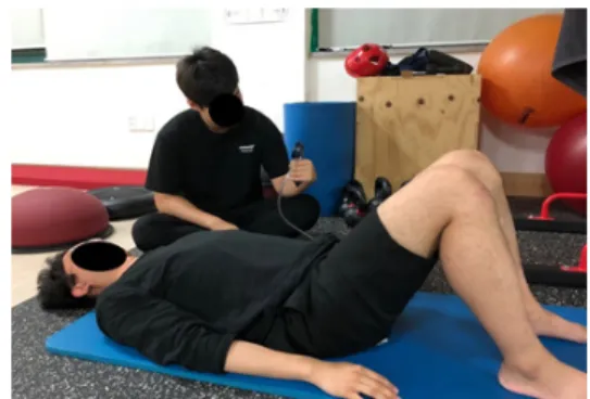
This experiment was conducted after 15 minutes of abdominal drawing-in training using a pressure biofeedback unit before the experiment, and the difference in the muscle activity of the trunk and legs during flat walking with or without an abdominal drawing-in technique was investigated.

Surface electromyography was used, and the electrode attachment site was the right sternocleidomastoid muscle,

†Corresponding Author : Su-Kyoung Lee

[email protected], https://orcid.org/0000-0002-4916-2188 This is an Open Access article distributed under the terms of the Creative Commons Attribution Non-Commercial License (http://creativecommons.org/licenses/by-nc/3.0) which permits unrestricted non-commercial use, distribution, and reproduction in any medium, provided the original work is properly cited.

splenius capitis muscle, rectus abdominis muscle, external abdominal oblique muscle, transverse abdominis muscle, erector spinae muscle, vastus medialis muscle, and vastus lateralis muscle (TM DTS, Noraxon, USA). The data were analyzed statistically using a paired t-test on SPSS version 18.0 (IBM).

RESULTS: The muscle activity of the rectus abdominis muscle, external abdominal oblique muscle, transverse abdominis muscle, vastus medialis muscle were increased significantly and maintained more than walking without maintaining an abdominal drawing-in maneuver (p < .05).

Moreover, muscle activity of the erector spinae muscle was decreased significantly and maintained more than walking without maintaining an abdominal drawing-in maneuver (p <

.05).

CONCLUSION: Maintaining an abdominal drawing-in maneuver during flat walking is more effective during walking training.

Key Words: Abdominal drawing-in maneuver, Biofeedback unit, Flat walking, Muscle activity

Research Article Open Access

복부 드로잉-인 기법이 평지 보행 시 몸통과 다리의 근 활성도에 미치는 효과

안수홍⋅이수경 1† ⋅조현대 2

동의대학교 보건의과학과 물리치료전공, 1 동의대학교 물리치료학과, 2 곤지암중학교

Effects of Abdominal Drawing-in Maneuver on Muscle Activity of the Trunk and Legs during Flat Walking

Su-Hong Ahn, PT, MS⋅Su-Kyoung Lee, PT, PhD 1† ⋅Hyun-Dai Jo, PhD 2 Department of Biomedical Health Science, Graduate School, Dong-Eui University

1 Department of Physical Therapy, College of Nursing, Healthcare Sciences, Dong-Eui University

2 Major of Physical Education, Gonjiam Middle School

Received: November 16, 2019 / Revised: November 28, 2019 / Accepted: January 3, 2020

ⓒ 2020 J Korean Soc Phys Med

(2)

Ⅰ. 서 론

안정화 운동은 신체의 움직임 조절능력을 회복시켜 주며, 신체정렬 유지 및 허리통증의 예방 및 치료에 큰 역할을 하고[1] 고유수용성 감각 조절능력과 균형 및 지구력을 향상시켜주는 역할을 한다[2]. 또한, 몸통 근육 의 수축을 조절하여 분절의 불안정성과 미세손상의 축 적을 줄여 주는 역할을 하며, 신체의 자세변화와 움직임 동안 척추와 골반의 안정성을 증가시킨다[3-5]. 안정화 를 담당하는 근육은 크게 광역 근육계와 국소 근육계로 나뉘며, 광역 근육계는 배곧은근, 배바깥빗근 등으로 구 성되어 몸통의 회전력을 발생시키고 전반적인 몸통의 안정성에 기여한다. 국소 근육계는 배가로근, 배속빗근 등으로 구성되어 있으며, 척추의 분절을 안정화시켜 움 직임 시 허리뼈에 직접적인 안정성을 제공하는 역할을 한다[6,7]. 안정화 운동 중 하나인 복부 드로잉-인 기법 (Abdominal drawing-in maneuver)은 배 근육들을 기능적 으로 자극하여[8] 배벽을 안쪽으로 당겨 배근육의 동시 수축을 유도하여 배 안쪽의 압력을 증가시키고 근육의 동시수축을 통해 허리 안정화(Lumbar stabilization)를 효 과적으로 수행하게 하며 동시에 과도한 허리의 전만이 나 골반의 전방 경사를 감소시켜준다[9]. 또한, 골반을 중력 중심선에 가까운 위치로 회복시키며 허리의 중립 자세를 유지시켜 허리에 가해지는 스트레스를 줄여준다 [10]. 즉, 복부근육의 조절은 몸통안정화 및 자세조절을 통해 정상적인 자세를 유지할 수 있도록 하는 매우 중요 한 역할을 한다[11]. 정상적인 자세는 척추가 정상적인 곡선을 이루고, 인체 분절들이 수직 정렬 내에 있으며, 체중 분산이 각 관절들의 밑면에 골고루 분산되어 근육 과 관절에 최소한의 압력을 받는 상태라고 하였다 [12,13]. 그러나 현대인들은 뼈대가 무너지는 장시간 오래 앉아있는 자세, 비만, 운동부족 등으로 인하여 자세와 관 련된 문제점들이 많다[14]. 이처럼 자세정렬에 변화가 있 으면 근육뼈대계에 문제가 발생할 수 있고 정상적인 움직 임이나 운동에도 제한이 올 수 있으며, 더 나아가 인간의 이동 수단인 보행에도 영향을 미친다고 하였다[12].

보행이란 두 다리를 반복적으로 이용하여 한 장소에 서 또 다른 장소로 이동하는 것을 의미하며[15], 인간이

일상생활 및 독립적인 생활을 영위하는데 있어서 가장 기본이 되고 중요한 역할을 하며[16], 100여개 이상의 팔다리의 뼈대가 모든 관절에서 상호 협력하여 복잡한 신체활동을 가능하게 하며[17], 동시에 신경과 근육 등 이 사용되어 몸을 앞으로 계속 움직이게 하는 반복적인 동작이라고 하였다[18]. 즉, 보행은 머리, 몸통, 팔다리 가 상호 연관성이 있으며, 체중부하를 통해 신체를 앞 으로 추진시키는 동안 안정성과 균형을 유지하여 이동 에 필요한 기본적인 운동을 제공하며[19], 복부 근육들 을 활성화시켜 몸통 근육들을 조절하는 몸통 안정화 운동과도 관련이 있다고 하였다[20,21]. 이처럼 보행과 몸통 안정화 운동은 서로 상관관계가 있기 때문에 다양 한 방법을 통해 신체에 운동 효과를 극대화시킬 수 있는 기초적인 자료를 제시할 필요가 있다. 따라서 본 연구 에서는 몸통안정화 운동 중 하나인 복부 드로잉-인 기 법과 보행을 동시에 적용하여 몸통 및 다리의 근 활성도 에 미치는 영향에 대해 알아보고자 한다.

Ⅱ. 연구방법

1. 연구대상자

본 연구는 부산에 소재한 D대학교에 재학중인 건강 한 20대 성인 남자 15명 여자 8명을 대상으로 실시하였 다. 본 연구를 시작하기에 앞서 연구대상자에게 연구의 목적과 절차에 대하여 설명하고 이를 충분히 숙지시킨 다음 실험에 참여하도록 하였고 연구 대상자의 선정기 준은 최근 6개월 이내에 근육뼈대계 질환의 진단을 받 지 아니한 자, 복부와 다리의 근력약화가 없는 자, 정상 적인 보행이 가능한 자로 선정하였다.

2. 측정도구 1) 표면근전도

평지 보행 시 복부 드로잉-인 기법 유무에 따른 몸통

및 다리의 근 활성도를 알아보기 위해 표면 근전도

(Myosystem TM DTS, Noraxon Inc., USA) 를 사용하여

배곧은근(Rectus Abdominal, RTA), 배바깥빗근(External

oblique ,EO), 배가로근(Transverse abdominal, TRA), 척

(3)

추세움근(Elector Spinae, ES), 안쪽넓은근(Vastus medialis oblique, VMO), 가쪽넓은근(Vastus lateralis oblique, VLO) 을 측정하였다. 전극을 부착하기 전 피부에 대한 전기저 항을 줄이기 위해 알콜솜을 이용하여 각질을 제거한 뒤 Ag/Agcl 재질의 일회용 표면 전극을 사용하였다. 전극은 6채널을 사용하였고 부착부위는 오른쪽이였으며 다음과 같다. 배곧은근은 칼돌기 외측을 따라 3 cm 지점에서 아래 5 cm에 위치, 배바깥빗근은 두덩뼈결절과 8번째 갈비뼈를 이은 가상선에서 8번째 갈비뼈의 아래 2 cm에 위치, 배가로근은 엉덩뼈 능선의 위에서부터 안쪽방향으 로 2 cm 아래로 떨어진 위치, 척추세움근은 1번 허리뼈의 가시돌기에서 가쪽으로 2 cm 떨어진 힘살부위에 척추와 평행하게 2 cm 떨어진 위치, 안쪽넓은근은 위앞엉덩뼈가 시에서 무릎의 안쪽 20% 사이의 거리에 위치, 가쪽넓은 근은 위앞엉덩뼈가시에서 무릎의 바깥쪽 25% 사이의 거리에 부착하였다[22]. 근활성도 측정은 10 m의 구간을 각 대상자가 평소에 걷는 속도로 복부 드로잉-인 기법을 적용하고 보행하는 것과 복부 드로잉-인 기법을 적용하 지 않고 보행 시 처음과 마지막 데이터를 제외한 3보의 데이터를 결과 값으로 사용하였으며, 총 3회 측정하여 평균화하였다(Fig. 1)[23,24]. 근전도 주파수는 2000 Hz로 샘플링하여 기록하였고 대역필터(Band pass filter)는 20-450 Hz 주파수 대역을 사용하여 노이즈 제거 후, 평균 제곱근(Root mean square, RMS)의 값을 도출하였다. 기 타 신호는 Rectification과 Smoothing으로 제거하였다.

2) 압력 생체 피드백 장치

본 연구실험 전 허리의 안정성을 높이기 위해 운동의

질과 정확도의 피드백을 제공해주는 압력 생체 피드백 장치(Stabilizer, Chattanooga Group Inc, USA)를 통해 복 부 드로잉-인 기법을 교육하였다. 대상자는 바로 누운 자세에서 무릎관절은 90° 굽히고 압력 생체 피드백 장 치를 대상자의 허리뼈 부위에 위치시켰다. 대상자는 압력 생체 피드백 장치에 연결되어 있는 압력계를 40 mmHg인 상태에서 10 mmHg를 증가시킨 50 mmHg 상 태를 눈으로 확인시켜 유지하도록 교육하였고, 이때 연구대상자에게 척추와 갈비뼈 그리고 골반을 고정시 킨 상태에서 복부가 허리방향으로 들어가도록 배꼽을 위쪽과 밑으로 천천히 당기면서 유지하라고 지시하였 다[25]. 복부 드로잉-인 기법을 하는 동안 정상적인 호 흡을 위해서는 적당한 강도로 배꼽을 넣어야 하기 때문 에 소변을 참듯이 아래 배에 천천히 힘을 주세요라는 구두명령을 지시하였다(Fig. 2)[26]. 복부 드로잉-인 기 법의 교육은 10초를 유지하였고 1회 실시 후 5초 동안 휴식을 취하였다. 총 10회씩 반복하여 5세트 실시하였 으며[27], 복부드로잉-인 기법 유무에 따른 평지 보행 또한 3분의 연습을 통하여 충분히 숙지한 뒤 본 실험을 실시하였다. 보행구간은 10 m로 설정하였고 보행속도 는 연구의 결과에 영향을 미치지 않도록 각 대상자가 평소에 걷는 속도로 설정하였다[24,25].

3. 실험절차

대상자에게 실험절차와 방법에 대해 설명을 하였고, 실험 전 복부 드로잉-인 기법 교육을 먼저 실시하여 충분히 학습한 뒤 실험자의 지시에 따라 평지 보행 시 복부 드로잉-인 기법 유무에 따른 근활성도 차이를 알

Fig. 1. EMG attachment site. Fig. 2. Abdominal drawing-in maneuver.

(4)

아보기 위해 근전도 신호량을 측정하였다. 실험 순서는 각 대상자에게 무작위 배정을 통해 복부 드로잉-인 기 법을 유지한 상태와 유지하지 않은 상태의 순서를 정하 였다. 순서의 결과는 복부 드로잉-인 기법을 먼저 유지 하고 실험을 실시한 대상자가 12명이였으며, 복부 드로 잉-인 기법을 유지하지 않고 실험을 먼저 실시한 대상 자가 11명으로 무작위 배정을 통해 나타난 실험 순서의 비율은 비슷하였다. 측정방법은 복부 드로잉-인 기법 유무에 따라서 각 대상자가 평소에 걷는 속도로 10 m 평지 구간 보행 시 근 활성도를 측정하여 비교하였다.

모든 근전도 자료는 처음과 마지막 데이터를 제외한 3보의 데이터를 결과 값으로 사용하였고 3회 반복 측정 하였으며, 3번 측정된 결과 값들을 평균화였다. 정량화 를 위해 평지 보행전에 특정 동작인 해부학적 자세를 5초간 유지하여 표준 근육 수축 값을 측정하였고, 근전 도 신호를 표준화하는 %RVC (Reference voluntary contraction) 를 통해 근전도 신호를 표준화하였다.

4. 자료분석

본 연구에서 수집된 결과 값은 평균 ± 표준오차 (Mean ± SE) 로 기술하였고 SPSS version 18.0을 이용하여 통계처리를 실시하였으며, 연구대상자의 일반적 특성은 기술통계를 사용하였다. 또한, 복부 드로잉-인 기법 유 무에 따른 평지 보행 시 몸통 및 다리 근육의 근활성도 차이를 알아보기 위하여 비모수 통계검정인 윌콕슨 부 호순위 검정(Wilcoxon signed rank test)을 사용하여 분석 하였다. 이때 통계학적 유의수준은 .05로 설정하였다.

Ⅲ. 연구결과

1. 일반적 특성

본 연구는 남자 15명 여자 8명으로 총 23명으로 실험 을 진행하였고, 대상자의 평균 나이는 22.57 ± 2.02세, 평균 신장은 169.57 ± 9.77 cm, 평균 체중은 63.57 ± 13.26 kg이었다(Table 1).

2. 평지 보행 시 복부 드로잉-인 기법 유무에 따른 근활성도 비교

평지 보행 시 근전도를 이용한 오른쪽 몸통 및 다리 의 근활성도를 측정한 결과 배곧은근, 배바깥빗근, 배 가로근, 안쪽넓은근은 복부 드로잉-인 기법을 유지하 지 않고 보행하는 것보다 유지하고 보행하는 것이 유의 하게 증가하였고(p < .05), 척추세움근은 유의하게 감소 하였다(p < .05)(Table 2).

Ⅳ. 고 찰

본 연구는 근전도 기기를 통하여 복부 드로잉-인 기

Variables Values

Sex (Male / Female) 15 / 8

Age (year) 22.57 ± 2.02

Height (cm) 169.57 ± 9.77 Weight (kg) 63.57 ± 13.26 Mmean (± Standard Deviation)

Table 1. General Characteristics of the Subjects

Muscle Pre Post Z P

RTA 2403.43 ± 354.40 2992.05 ± 549.86 -2.346 .019*

EO 2823.20 ± 372.25 4044.87 ± 816.45 -2.589 .010*

TRA 3654.55 ± 496.54 6505.63 ± 894.22 -3.806 .000*

ES 5533.96 ± 748.55 3611.94 ± 456.29 -2.589 .010*

VMO 12031.18 ± 1999.35 14047.14 ± 2386.83 -2.346 .019*

VLO 11440.51 ± 2214.20 11576.41 ± 2226.37 -3.330 .741

Mean (± Standard Error), *P ˂ .05

RTA: Rectus abdominis, EO: External abdominal oblique, TRA: Transverse abdominis, ES: Erector spinae, VM: Vastus medialis, VL: Vastus lateralis

Table 2. Comparison of Muscle Activity with Abdominal Drawing-in During Flat Walking (unit : % RVC)

(5)

법의 유무에 따라 평지 보행 시 몸통 및 다리의 근 활성도 의 차이를 알아본 결과 복부 드로잉-인 기법을 유지한 보행이 유지하지 않은 보행에 비하여 배곧은근, 배바깥 비근, 배가로근의 근 활성도가 유의하게 증가하였다.

몸통 안정화 운동은 다양한 환자들에게 적용가능하 며, 허리 통증의 감소와 보행능력과 유연성 그리고 근 력 및 균형능력을 향상시켜 삶의 질을 개선시켜준다 [28]. 몸통 안정화 운동 중 하나인 복부 드로잉-인 기법 은 복부근육을 활성화시키는데 특히 배가로근, 배곧은 근, 배바깥빗근과 같은 심부 근육을 많이 활성화시키며 [29], 복부의 수의적 움직임과 운동을 조절하고 몸통의 안정화에 기여한다[25]. Lee [30]는 경사로 보행 시 복부 드로잉 기법을 유지하는 것이 유지하지 않았을 때보다 복부의 근 활성도가 유의하게 증가한다 하였고, 계단 보행 또한 복부 드로잉-인 기법 수행이 수행하지 않았 을 때에 비하여 몸통의 안정성이 증가하여 척추의 동적 안정성에 기여하는 복부의 근 활성도가 유의하게 증가 한다고 하였다[31]. 본 연구에서도 경사로와 계단과 같 은 수직 보행 시의 선행연구 결과처럼 수평 보행에서도 복부 드로잉-인 기법을 유지하는 것이 복부의 근 활성 도가 유의하게 증가한다고 나왔는데 이는 기능적 활동 시 내부 복압을 증진시켜 몸통을 안정화시키며, 보행 시 자세조절을 통해 몸통을 안정화시키기 위해 복부의 역할이 크게 작용하기 때문이다[32,33].

반면에 척추세움근의 근 활성도는 유의하게 감소하 였는데 복부 드로잉-인 기법 시 몸통을 안정화시키는 복부 근육들이 활성화되어 공동 수축을 유도해 심부의 안정성을 증가시킨다. 따라서 허리의 내적 안정화에 기여하여 척추를 중립위치에 유지하도록 신체에 적절 한 반응 속도를 회복시켜 허리에 가해지는 압력을 감소 시키며[34-36], 신경근 조절을 통하여 사전에 허리의 통증을 예방한다[37,38]. 또한, 복부의 벽을 허리 쪽 방 향으로 당김으로써 복부 내압을 증가 및 복부 근육을 수축시켜 허리의 안정성을 증가시키며, 근육의 동시수 축을 통해 과도한 허리 및 골반 전방 경사를 줄여준다 [9]. Oh 등[39]은 복부 드로잉-인 기법을 유지하며 엎드 린 자세에서 엉덩관절 폄 운동 시 허리폄근의 근활성도 가 유의하게 감소한다고 하였고, Park 등[40]의 연구에

서도 복부 드로잉-인 기법을 유지하는 것이 허리폄돌림 증후군이 있는 환자의 허리폄근의 근활성도가 감소하 여 허리통증이 줄어들었다고 하였다. 따라서, 본 연구 에서 복부드로잉인 기법으로 인해 복부의 근육들이 몸 통을 안정화시키고 허리의 전방경사를 줄여주어 허리 의 폄근의 작용이 감소하여 척추세움근의 활성도가 감 소하였다고 볼 수 있다.

안쪽넓은근은 무릎관절의 기능적인 움직임과 안정 성을 동시에 제공하며[41], 보행 시 신체를 앞으로 이동 시키는 체중부하구조로서 이동에 중요한 역할을 한다 [42]. 본 연구에서 안쪽넓은근의 근 활성도는 복부 드로 잉-인 기법을 유지한 상태에서 유의하게 증가하였는데, 배근육의 활성화는 골반을 안정화시키며, 골반이 안정 화된 상태는 몸통에 미치는 힘들이 효율적으로 다리로 전달이 되며[43], 또한, 배 근육의 수축을 통해 몸통을 안정화시키고 다리의 가동성을 증진시킨다[44,45]. 이 처럼 배 근육의 자극은 방산효과를 통해 다리를 근 수축 시켜 다리의 근력을 증가시키며[46], 근육의 협력 작용 을 통해 증가된 다리의 근활성도는 척추 안정화에 기여 하는 배 근육들의 활성도 또한 증가시키기 때문에 본 실험의 연구결과를 뒷받침해 준다[47]. 따라서 보행 시 복부 드로잉-인 기법을 유지하는 것이 몸통의 심부근육 들을 활성화시켜 몸통을 안정화시키고 복부 근육의 동 시 수축을 통해 허리의 전만을 줄여준다. 또한, 골반을 안정화시켜 몸통에 미치는 힘들이 효율적으로 다리에 전달되어 다리의 근육이 활성화되기 때문에 몸통의 기 능개선 및 다리의 근력강화 운동이 필요한 사람들에게 효과적일 것이라고 판단된다. 더 나아가 허리의 통증이 나 자세의 기능개선에도 긍적적인 효과가 있으므로 허 리통증이나 자세 등에 문제가 있는 사람들에게 적용하 면 효과적일 것이라고 생각된다. 향후 허리 통증 환자 나 자세 등에 문제가 있는 환자를 대상으로 진행하는 다양한 연구가 필요할 것으로 사료된다.

Ⅴ. 결 론

본 연구는 건강한 20대 남녀 23명을 대상으로 평지

보행 시 복부 드로잉-인 기법 유무에 따라서 몸통 및

(6)

다리의 근 활성도에 미치는 영향을 비교하였다. 복부 드로잉-인 기법을 유지하며 보행하는 것이 배곧은근, 배 바깥빗근, 배가로근, 안쪽넓은근의 근활성도를 증가시 켜 유의하게 증가하였고 반면에 척추세움근의 근활성도 를 감소시켜 유의하게 감소하였다. 본 연구결과 복부 드로잉-인 기법을 유지하고 보행하는 것이 복부와 다리 의 근활성도를 증가시키기 때문에 몸통을 안정화시키고 다리 근력강화에도 효과적인 방법이라고 판단된다.

References

[1] Lehman GJ, Hoda W, Oliver S. Trunk muscle activity during bridging exercises on and off a Swissball. Chiropr Osteopat. 2005;13(14):1-8.

[2] Macedo LG, Maher CG, Latimer J, et al. Motor control exercise for persistent, nonspecific low back pain: a systematic review. Phys Ther. 2009;89(1):9-25.

[3] O'sullivan PB, Twomey L, Allison GT. Dynamic stabilization of the lumbar spine. Critical Reviews of Phys and Rehab Med. 1997;9(3-4):315-30.

[4] Richardson CA, Hodeg PW, Hides JA. Therapeutic exercise for spinal segmental stabilization in low back pain: scientific basis and clinical approach. Edinburgh, Churchill Livingstone. 1999.

[5] Richardson CA, Snijders CJ, Hides JA, et al. The relation between the transversus abdominis muscle, sacroiliac joint mechanics, and low back pain. Spine. 2002;27(4):399-405.

[6] Moseley L. Combined physiotherapy and education is efficacious for chronic low back pain. Aust J Physiother.

2002;48(4):297-302.

[7] Panjabi MM. The stabilizing system of the spine. Part I. Function, dysfunction, adaptation, and enhancement.

J Spinal Disord. 1992;5(4):383-9.

[8] Richardson CA., Hodges P, Hides JA. Therapeutic exercise for lumbopelvic stabilization: a motor control approach for the treatment and prevention of low back pain. (2nd ed). Edinburgh, Churchill Livingstone. 2004.

[9] Kisner C, Colby LA. Therapeutic exercise: foundations

and techniques (4th ed). Philadelphia, FA Davis. 2002.

[10] Shin MG. The effect of abdominal muscle contraction on center of gravity moving and lumbar lordosis : In low back pain patients with pelvic anterior shift. Master’s Degree. Kyonggi University. 2010.

[11] Bjerkefors A, Ekblom MM, Josefsson K, et al. Deep and superficial abdominal muscle activation during trunk stabilization exercises with and without instruction to hollow. Man Ther. 2010;15(5):502-7.

[12] Choi JW. The influence of different types of trunk rotation exercise on posture and gait in the individuals with malalignment syndrome. Doctor’s Degree. Daegu University. 2009.

[13] Norkin CC, Levangie PK. Joint structure & function :Comprehensive analysis. (2nd ed). Philadelpia, FA Davis.

1992.

[14] Kang SY. Correlation between change in curve and flexibility in girls with mild scoliosis. The Journal of Korean Aerobic Exercise. 2003;7(1):109-15.

[15] Cho SH. The comparative analysis of EMG and Gait patterns depending on variations of speed and ways to walk. Doctor’s Degree. Kangnung National University.

2007.

[16] Han JT, Bae SS. Gait analysis methods and walking pattern of hemiplegic patients after stroke. PNF and Movement. 2007;5(1):37-47.

[17] Whittle M. Gait analysis: an introduction. Boston, Butterworth-Heinemann. 2002.

[18] Kim JT, Park SH. The displacement of Center of Pressure during Adult Female Gait based on the Body Mass.

Changwon National University Sport Science Institute.

2005;10(1):59-65.

[19] Galley PM, Foster AL. Human movement. New York, Churchill Livingstone. 1987.

[20] Kim YH. The effects of trunk stability exercise on the balance and Gait in the patients with stroke. Master’s Degree. Daegu University. 2010.

[21] Hodges PW, Cresswell AG, Daggfeldt K, et al. Three

(7)

dimensional preparatory trunk motion precedes asymmetrical upper limb movement. Gait Posture. 2000;

11(2):92-101.

[22] Hermens HJ, Freriks B, Disselhorst-Klug C, et al.

Development of recommendations for SEMG sensors and sensor placement procedures. J Electromyogr Kinesiol.

2000;10(5):361-74.

[23] Shin JH. Effects of foot strength exercise and functional insole on range of motion, muscle activity and foot plantar pressure in the elderly women. Doctor’s Degree. Silla University. 2018.

[24] Heo BS, Kim YJ, Mun CH, et al. Analysis of surface muscle activity of lower and upper musculus erector spinae according to walkers form. JFMSE. 2014;26(2):

308-15.

[25] Sim YH. The effects of abdominal muscle drawing-In exercise and MFR application on back pain, flexibility and balance in elderly women. Master’s Degree. Hanyang University. 2015.

[26] Lee HO, Park DJ. Understanding and clinical application of abdominal hollowing exercise: a literature review. PNF and Movement. 2011;9(2):9-19.

[27] Lee JM, Lee CH, Kwon OY, et al. The effect of lumbar stabilization exercise for caregivers with chronic low back pain. Phys Ther Korea. 2011;18(2):9-17.

[28] Choi SH, Lim JH, Cho HY, et al. The effects of trunk stabilization exercise using swiss ball and core stabilization exercise on balance and gait in elderly women. J Korean Soc Phys Med. 2012;7(1):49-58.

[29] Park SD, Yu SH. The effects of abdominal draw-in maneuver and core exercise on abdominal muscle thickness and oswestry disability index in subjects with chronic low back pain. J Exerc Rehabl. 2013;9(2):286-91.

[30] Lee SK. The Effects of Abdominal Drawing-in on Muscle Activity in the Trunk and Legs during Ramp Walking.

PNF and Movement. 2019;17(1):137-44.

[31] Lee AY, Kim EH, Cho YW, et al. Effects of abdominal hollowing during stair climbing on the activations of

local trunk stabilizing muscles: a cross-sectional study.

Ann Rehabil Med. 2013;37(6):804-13.

[32] McNeil W. Core stability is a subset of motor control.

J. bodyw Mov Ther. 2010;14(1);80-3.

[33] Zazulak B, Cholewicki J, Reeves NP. Neuromuscular control of trunk stability: clinical implications for sports injury prevention. J Am Acad Orthop Surg. 2008;16(9):

497-505.

[34] Sung SW. The spinal flexibility and response time of erector spinae muscle following stabilization exercise.

Korean J Orthop Manu Ther. 2000;6(1):35-49.

[35] Choi HS, Kwon OY, Yi CH, et al. The comparison of trunk muscle activities during sling and mat exercise.

Phys Ther Korea. 2005;12(1):1-10.

[36] Stevens VK, Bouche KG, Mahieu NN, et al. Trunk muscle activity in healthy subjects during bridging stabilization exercises. BMC Musculoskelet Disord. 2006;7(75):1-8.

[37] Macedo LG, Maher CG, Latimer J, et al. Motor control exercise for persistent, nonspecific low back pain: a systematic review. Phys ther. 2009;89(1):9-25.

[38] von Garnier K, Köveker K, Rackwitz B, et al. Reliability of a test measuring transversus abdominis muscle recruitment with a pressure biofeedback unit. Physio.

2009;95(1):8-14.

[39] Oh JS, Cynn HS, Won JH, et al. Effects of performing an abdominal drawing-in maneuver during prone hip extension exercises on hip and back extensor muscle activity and amount of anterior pelvic tilt. J Orthop Sports Phys Ther. 2007;37(6):320-4.

[40] Park KN, Cynn HS, Kwon OY, et al. Effects of the abdominal drawing-in maneuver on muscle activity, pelvic motions, and knee flexion during active prone knee flexion in patients with lumbar extension rotation syndrome. Arch Phys Med Rehabil. 2011;92(9):1477-83.

[41] Hanten WP, Schulthies SS. Exercise effect on electromyographic activity of the vastus medialis oblique and vastus lateralis muscles. Phys Ther. 1990;70(9):561-5.

[42] Yun NS, Lee KO, Kim JY. Kinematic comparison of

(8)

walking on various inclined walkways. JKPEAGW.

1999;3(1):89-101.

[43] Neumann DA. Kinesiology of the musculoskeletal system: foundations for physical rehabilitation. (2nd ed). Singapore. Mosby Elsevier. 2010.

[44] McGill SM. Low back stability: from formal description to issues for performance and rehabilitation. Exerc Sport Sci Rev. 2001;29(1):26-31.

[45] McGill SM, Grenier S, Kavcic N, et al. Coordination of muscle activity to assure stability of the lumbar spine.

J Electromyogr Kinesiol. 2003;13(4):353-9.

[46] Hwang BJ, Kim JW. Effects of lumbar stabilzation exercise on lumbar and lower extremity strength of the elderly women. J of the korean society of physical medicine.

2011;6(3):267-75.

[47] Choi SA, Cynn HS, Yi CH, et al. Isometric hip abduction using a thera-band alters gluteus maximus muscle activity and the anterior pelvic tilt angle during bridging exercise.

J Electromyogr Kinesiol. 2015;25(2):310-5.

수치

Fig. 1. EMG attachment site. Fig. 2. Abdominal drawing-in maneuver.
Table  2.  Comparison  of  Muscle  Activity  with  Abdominal  Drawing-in  During  Flat  Walking                                    (unit  :  %  RVC)

참조

관련 문서

Results: A statistically significant correlation was found between the internal oblique, transverse abdominis, and erector spinae muscle activity and gait velocity,

Surface electromyography was employed to measure the onset times of trunk muscle contractions in the right anterior deltoid, rectus abdominis, external oblique

The aim of this study is to compare the effect of abdominal drawing-in maneuver (ADIM) on lower trapezius (LT), serratus anterior (SA), and erector spinae (ES) muscle

Background: The purpose of this study was to determine if there is a difference in activation of the rectus femoris, vastus medialis oblique, vastus lateralis,

This study was designed to identify the effect of medial, lateral wedge and difference of Quadriceps angle (Q-angle) on vastus medialis oblique/vastus lateralis muscle

Comparison of bilateral rectus abdominis muscle activity The TSP with hip abduction (HAB)30 condition showed significantly greater muscle activity than TSP with HAB0 and

Changes in transversus abdominis (TrA), internal obli- que (IO), and external oblique (EO) thickness according to the po- sitions (crook lying, standing, and saddle

The external oblique, tibialis anterior, and rectus femoris muscles showed significant difference in 0˚(43cm) and 15˚(58cm), erector spinae and vastus medialis muscles