Comparisons of Trunk Muscle Activity During Arm Lift in Prone and Standing Positions With and Without Abdominal Drawing-in Maneuver
Ki-song Kim1, MPH, PT, One-bin Lim2, BHSc, PT, Chung-hwi Yi3, PhD, PT, Heon-seock Cynn3, PhD, PT
1Dept. of Physical Therapy, Gangnam Severance Hospital Yonsei University, College of Medicine,
2Dept. of Physical Therapy, The Graduate School, Yonsei University,
3Dept. of Physical Therapy, College of Health Science, Yonsei University, Dept. of Ergonomic Therapy, The Graduate School of Health Science, Yonsei University
Abstract
1)The aim of this study is to compare the effect of abdominal drawing-in maneuver (ADIM) on lower trapezius (LT), serratus anterior (SA), and erector spinae (ES) muscle activity during arm lifts in prone and standing positions. Twenty healthy subjects were recruited, and NoraxonTeleMyo 2400T was used to collect electromyographic signals from the LT, SA, and ES muscles. A two-way repeated analysis of variance (ANOVA) used a significance level of .05. If a significant interaction was found, pairwise comparisons were performed with a Bonferroni adjustment (.05/4=.013). The results of the study were as follows: 1) In LT, no significant ADIM by position interaction was found (F1,19=.356, p=.558). There was a significant main effect for ADIM. LT muscle activity with ADIM was significantly greater compared with muscle activity without ADIM (F1,19=82.863, p<.001). There was also a significant main effect for position.
LT muscle activity in the prone position was greater compared with muscle activity in the standing position (F1,19=116.401, p<.001). 2) In SA, significant ADIM by position interaction was found (F1,19=8.687, p=.008). There were significant differences in all pairwise comparisons. The greatest SA muscle activity was observed in the standing position with ADIM. 3) In ES, significant ADIM by position interaction was found (F1,19=122.473, p<.001). The lowest ES muscle activity was elicited in the standing position with ADIM. Based on these results, ADIM is advocated in the prone position to increase LT muscle activity. In addition, it is concluded that arm lifts in the standing position with ADIM offer the most favorable combination for reducing ES muscle activity and increasing SA muscle activity.
[Ki-song Kim, One-bin Lim, Chung-hwi Yi, Heon-seock Cynn. Comparisons of Trunk Muscle Activity During Arm Lift in Prone and Standing Positions With and Without Abdominal Drawing-in Maneuver.
Phys Ther Kor. 2012;19(4):38∼45.]
Key Words: Abdominal drawing-in maneuver; Arm lift; Erector spinae; Lower trapezius; Muscle activity; Serratus anterior.
Introduction
The arm lift has been utilized to activate the low- er trapezius (LT) and the serratus anterior (SA) muscles in both prone and standing positions (Arlotta et al, 2011; Ebaugh et al, 2005; Hardwick et al, 2006). The exercise for inducing muscle activities
of LT and SA muscles is helpful to increase range of motion during arm lift (Ekstrom et al, 2003). LT and SA move scapular to tilt posteriorly and rotate upwardly during arm lift to prevent impingement of supraspinatus tendon between acromial process and greater tuberosity. However, various compensations can occur during arm lift. In particular, erector spi- Corresponding author: Heon-seock Cynn [email protected]
Characteristics Mean±SDb
Age (yrs) 23.9±3.3
Height (m) 1.7±4.6
Weght (㎏) 72.5±10.7
BMIa 23.8±3.4
Right handed (n) 20
Left handed (n) 0
abody mass index, bmean±standard deviation.
Table 1. General characteristics for this subjects (N=20) nae (ES) can cause excessive lumbar lordosis
(Marks et al, 2009; Vedantam et al, 2000) and/or an- terior pelvic tilt, compromising the quality of the arm lift (Aota et al, 2011).
The abdominal drawing-in maneuver (ADIM) us- ing a pressure biofeedback unit is commonly per- formed to stabilize the lumbopelvic area during arm and leg exercises. Previous studies used ADIM in supine, prone, and side-lying positions (Gill and Callaghan, 1998; Jull et al, 1993; Urquhart and Hodges, 2005). A pressure biofeedback unit was used in various positions to stabilize the lumbopelvic area in other previous studies (Cynn et al, 2006; Hodges and Richardson, 1996; Park et al, 2011; von Garnier et al, 2009).
A recent study reported that the backward rocking diagonal arm lift induced greater activity in the LT than the backward rocking arm lift and wall facing arm lift, and the backward rocking arm lift induced greater activity in the SA than other exercises did (Ha et al, 2012). However, no previous studies re- ported the effect of ADIM on trunk muscle activity during arm lifts in prone and standing positions.
Thus, the aim of this study is to compare the ef- fect of ADIM on trunk muscle activity during arm lifts in prone and standing positions with and with- out ADIM. We hypothesized that ADIM would in- crease the LT and SA muscle activity and decrease the ES activity during the arm lift and that muscle activity during arm lifts would differ in prone and standing positions.
Methods
Subjects
Power analysis was used to calculate the appro- priate sample size at an alpha level of .05, power of .8, and effect size of .28. The general characteristics of the subjects are presented in Table 1. The in- clusion criteria were as follows: 1) the subject was
able to perform full flexion comfortably in the sag- ittal plane, full abduction in the frontal plane, and full scaption in the scapular plane. 2) the pectoralis minor, levator scapulae, and rhomboid muscles were within normal length, as determined by tests for muscle length (Page et al, 2010; Sahrmann, 2011).
The exclusion criteria included current shoulder pain or shoulder surgery and a history of neurological, musculoskeletal, or cardiopulmonary disease that could interfere with shoulder motion in the testing positions. Each subject signed an informed written consent. This study was approved by the Human Studies Committee of Yonsei University Wonju Campus.
Instruments2)
Noraxon TeleMyo 2400T1) was used to collect electromyographic signals from the LT, SA, and ES muscles. The electrode sites were shaved and cleaned with rubbing alcohol. The surface electrode pairs were positioned at an interelectrode distance of 2 ㎝. The reference electrode was placed on the ipsi- lateral clavicle. Electrmyographic (EMG) data were collected on the LT muscle (electrodes were placed at an oblique vertical angle, with one electrode supe- rior and one inferior to a point 5 ㎝ inferomedial from the root of the spine of the scapula), the SA muscle (electrodes were placed vertically along the mid-axillary line at rib levels 6-8), and the ES mus- cle (electrodes were placed over the muscle belly
1) Noraxon TeleMyo 2400T, Noraxon Inc., Scottsdale, AZ, U.S.A.
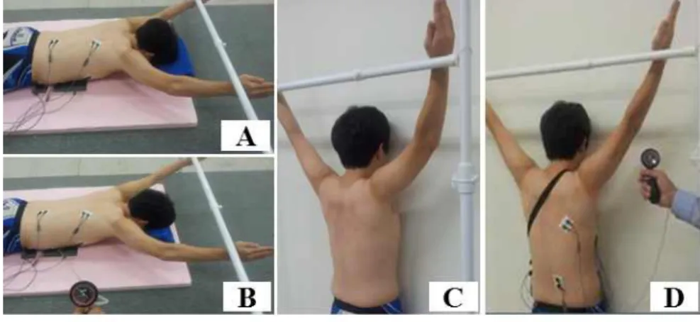
Figure 1. Arm lift in each position with or without abdominal drawing-in maneuver (ADIM) (A: prone without ADIM, B: prone with ADIM, C: standing without ADIM, D: standing with ADIM).
parallel to the spine, approximately 2 ㎝ lateral to the spinous process of L3) (Cram et al, 1998). The sampling rate was 1000 ㎐. A bandpass filter be- tween 20 and 300 ㎐ was used. EMG data were processed into the root mean square (RMS) value, which was calculated from 50 ㎳ window data points.
Procedures
1) Preferred arm lift
The preferred arm lift was performed first. The principal investigator explained the procedure to the subjects in detail prior to the experiment. The sub- ject was asked to lift both arms in prone and stand- ing positions in random order. A computer-generated random number was used to eliminate the effects of the order and training between the prone and stand- ing arm lifts.
For the prone arm lift, the subject was placed in the prone position. Using a goniometer, the humerus was aligned diagonally overhead with a shoulder ab- duction of 145°; the forearm was in a neutral position. The subjects were asked to keep the fore- head on the folded towel (3 ㎝ in height) to stabilize the neck and thoracic spine. The subject was then instructed to lift both arms with elbows extended until the radial border of the wrist slightly touched
the target bar without pushing it while maintaining the arm position. The distance to the target bar was determined by extending a vertical line from the floor to the subject’s earlobe in the prone position (Figure 1).
For the standing arm lift, the subject was required to stand facing the wall, touching from the nose to the knees with the feet shoulder-width apart. In the starting position, the ulnar border of the forearms and medial side of the humerus were in contact with the wall, and the shoulder abducted 90° with elbows flexed at 90°. The subjects were instructed to slide their arms up the wall. The sliding movement ended when the shoulder reached 145° of abduction. The subject was then instructed to lift both hands with elbows extended until the radial border of the wrist slightly touched the target bar without pushing it while maintaining the arm position. Target bar was place placed at the same distance in arm lift in prone (Figure 1).
2) ADIM familiarization3)
After the collection of data on the preferred arm lift in prone and standing positions, the subjects were familiarized with the use of the pressure bio- feedback unit2). With the subject in the prone posi- tion, a pressure biofeedback unit was placed between the pad of the therapeutic table and the subject's
2) Stabilizer, Chattanooga group Inc., Hixson, U.S.A.
lower abdomen to monitor abdominal muscle action.
The elastic bag of the pressure biofeedback unit was inflated to 70 ㎜Hg, and the subject was instructed to draw in the abdomen and hold this position. The subject was asked to maintain a pressure of 60 ㎜Hg by visual feedback from an analog pressure gauge during hip extension (Richardson and Jull, 1995).
Data within pressure changes of 65 ㎜Hg were used for the statistical analysis. In the standing position, the familiarization procedure was identical to the prone position, except that in this condition, a pres- sure biofeedback unit was placed between the wall and the subject’s lower abdomen, and the inelastic bag was inflated to 40 ㎜Hg.
3) Arm lift with ADIM
The order of arm lifts with ADIM in prone and standing positions was randomized in the same man- ner as in preferred arm lift. For the arm lift in the prone position, the pressure biofeedback unit was placed under the abdomen. For arm lifts against the wall in the standing position, the pressure biofeed- back unit was placed on an adjusted point of the subject's abdominal height. A different tester moni- tored the gauge of the pressure biofeedback unit and gave verbal cues to the subject in order to maintain the performance quality of ADIM during the arm lift with ADIM against the wall in the standing position.
Data collection
The EMG data were normalized by calculating the mean RMS of three trials of maximal voluntary iso- metric contraction (MVIC) for each muscle. We used the manual muscle testing positions recommended by Kendall et al (2005) for measuring MVIC. LT muscle activity was tested in the prone position; both arms were placed diagonally overhead in line with the lower fibers of the trapezius muscle during external rotation while resistance was applied distal to the elbow. The SA muscle was tested while the subject was seated on a treatment table with no back support. The shoulder was internally rotated and ab-
ducted to 125° in the scapular plane while resistance was applied proximal to the subject’s elbow by the principal investigator. Each contraction was held for 6 sec with maximal effort against manual resistance.
The first and last second of the EMG data from each MVIC trial were discarded, and the remaining 4 sec of data were used (De Oliveira et al 2008;
Vezina and Hubley-Kozey, 2000). Three repetitions of each test were performed, with a 2 min rest interval between repetitions to minimize muscle fatigue (Vera-Garcia et al, 2010). The mean MVIC value of the three trials was calculated. Each isometric arm-lifting exercise was performed for 6 sec; the first and last second of each exercise trial were dis- carded, and the remaining 4 sec of EMG data were used (De Oliveira et al, 2008; Vezina and Hubley-Kozey, 2000). The mean of three trials for each arm-lifting exercise was analyzed. The partic- ipants were allowed to rest for 2 min between trials and for 3 min between the different exercise posi- tions (De Mey et al, 2009; Lehman et al, 2006). The data for each trial were expressed as a percentage of the calculated mean RMS of the MVIC (%MVIC).
The mean %MVIC of the three trials was used in the analysis.
Statistical Analysis
A two-way (2 by 2) repeated analysis of variance (ANOVA) was used with a significance level of .05.
If a significant interaction was found, a paired t-test was performed with a Bonferroni adjustment (.05/4=.013). SPSS (Statistical Package for the Social Sciences) ver. 12.0 was used for the statistical analysis.
Results
The descriptive statistics of mean %MVIC in each exercise condition are presented in Table 2. In LT, no significant ADIM by position interaction was found (F1,19=.356, p=.558). There was a significant
Variables Exercise conditions compared Difference (95% CI) p
%MVICa of SAb
ADIMd prone vs. ADIM standing 8.2(6.2, 10.1) <.001 noADIMe prone vs. noADIM standing 4.7(3.3, 6.2) <.001 ADIM prone vs. noADIM prone -7.7(-11.6, -3.8) <.001 ADIM standing vs. noADIM standing -11.1(-14.6, -7.7) <.001
%MVIC of ESc
ADIM prone vs. ADIM standing 9.8(7.4, 12.2) <.001 noADIM prone vs. noADIM standing 27.9(24.9, 30.8) <.001 ADIM prone vs. noADIM prone -14.5(-17.5, -11.5) <.001 ADIM standing vs. noADIM standing 3.6(2.1, 5.0) <.001
apercentage of reference voluntary contraction, bserratus anterior, cerector spinae, dabdominal drawing-in maneuver
econdition of do not abdominal drawing-in maneuver.
Table 3. Post hoc comparisons of the differences in the muscle activity of the SA and ES
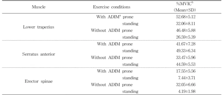
Muscle Exercise conditions %MVICb
(Mean±SD) Lower trapezius
With ADIMa prone 52.68±5.12
standing 32.06±8.11
Without ADIM prone 46.48±5.88
standing 26.59±5.39
Serratus anterior
With ADIM prone 41.67±7.28
standing 49.33±6.34
Without ADIM prone 33.47±5.96
standing 44.59±5.53
Erector spinae
With ADIM prone 17.55±5.56
standing 7.44±3.71
Without ADIM prone 32.05±6.66
standing 4.19±1.98
aabdominal drawing-in maneuver, bpercentage of reference voluntary contraction.
Table 2. Descriptive statistics of mean %MVIC in each exercise conditions
main effect for ADIM. LT muscle activity with ADIM was significantly greater compared with mus- cle activity without ADIM (F1,19=82.863, p<.001).
There was also a significant main effect according to position. LT muscle activity in the prone position was greater than muscle activity in the standing po- sition (F1,19=116.401, p<.001). In SA, a significant ADIM by position interaction was found (F1,19=8.687, p=.008). There were significant differences in all pairwise comparisons (Table 3). The greatest SA muscle activity was observed in the standing posi- tion with ADIM. In ES, significant ADIM by position interaction was found (F1,19=122.473, p<.001). There
were significant differences in all pairwise compar- isons (Table 3). The lowest ES muscle activity was elicited in the standing position with ADIM.
Discussion
This present study was undertaken to compare the effect of ADIM on trunk muscle activity during arm lifts in prone and standing positions. To our knowl- edge, this is the first study to investigate and com- pare trunk and abdominal muscle activity during prone and standing arm lifts. The findings of this
study supported our research hypothesis that ADIM increases LT and SA muscle activity and decreases ES activity during arm lift, and LT muscle activity increases in the prone position, whereas in the standing position, SA activity increases and ES ac- tivity decreases.
In LT, no significant ADIM by position interaction was found. There was a significant main effect for ADIM. LT muscle activity with ADIM was sig- nificantly greater than muscle activity without ADIM.
There was also a significant main effect for position.
LT muscle activity in the prone position was greater than muscle activity in the standing position. The findings of this study are in agreement with a pre- vious study that reported increased LT activation during arm lifts in prone and backward rocking quadruped positions compared with arm lifts in the standing position (Ha et al, 2012). The LT muscle tilts the scapular backward and assists complete shoulder flexion in the terminal phase. When the subject lifts the arm in the prone position, the arm reaches out from the proximal trunk; hence, this po- sition could be affected by gravity more than by the standing position.
In SA, significant ADIM by position interaction was found. There were significant differences in all pairwise comparisons. The greatest SA muscle activ- ity was observed in the standing position with ADIM. This result could be because the flexion tor- que in the axial skeleton could be increased by the muscular action of the transvers abdominis (Neumann, 2010), and the flexion momentum in the standing position might be greater than that in the prone position. This explanation should be supported by supplementary data on other kinetic and kine- matic activity. However, the result of a previous study might support that there was no significant difference in the muscle activity of SA between the arm lift against the wall in the standing position and the prone position. Moreover, the muscle activity in the wall standing position was greater than that in the prone position (Ha et al, 2012).
Increased SA and LT activity with ADIM can be also explained by enhanced lumbopelvic stability.
Because arm lifts can be a destabilizing activity, en- hanced lumbopelvic stability in the ADIM condition may have provided a stable base whereby the proper upward rotation of the scapula was possible and the SA and LT were activated.
In ES, significant ADIM by position interaction was found. There were significant differences in all pairwise comparisons. The lowest ES muscle activity was elicited in the standing position with ADIM.
Reduced ES activity during arm lifts in the standing position compared with arm lifts in the prone posi- tion could be explained by different degrees of anti- gravity arm lift movement between the standing and prone positions. Obviously, the arm lift in the prone position involves more antigravity movement than in the arm lift in the standing position, thus requiring further ES muscle recruitment. The greater ES mus- cle activity with ADIM can be explained by the ef- fect of lumbopelvic stability, which was ensured by the use of the pressure biofeedback unit. The results of previous studies also support our positive effect of lumbosacral stabilization in which core stability eli- cited the muscle activity of the primary mover and decreased that of erector spinae (Cynn et al, 2006;
Park et al, 2011). By applying ADIM, deep segmental stabilizers of the abdominal muscles, transversus ab- dominis and internal oblique, and the multifidus muscle of the back extensors may have been acti- vated; thus, ES activity was decreased by inhibition of extrinsic muscular stabilizer and increased by multifidus activation as a role of intrinsic muscular stabilizer (Neumann, 2010). Clinically excessive ES activation is associated with excessive lumbar lordo- sis, creating microtrauma and pain (Holmstrom et al, 1992; Kuramoto et al, 2011). In particular, arm lifts with increased lumbar lordosis can decrease the movements of the scapular and shoulder joints while overusing lumbar extension. The results of our study showed that by using ADIM, this unwanted compen- sation could be successfully prevented by favoring
the use of ADIM during arm lift exercises. This is the first study that reported the effect of ADIM on trunk muscle activity during arm lifts in prone and standing positions. Hence, these result of favorable combination with ADIM and position could provide effective methods to increase strength of targeted LT and SA muscles during arm lift exercise for the pa- tients with limited motion of shoulder joint and low back pain.
This study has several limitations. First, kinematic data for scapular upward rotation, posterior tilt, and adduction would have indicated the degree of optimal scapular movement during arm lift. Second, even though authors in this study attempted to control head movements and eye gaze, the influence of sub- tle head and eye movements might have affected arm and trunk movements. Third, since this is a cross-sectional study, the training effect of ADIM during arm lifts in prone and standing position can- not be ascertained from the results of this study.
Thus, a further study is required to examine the long-term effects of ADIM by recruiting a patient population for generalization.
Conclusion
This study compared trunk muscle activity during arm lifts in prone and standing positions with and without ADIM in healthy subjects using surface EMG. LT muscle activity with ADIM was sig- nificantly greater than muscle activity without ADIM, and LT muscle activity in the prone position was greater than muscle activity in the standing position.
The greatest SA muscle activity was observed in the standing position with ADIM. The lowest ES muscle activity was elicited in the standing position with ADIM. Therefore, these findings indicate that ADIM can effectively increase LT muscle activity in the prone position and that arm lifts in the standing position with ADIM offer the most favorable combi- nation for reducing ES muscle activity and increas-
ing SA muscle activity.
References
Aota Y, Saito T, Uesugi M, et al. Optimal arm posi- tion for evaluation of spinal sagittal balance. J Spinal Disord Tech. 2011;24(2):105-109.
Arlotta M, LoVasco G, McLean L. Selective recruit- ment of the lower fibers of the trapezius muscle.
J Electromyogr Kinesiol. 2011;21:403-410.
Cram JR, Kasman GS, Holtz J. Introduction to Surface Electromyography. Gaithersburg, MD, Aspen Publishers Inc, 1998:277-282.
Cynn HS, Oh JS, Kwon OY, Yi CH. Effects of lum- bar stabilization using a pressure biofeedback unit on muscle activity and lateral pelvic tilt during hip abduction in sidelying. Arch Phys Med Rehabil. 2006;87(11):1454-1458.
De Mey K, Cagnie B, Danneels LA, et al. Trapezius muscle timing during selected shoulder re- habilitation exercises. J Orthop Sports Phys Ther. 2009;39:743-752.
De Oliveira AS, De Morais Carvalho M, De Brum DP. Activation of the shoulder and arm muscles during axial load exercises on a stable base of support and on a medicine ball. J Electromyogr Kinesiol. 2008;18:472-479.
Ebaugh DD, McClure PW, Karduna AR. Three-di- mensional scapulothoracic motion during active and passive arm elevation. Clin Biomech (Bristol, Avon). 2005;20(7):700-709.
Ekstrom RA, Donatelli RA, Soderberg GL. Surface electromyographic analysis of exercises for the trapezius and serratus anterior muscles. J Orthop Sports Phys Ther. 2003;33:247-258.
Gill PK, Callaghan JM. The measurement of lumbar propriception in individuals with and without low back pain. Spine (Phila Pa 1976).
1998;23(3):371-377.
Ha SM, Kwon OY, Cynn HS, et al. Comparison of electromyographic activity of the lower trapezius
This article was received September 19, 2012, was reviewed September 20, 2012, and was accepted October 6, 2012.
and serratus anterior muscle in different arm-lifting scapular posterior tilt exercises. Phys Ther Sport. 2012;13(4):227-232.
Hardwick DH, Beebe JA, McDonnell MK, et al. A comparison of serratus anterior muscle activa- tion during a wall slide exercise and other tra- ditional exercises. J Orthop Sports Phys Ther.
2006;36:903-910.
Hodges PW, Richardson CA. Inefficient muscular stabilization of the lumbar spine associated with low back pain. A motor control evaluation of transversus abdominis. Spine (Phila Pa 1976).
1996;21(22):2640-2650.
Holmstrom E, Moritz U, Andersson M. Trunk muscle strength and back muscle endurance in con- struction workers with and without low back disorders. Scand J Rehabil Med. 1992;24:3-10.
Jull GA, Richardson C, Toppenberg R, et al.
Towards a measurement of active muscle con- trol for lumbar stabilization. Aust J Physiother.
1993;39(3):187-193.
Kendall FP, McCreary EK, Provance PG, et al.
Muscles: Testing and Function with Posture and Pain. 5th ed. Baltimore, Lippincott Williams &
Wilkins, 2005:330-333.
Kuramoto A, Chang L, Graham J, et al. Lumbar spi- nal stenosis with exacerbation of back pain with extension: A potential contraindication for supine MRI with sedation. J Neuroimaging. 2011;21(1):
92-94.
Lehman GJ, MacMillan B, MacIntyre I, et al.
Shoulder muscle EMG activity during push up variations on and off a Swiss ball. Dyn Med.
2006;9:7.
Marks M, Stanford C, Newton P. Which lateral ra- diographic positioning technique provides the most reliable and functional representation of a patient's sagittal balance? Spine (Phila Pa 1976).
2009;34(9):949-954.
Neumann DA. Kinesiology of the Musculoskeletal System: Foundations for rehabilitation. 2nd ed.
St. Louis, Mosby, 2010;383-395.
Page P, Franck C, Lardner R. Assessment and Treatment of Muscle Imbalance: The Janda approach. Champaign, IL., Humen Kinetics.
2010:43-55.
Park KN, Cynn HS, Kwon OY, et al. Effects of the abdominal drawing-in maneuver on muscle ac- tivity, pelvic motions, and knee flexion during active prone knee flexion in patients with lum- bar extension rotation syndrome. Arch Phys Med Rehabil. 2011;92(9):1477-1483.
Richardson CA, Jull GA. Muscle control-pain control.
What exercises would you prescribe? Man Ther.
1995;1:2-10.
Sahrmann SA. Movement System Impairment Syndromes of the Extremities, Cervical and Thoracic Spines. St. Louis, MO, Mosby, 2011:392-395.
Urquhart DM, Hodges PW. Differential activity of regions of transversus abdominis during trunk rotation. Eur Spine J. 2005;14(4):393-400.
Vedantam R, Lenke LG, Bridwell KH, et al. The ef- fect of variation in arm position on sagittal spi- nal alignment. Spine (Phila Pa 1976).
2000;25(17):2204-2209.
Vera-Garcia FJ, Moreside JM, McGill SM. MVC techniques to normalize trunk muscle EMG in healthy women. J Electromyogr Kinesiol.
2010;20:10-16.
Vezina MJ, Hubley-Kozey CL. Muscle activation in therapeutic exercises to improve trunk stability.
Arch Phys Med Rehabil. 2000;81:1370-1979.
von Garnier K, Köveker K, Rackwitz B, et al.
Reliability of a test measuring transversus ab- dominis muscle recruitment with a pressure bio- feedback unit. Physiotherapy. 2009;95(1):8-14.