ISSN: 2233-601X (Print) ISSN: 2093-6516 (Online)
− 126 −
Received: July 13, 2016, Revised: December 3, 2016, Accepted: December 13, 2016, Published online: April 5, 2017
Corresponding author: Jhingook Kim, Department of Thoracic and Cardiovascular Surgery, Samsung Medical Center, Sungkyunkwan University School of Medicine, 81 Irwon-ro, Gangnam-gu, Seoul 06351, Korea
(Tel) 82-2-3410-3483 (Fax) 82-2-3410-6986 (E-mail) [email protected]
© The Korean Society for Thoracic and Cardiovascular Surgery. 2017. All right reserved.
This is an open access article distributed under the terms of the Creative Commons Attribution Non-Commercial License (http://creativecommons.org/
licenses/by-nc/4.0) which permits unrestricted non-commercial use, distribution, and reproduction in any medium, provided the original work is properly cited.
Circulating Aneuploid Cells Detected in the Blood of Patients with Infectious Lung Diseases
Hongsun Kim, M.D. 1 , Jong Ho Cho, M.D., Ph.D. 1 , Chung-Hee Sonn, Ph.D. 2 , Jae-Won Kim, M.S. 2 , Yul Choi, P.N. 1 , Jinseon Lee, Ph.D. 2 , Jhingook Kim, M.D., Ph.D. 1,2
1
Department of Thoracic and Cardiovascular Surgery and
2Samsung Biomedical Research Institute, Samsung Medical Center, Sungkyunkwan University School of Medicine
The identification of circulating tumor cells (CTCs) is clinically important for diagnosing cancer. We have pre- viously developed a size-based filtration platform followed by epithelial cell adhesion molecule immuno- fluorescence staining for detecting CTCs. To characterize CTCs independently of cell surface protein ex- pression, we incorporated a chromosomal fluorescence in situ hybridization (FISH) assay to detect abnormal copy numbers of chromosomes in cells collected from peripheral blood samples by the size-based filtration platform. Aneuploid cells were detected in the peripheral blood of patients with lung cancer. Unexpectedly, aneuploid cells were also detected in the control group, which consisted of peripheral blood samples from patients with benign lung diseases, such as empyema necessitatis and non-tuberculous mycobacterial lung disease. These findings suggest that chromosomal abnormalities are observed not only in tumor cells, but al- so in benign infectious diseases. Thus, our findings present new considerations and bring into light the pos- sibility of false positives when using FISH for cancer diagnosis.
Key words: 1. Circulating neoplastic cells 2. Fluorescent in situ hybridization 3. Lung neoplasms
Case report
The identification and characterization of circulat- ing tumor cells (CTCs) in the blood of cancer pa- tients can help to diagnose cancer more accurately.
Epithelial cell adhesion molecule (EpCAM)-based plat- forms are usually used to capture and identify CTCs [1-3]. CTCs can be successfully characterized by fluo- rescence in situ hybridization (FISH) following en- richment using the EpCAM-positive selection plat- forms [4]. However, as CTCs undergoing epithelial- mesenchymal transition frequently lose the expres- sion of EpCAM, technologies targeting only EpCAM-
positive cells may underestimate the actual number of CTCs [5,6]. Once CTCs are enumerated, they have to be properly characterized to ensure the sensitivity and reliability of the CTC enumeration platform used.
We have previously developed a highly sensitive method for detecting CTCs that employs size-based filtration and EpCAM immunofluorescence staining [7]. CTCs of epithelial origin were distinguished from blood cells as all 4',6-diamidino-2-phenylindole (DAPI)- positive, leukocyte common antigen (CD45)-negative, and EpCAM-positive cells. To characterize CTCs in- dependently of cell surface protein expression, we in- corporated a chromosomal FISH assay to detect ab-
Korean J Thorac Cardiovasc Surg 2017;50:126-129 □ CASE REPORT □
https://doi.org/10.5090/kjtcs.2017.50.2.126
Pitfalls of FISH for Cancer Diagnosis
− 127 −
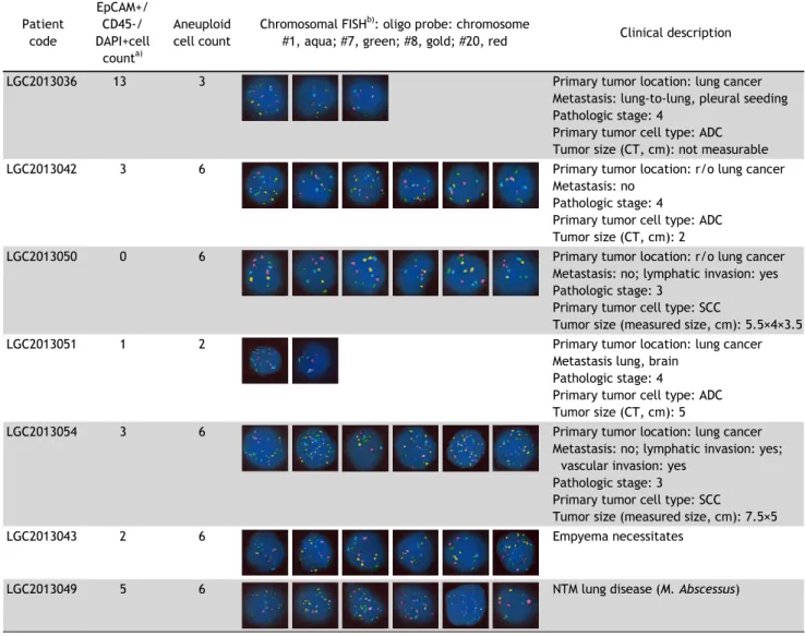
Table 1. Detection of aneuploid cells in the blood of patients with lung diseases
Patient code
EpCAM+/
CD45-/
DAPI+cell count
a)Aneuploid cell count
Chromosomal FISH
b): oligo probe: chromosome
#1, aqua; #7, green; #8, gold; #20, red Clinical description
LGC2013036 13 3 Primary tumor location: lung cancer
Metastasis: lung-to-lung, pleural seeding Pathologic stage: 4
Primary tumor cell type: ADC Tumor size (CT, cm): not measurable
LGC2013042 3 6 Primary tumor location: r/o lung cancer
Metastasis: no Pathologic stage: 4
Primary tumor cell type: ADC Tumor size (CT, cm): 2
LGC2013050 0 6 Primary tumor location: r/o lung cancer
Metastasis: no; lymphatic invasion: yes Pathologic stage: 3
Primary tumor cell type: SCC
Tumor size (measured size, cm): 5.5×4×3.5
LGC2013051 1 2 Primary tumor location: lung cancer
Metastasis lung, brain Pathologic stage: 4
Primary tumor cell type: ADC Tumor size (CT, cm): 5
LGC2013054 3 6 Primary tumor location: lung cancer
Metastasis: no; lymphatic invasion: yes;
vascular invasion: yes Pathologic stage: 3
Primary tumor cell type: SCC
Tumor size (measured size, cm): 7.5×5
LGC2013043 2 6 Empyema necessitates
LGC2013049 5 6 NTM lung disease (M. Abscessus)
ADC, adenocarcinoma; CT, computed tomography; SCC, squamous cell carcinoma; NTM, non-tuberculous mycobacteria; CD45, leukocyte common antigen; mAb, monoclonal antibody; EpCAM, epithelial cell adhesion molecule; DAPI, 4',6-diamidino-2-phenylindole; FISH, fluorescence in situ hybridization.
a)